

Hepatobiliary System
Liver
Narrative
The histomorphology of the normal liver reflects its many physiological functions. Residual hematopoiesis may be present in the liver of young rodents, and hepatodiaphragmatic nodules protruding into the diaphragm may occur during liver growth early in development. Documenting these changes may be important in studies that include dosing during gestation and early development. Varying degrees of glycogen storage within hepatocytes will be present in histological sections obtained at different times throughout the day (for examples, see Liver, Hepatocyte – Glycogen Accumulation). As rats and mice age, spontaneous occurrence of foci of cellular alteration (see Liver – Focus) and increased nuclear ploidy (see Liver, Hepatocyte – Karyomegaly) are normal aging changes. Since some of these changes may be exacerbated by treatment, resulting in increased multiplicity or decreased latency, they are usually diagnosed when above a threshold level or when there is an apparent treatment-related increase or decrease in their occurrence.
Unique artifacts specific to the liver are rare. Figure 1 and Figure 2 represent examples of the impression of a tissue cassette that squeezed down on the unfixed liver, leaving the cassette impression on the natural surface of the liver that became permanent during fixation. Figure 3 is a prominent example of postmortem vacuolation that can occur in rats that are not completely exsanguinated or are anoxic at necropsy (for details, see Li et al. Toxicol Pathol 2003;31:682–688; Sykes et al. Toxicol Appl Pharmacol 1976;36:31-39). Figure 4 and Figure 5 are examples of hematoxylin crystallizing out within hepatocytes during routine staining with unfiltered hematoxylin staining solution.
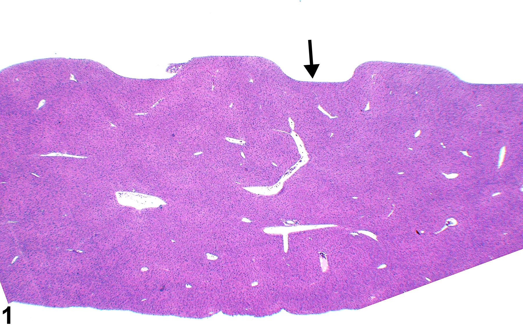
Figure 1. Artifact showing impression of tissue cassette on natural surface of liver (arrow).
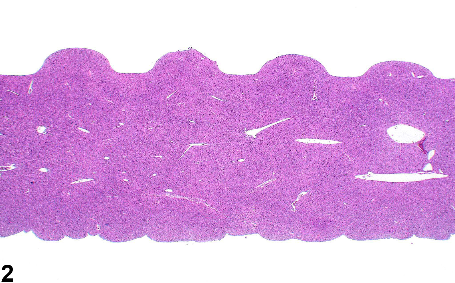
Figure 2. Artifact showing impression of tissue cassette on natural surfaces. The impression is more prominent on the top surface while a more subtle effect is present on the bottom surface of the liver.
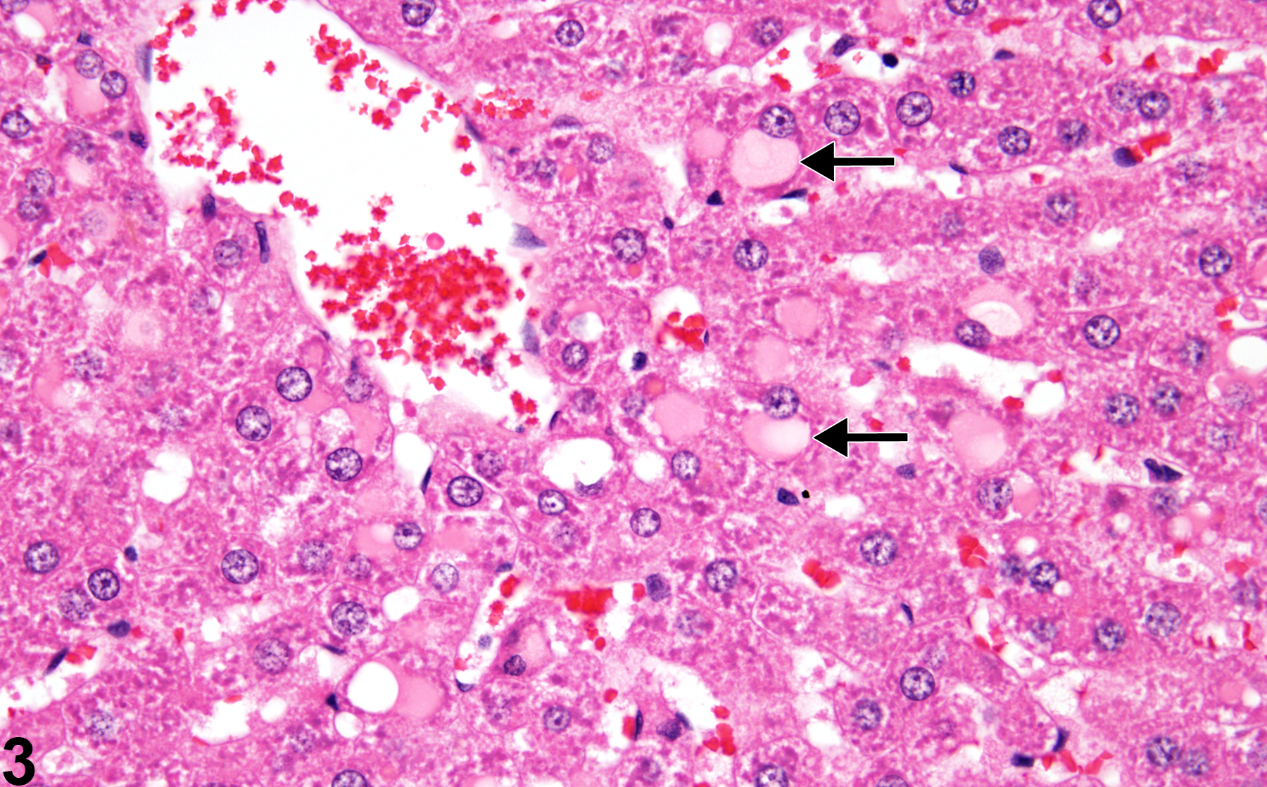
Figure 3. Postmortem occurring cytoplasmic vacuoles containing eosinophilic protein material (arrows) sometimes referred to as "plasma influx."
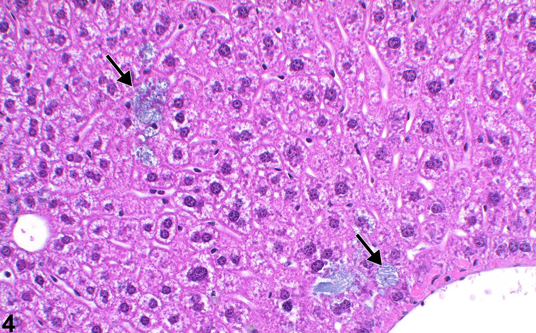
Figure 4. Hematoxylin crystals deposited within hepatocytes (arrows) during staining with unfiltered hematoxylin staining solution.
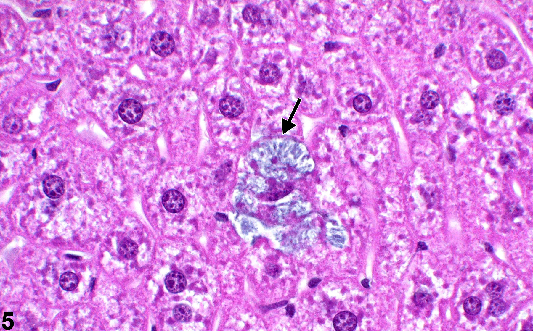
Figure 5. Hematoxylin crystals deposited within hepatocytes (arrow) during staining with unfiltered hematoxylin staining solution.