

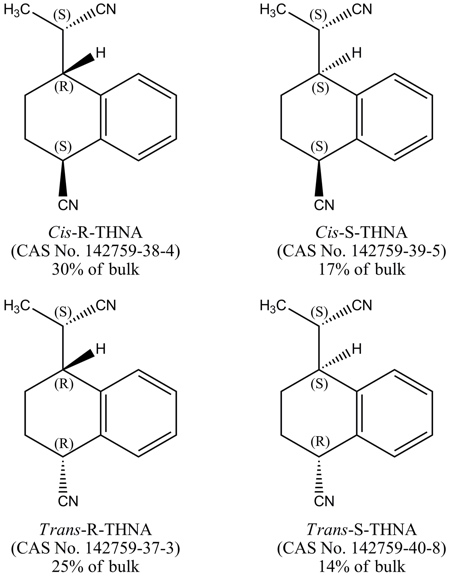
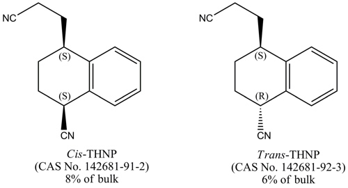
Styrene-acrylonitrile trimer (SAN Trimer) is a mixture of isomers formed by the condensation of two moles of acrylonitrile and one mole of styrene and has a molecular weight of 210. The mixture is composed of two structural forms: 4-cyano-1,2,3,4-tetrahydro-a-methyl-1-naphthaleneacetonitrile (THNA, CAS No. 57964-39-3) and 4-cyano-1,2,3,4-tetrahydro-1-naphthalenepropionitrile (THNP, CAS No. 57964-40-6).
The THNA form consists of four stereoisomers
The THNP form consists of two stereoisomers
SAN Trimer is a by-product of the production of acrylonitrile styrene plastics and is created in specific manufacturing processes for polymers of acrylonitrile and styrene. In June 1998, due to community concerns about the toxicity of SAN Trimer, it was nominated to the NTP for carcinogenicity testing by a member of Congress.
Male and female F344/N rats were exposed to SAN Trimer in feed in perinatal and postnatal studies for 7 weeks, 18 weeks, or 2 years. Genetic toxicology studies were conducted in Salmonella typhimurium and Escherichia coli, and in rat reticulocytes, leukocytes, liver cells, and brain cells. In vivo comet and micronucleus assays were performed in the juvenile rats.
Seven-week study in rats
Groups of 10 male and 10 female rats were fed diets containing 0, 250, 500, 1,000, 2,000, or 4,000 ppm SAN Trimer (equivalent to average daily doses of approximately 50, 90, 175, 270, or 410 mg SAN Trimer/kg body weight to males and 45, 90, 185, 295, or 430 mg/kg to females) for 2 weeks postweaning; the dams of these rats were fed the same concentrations of SAN Trimer from gestation day 7 until the pups were weaned. One 4,000 ppm male rat died 3 days after weaning; all other rats that started the postweaning phase survived to the end of the study. Mean body weights of 1,000, 2,000, and 4,000 ppm males and 2,000 and 4,000 ppm females were significantly less than those of the controls; weaning mean body weights were reduced in 4,000 ppm males and females and in 2,000 ppm females. Feed consumption by 2,000 and 4,000 ppm males and females was less than that by the control groups. Thinness in 4,000 ppm male rats was the only clinical finding related to SAN Trimer exposure. Nonneoplastic lesions were observed in the brain, thymus, spleen, liver, kidney, and reproductive organs of males and females and were considered due to overt toxicity.
Eighteen-week study in rats
Groups of 10 male and 10 female rats were fed diets of 0, 100, 200, 400, 800, or 1,600 ppm SAN Trimer (equivalent to average daily doses of 10, 20, 40, 80, or 150 mg/kg to males and females) for 3 months postweaning; the dams of these rats were fed the same concentrations from gestation day 7 until the pups were weaned. All rats survived to the end of the study. Mean body weights of 1,600 ppm males and females exposed to 200 ppm or greater were significantly less than those of the controls. At termination, brown staining of the urogenital fur was observed in females exposed to 200 ppm or greater. The liver weights of all exposed groups of males and the spleen weights of 800 and 1,600 ppm males and 1,600 ppm females were significantly greater than those of the controls. There were no significant differences in sperm parameters of male rats or the estrous cyclicity of female rats administered 400, 800, or 1,600 ppm in the diet when compared to the control groups. No exposure-related histopathologic lesions were observed.
Two-year study in rats
Groups of 50 male and 50 female core study rats were fed diets of 0, 400, 800, or 1,600 ppm SAN Trimer (equivalent to average daily doses of approximately 20, 40, or 75 mg/kg to males and 20, 40, or 85 mg/kg to females) for 2 years. Special study groups of 20 males and 20 females were fed the same exposure concentrations and were evaluated at 27, 52, and 78 weeks for hematology and clinical chemistry or at 26, 51, and 77 weeks for urinalysis. The dams of core and special study rats were fed the same concentrations from gestation day 7 until the pups were weaned. Mean body weights of 1,600 ppm males were less than 90% of the controls after week 1; mean body weights of 800 and 1,600 ppm females were less than 90% of the controls after weeks 41 and 13, respectively. Feed consumption by exposed groups of males and females was generally similar to that by the control groups. Brown staining of the urogenital fur was observed in all exposed groups, and the number of animals affected increased with increasing exposure concentration.
Rare neoplasms were present in the central nervous system of male and female rats. In the original evaluation, the 800 and 1,600 ppm groups of male rats each had one astrocytoma and one granular cell tumor in the brain. Also in the brain, one 400 ppm female had a granular cell tumor and one control, one 400 ppm, and one 800 ppm female had a mixed cell glioma. In the spinal cord, one astrocytoma was noted in a 1,600 ppm male in the original evaluation. In the expanded review of the spinal cord, one granular cell tumor was found in a 400 ppm male and one meningioma was found in an 800 ppm female.
There were statistically significant increases in the incidence of spinal nerve root degeneration in 1,600 ppm males and the incidences of sciatic nerve degeneration in 800 and 1,600 ppm females. More importantly, there were increases in the severities of both nerve lesions in males and in the severity of spinal nerve root degeneration in females.
The incidences of bone marrow hyperplasia were significantly increased in 1,600 ppm males and females and 800 ppm females. Incidences of bone marrow granulomatous inflammation were increased in 1,600 ppm males and 800 and 1,600 ppm females, and the increase in the 800 ppm females was significant. Because this lesion is very rare and did not occur in control animals, it should be considered biologically significant. In the liver, the incidence of eosinophilic focus was significantly increased in 1,600 ppm males and the incidences of mixed cell focus were significantly increased in 400 and 1,600 ppm males. Incidences of mixed cell focus were increased in the liver of all exposed groups of females, and the increase was significant in the 1,600 ppm group. The incidence of transitional epithelial hyperplasia of the urinary bladder in 1,600 ppm females was significantly greater than that in the controls.
There were significant decreases in the incidences of pituitary gland pars distalis adenoma in 1,600 ppm males and females, and the incidences in both sexes occurred with negative trends. The incidences of mammary gland fibroadenoma occurred in females with a negative trend, and the incidences in 800 and 1,600 ppm females were significantly less than that in the control group. The incidences of mononuclear cell leukemia in all exposed groups of males and females were significantly less than those in the controls.
Genetic toxicology
SAN Trimer (Batch 3) was not mutagenic in Salmonella typhimurium strains TA98 or TA100 or in Escherichia coli strain WP2 uvrA/pKM101 in tests conducted with and without exogenous metabolic activation.
In vivo, however, results of a comet assay indicated significantly increased levels of DNA damage in brain cells of male and female juvenile rats following administration of SAN Trimer (Batch 3) by oral gavage. Dose-related increases in DNA damage in liver cells of these rats were also observed, but the increases were smaller than those observed in brain cells and were judged to be equivocal in both males and females. Indications of DNA damage following exposure to SAN Trimer were also seen in leukocytes of male and female rats. Increases in male rats were significant, but in females, observed levels of DNA damage did not correlate with dose. Therefore, the results were judged to be positive in males and equivocal in females. In addition to the positive comet assay results, significant increases in the frequencies of micronucleated reticulocytes were observed in peripheral blood of male and female juvenile rats dosed with SAN Trimer.
Conclusions
Under the conditions of this 2-year feed study preceded by perinatal exposure, there was no evidence of carcinogenic activity of SAN Trimer in male and female F344/N rats given feed containing 400, 800, or 1,600 ppm SAN Trimer.
Exposure to SAN Trimer resulted in increased incidences and/or severities of peripheral nerve degeneration in male and female F344/N rats, increased incidences of nonneoplastic lesions of the bone marrow and liver in male and female F344/N rats, and of nonneoplastic urinary bladder lesions in female F344/N rats.
The incidences of pituitary gland adenoma and mononuclear cell leukemia in male and female F344/N rats and mammary gland fibroadenoma in female F344/N rats were decreased.