Note on Accessibility: Persons using mobile devices may find some tables are not fully accessible. Note that you can view tables on a larger screen or in the PDF version of the report/monograph. If you need additional assistance, email us or use our contact form and identify the tables for which access is required. We will assist you in accessing the content. NIH has helpful information on accessibility.
Technical Report 605
NTP Technical Report on the Toxicology and Carcinogenesis Studies of Sulfolane (CASRN 126-33-0) Administered in Drinking Water to Sprague Dawley (Hsd:Sprague Dawley SD) Rats and B6C3F1/N Mice

Abstract
Sulfolane is commonly used as an industrial solvent in liquid-liquid and liquid-vapor extractions, in natural gas and petroleum refineries, and in the extraction of acidic components from sour gas streams. Sulfolane was detected in contaminated groundwater and drinking water wells near a petroleum refinery in North Pole, Alaska, and at additional sites in Canada and the United States. Because of concerns about potential long-term human exposure via contaminated drinking water and the lack of toxicity data, chronic exposure studies in rats and mice were conducted to evaluate the potential toxicity and carcinogenic activity of sulfolane.
Time-mated female Sprague Dawley (Hsd:Sprague Dawley SD) rats were exposed to sulfolane in drinking water at 0, 30, 100, 300, or 1,000 mg/L beginning at gestation day (GD) 6 through lactation. On postnatal day (PND) 28, offspring (50/sex/group) were continued on study and provided dosed water containing the same sulfolane concentration as their respective dam for 2 years. In B6C3F1/N mice, groups of 50 mice per sex, aged 6 weeks at study start, were provided dosed water containing 0, 30, 100, 300, or 1,000 mg/L sulfolane for 2 years. Interim evaluations of hematology, clinical chemistry (rats only), vaginal cytology, and internal concentration assessment were conducted at 3 months in rats and mice. At study termination, toxicity (e.g., survival, body weights) and the incidences of neoplasms and exposure-related histopathological changes were evaluated in rats and mice.
Two-year Study in Rats
Sulfolane exposure to dams had no toxicologically relevant effects on maternal measurements during gestation or lactation. Slight but significantly decreased (within 5%) body weights were observed in the 1,000 mg/L group during lactation but were not considered exposure related. Daily sulfolane intake during GD 6–21 was estimated to be 4, 12, 37, and 126 mg/kg/day for the 30, 100, 300, and 1,000 mg/L groups, respectively; during lactation days 1–14, intake was estimated to be 6, 20, 59, and 206 mg/kg/day for the 30, 100, 300, and 1,000 mg/L groups, respectively. There were no significant effects on littering parameters and offspring survival through lactation. Pup body weights were significantly decreased by up to 9% in males and 11% in females exposed to 1,000 mg/L sulfolane during lactation. In female offspring, vaginal opening was significantly delayed in the 300 and 1,000 mg/L groups, with and without adjustment for body weight at weaning.
Interim evaluation at 3 months showed marginal effects in male rats, with reduced body weights that were within 10% of control values. Clinical chemistry, hematology, and organ weights were unaffected by sulfolane exposure. Sulfolane plasma concentrations were consistent between the sexes and showed metabolic saturation at higher exposure concentrations.
Following exposure to sulfolane for 2 years, there were no significant differences in the survival of rats exposed to sulfolane. While body weights were significantly decreased in the 1,000 mg/L male and female groups throughout the 2-year exposure, the decrease was not significant at the end of the 2-year study in males. Water consumption in the sulfolane-exposed groups was comparable to that of the control groups throughout the study. Daily PND 28 to 119 sulfolane intake was estimated to be 3, 8, 25, and 80 mg/kg/day for males and was estimated to be 3, 11, 31, and 104 mg/kg/day for females in the 30, 100, 300, and 1,000 mg/L groups, respectively. For the remainder of the study (study day 21 to 616), sulfolane intake was estimated to be 1, 5, 13, and 49 mg/kg/day for males and 2, 8, 21, and 80 mg/kg/day for females in the 30, 100, 300, and 1,000 mg/L groups, respectively.
Histopathological evaluations identified significant increases in the incidence of mammary gland adenoma in females exposed to 100 mg/L sulfolane.
Two-year Study in Mice
In male and female mice exposed to sulfolane for 2 years, no significant changes were observed in survival or body weights, with intermittent changes in water consumption. Daily sulfolane intake from study day 0 to 707 was estimated to be 3, 9, 28, and 89 mg/kg/day for males in the 30, 100, 300, and 1,000 mg/L groups, respectively; for females, intake was estimated to be 2, 7, 20, and 65 mg/kg/day in the 30, 100, 300, and 1,000 mg/L groups, respectively. Interim evaluation at 3 months showed no exposure-related effects on body weights, hematology, or organ weights, and sulfolane plasma concentrations showed metabolic saturation in female mice. Histopathological evaluations identified positive trends and significant increases in the incidences of hemangiosarcomas of all organs (systemic) and of the liver of male mice exposed to 1,000 mg/L sulfolane. A significantly increased incidence of hemangiosarcoma was observed in the spleen of males exposed to 30 mg/L sulfolane. In female mice, a positive trend and a significant increase in the incidence of hepatocellular carcinoma were observed in the 1,000 mg/L group. Additionally, the incidence of mixed cell focus of the liver in males was significantly increased in the 30, 100, and 300 mg/L groups. In females, a positive trend in the incidence of clear cell focus of the liver was observed, and the incidence of focal fatty change of the liver was significantly increased in the 100 and 300 mg/L groups. The incidence of follicle cysts of the ovary was significantly increased in the 30, 300, and 1,000 mg/L groups.
Genetic Toxicology
In the in vivo rodent peripheral blood micronucleus assay, no increases in micronucleated erythrocytes were observed in male or female rats or mice administered sulfolane via dosed water for 3 months. A positive trend in the percentage of reticulocytes was observed in female rats, but the increase was within the historical control 95% confidence interval.
Conclusions
Under the conditions of these 2-year drinking water studies, there was no evidence of carcinogenic activity of sulfolane in male Hsd:Sprague Dawley SD rats at exposure concentrations of 30, 100, 300, or 1,000 mg/L. There was equivocal evidence of carcinogenic activity of sulfolane in female Hsd:Sprague Dawley SD rats based on the increased incidence of mammary gland adenoma.
There was clear evidence of carcinogenic activity of sulfolane in male B6C3F1/N mice based on the increased incidence of hemangiosarcoma (all organs), which predominately occurred in the liver. There was equivocal evidence of carcinogenic activity of sulfolane in female B6C3F1/N mice based on the increased incidence of hepatocellular carcinoma.
Exposure to sulfolane resulted in increased incidences of nonneoplastic lesions of the liver in male and female mice and the ovary in female mice. Sulfolane exposure also induced a delay in vaginal opening in female rats.
Synonyms: 2,3,4,5-tetrahydrothiophene-1,1-dioxide, tetramethylene sulfone, tetrahydrothiophene-1,1-dioxide
2,3,4,5-tetrahydrothiophene-1,1-dioxide, tetramethylene sulfone, tetrahydrothiophene-1,1-dioxide
Summary of the Perinatal and Two-year Carcinogenesis and Genetic Toxicology Studies of Sulfolane
| Male Sprague Dawley Rats | Female Sprague Dawley Rats | Male B6C3F1/N Mice | Female B6C3F1/N Mice | |
|---|---|---|---|---|
| Concentrations in drinking water | 0, 30, 100, 300, or 1,000 mg/L | 0, 30, 100, 300, or 1,000 mg/L | 0, 30, 100, 300, or 1,000 mg/L | 0, 30, 100, 300, or 1,000 mg/L |
| Survival rates | 16/50, 17/50, 15/50, 17/50, 10/50 | 21/50, 26/50, 26/50, 27/50, 19/50 | 34/50, 28/50, 35/50, 32/50, 30/50 | 35/50, 39/50, 34/50, 34/50, 37/50 |
| Body weights | F1 generation: Lactation: ↓ Study termination: no effect | F0 generation: Gestation: exposed groups within 10% of the control group Lactation: exposed groups within 10% of the control group F1 generation: Lactation: ↓ Study termination: ↓ (1,000 mg/L group 15% less than the control group) | No effect | No effect |
| Pubertal endpoints | – | Vaginal opening: delayed (300 mg/L and 1,000 mg/L) | – | – |
| Nonneoplastic effects | Nonea | None | Liver: mixed cell focus (6/50, 18/50, 15/50, 13/50, 7/50) | Liver: clear cell focus (4/50, 4/50, 6/50, 4/50, 10/50); fatty change, focal (1/50, 5/50, 8/50, 11/50, 2/50) Ovary: follicles, cyst (2/48, 9/46, 3/47, 11/48, 9/49) |
| Neoplastic effects | None | None | All organs (systemic): hemangiosarcoma (3/50, 6/50, 4/50, 3/50, 12/50) Liver: hemangiosarcoma (3/50, 5/50, 3/50, 2/50, 11/50) Spleen: hemangiosarcoma (0/49, 5/50, 1/50, 1/50, 3/50) | None |
| Equivocal findings | None | Mammary gland: adenoma (1/50, 4/50, 7/50, 2/50, 5/50) | None | Liver: hepatocellular carcinoma (2/50, 3/50, 5/50, 5/50, 8/50) |
| Level of evidence of carcinogenic activity | No evidence | Equivocal evidence | Clear evidence | Equivocal evidence |
| Genetic toxicology | ||||
| Micronucleated erythrocytes (in vivo) | ||||
| Rat peripheral blood: Negative in males and females exposed via dosed water for 3 months | ||||
| Mouse peripheral blood: Negative in males and females exposed via dosed water for 3 months | ||||
Introduction
Chemical and Physical Properties
Sulfolane is an organosulfur compound containing a sulfonyl group with a sulfur atom double-bonded to two oxygen atoms. The double bond is highly polar, whereas the carbon ring has high nonpolar stability. Sulfolane is a clear, colorless gel at ambient temperature (approximately 25°C) with a melting point in the range of 27.4°C to 27.8°C and a density of 1.261 g/cm3, liquid.6 Sulfolane is miscible in water, acetone, glycerol, and many oils. It is chemically stable in the presence of many chemical substances except sulfur and aluminum chloride.7 Sulfolane has a low vapor pressure (0.0062 mm Hg at 27.6°C).6
Production, Use, and Human Exposure
Sulfolane production occurs when sulfur dioxide and butadiene react together, forming sulfolene; sulfolene is then hydrogenated to form sulfolane.
Sulfolane is commonly used as an industrial solvent in liquid-liquid and liquid-vapor extractions, such as in the extraction of benzene, toluene, and xylene from mixtures with aliphatic hydrocarbons (e.g., in petroleum refining) and in the extraction of acidic components (e.g., hydrogen sulfide, carbon dioxide, carbonyl sulfide, carbon dioxide, mercaptans) from sour gas streams.8-10 It is also used in textile refinishing, in the fractionalization of wood tars, as a component of hydraulic fluid, and as a curing agent in epoxy resins.10 It is estimated that approximately 150 extraction units utilizing sulfolane are in use around the world, and sulfolane is considered a high production volume chemical in the United States, with an annual production of 10–50 million pounds.11
Release of sulfolane into the environment through various waste streams near refineries results in groundwater contamination. It is presumed that sulfolane does not break down easily in groundwater, likely because of low oxygen and nutrient levels, and while it does not accumulate in the aquatic food chain, it is taken up by plants.12,13 In North Pole, Alaska, contamination of groundwater from a nearby petroleum refinery led to detection of sulfolane in nearly 300 drinking water wells in the area since 2009, with measurements in 2015 ranging from 4 to 7 ppb in older supply wells14 and several measurements within 100–400 ppb in previous years.15,16 Sulfolane has been detected at additional sites in Canada and the United States near natural gas or petroleum refineries.17-20
Human exposure to sulfolane occurs through occupational (inhalation and/or dermal) and environmental (drinking water) sources; however, no exposure data are available.
Regulatory Status
Currently, no federal regulatory limits exist for sulfolane levels in drinking water. The Agency for Toxic Substances and Disease Registry’s (ATSDR’s) recommended public health action levels in drinking water are based on the Zhu research group identifying an oral no-observed-adverse-effect level (NOAEL) for guinea pigs of 0.25 mg sulfolane/kg body weight/day (mg/kg/day).21 Because of limited available research, ATSDR applied an uncertainty factor of 100 to the NOAEL of 0.25 mg/kg/day, resulting in a health guidance value dose of 0.0025 mg/kg/day (2.5 μg/kg/day). On the basis of average water intake, the public health action levels in drinking water for sulfolane are 20 ppb (infants), 32 ppb (children), and 70 ppb (adults).22 The U.S. Environmental Protection Agency (EPA) Provisional Peer-reviewed Toxicity Values for subchronic and chronic sulfolane exposure are 10 μg/kg/day and 1 μg/kg/day, respectively, and are based on decreased white blood cell (WBC) counts in female CD rats after a 90-day drinking water exposure.23
Absorption, Distribution, Metabolism, and Excretion
Experimental Animals
As part of the National Toxicology Program (NTP) research program on sulfolane, absorption, distribution, metabolism, and excretion studies, as well as toxicokinetic (TK) studies, were conducted following a single gavage administration or dermal application in male and female Sprague Dawley (Hsd:Sprague Dawley SD) rats and B6C3F1/N mice.2,3 Sulfolane was well absorbed following administration of 30, 100, or 300 mg/kg [2,5-14C]sulfolane in rats and mice and was excreted extensively in urine (rats, approximately 91%–93%; mice, approximately 64%–86%) with low tissue retention (<2%) at 48 hours following oral administration.2 Following dermal application of 100 mg/kg [2,5-14C]sulfolane to a covered application site, the absorption of sulfolane was lower in male and female rats (approximately 16%–19%) compared to that in male and female mice (approximately 70%–80%).2 There were no route, species, or sex differences in urinary radiochemical profiles, with sulfolane and 3-hydroxysulfolane analytes found to be the main analytes in urine.2 Other investigators have also reported 3-hydroxysulfolane as the primary metabolite of sulfolane in rats, mice, and rabbits.24 Following intraperitoneal (IP) injection of 100 mg of [35S]sulfolane/kg, 85% of the radioactivity excreted in the urine during the first 24 hours was associated with the metabolite 3-hydroxysulfolane.24
In studies investigating the TK behavior of sulfolane in male and female rats and mice, absorption of sulfolane was rapid following a single gavage administration of 10, 30, or 100 mg/kg of sulfolane, with the maximum plasma concentration (Cmax) reached at 1.47 hours in rats and at 0.55 hours in mice.3 In rats of both sexes, Cmax increased proportionally with the dose, whereas the increase in Cmax was greater than proportional in mice. The plasma elimination half-life increased with dose in rats (1.99–6.33 hours) and mice (0.299–1.25 hours), with shorter half-lives observed in mice compared with rats. Estimated oral bioavailability was higher in rats (81%–83%) than in mice (59%–63%) at 10 mg/kg; values were >100% at 30 and 100 mg/kg in both species and sexes, suggesting that saturation of the metabolism and clearance pathways of sulfolane may begin at doses of approximately 30 mg/kg.2
Humans
The literature contains no studies on the absorption, metabolism, distribution, or excretion of sulfolane in humans.
Toxicity
Experimental Animals
General Toxicity
In subchronic inhalation studies of aerosolized sulfolane (3% water) conducted in rats, guinea pigs, dogs, and squirrel monkeys, overt toxicity was observed in squirrel monkeys and dogs within the first 7 days of exposure to 200 mg/m3 sulfolane. Chronic lung inflammation was observed in all species following 27 days of exposure (8 hours/day, 5 days/week) to 495 mg/m3 sulfolane.25 At 60 days, the only remaining male dog had grossly elevated plasma aspartate aminotransferase, alanine aminotransferase, and lactate dehydrogenase activities. In guinea pigs, chronic pleuritis was noted in the lungs after 30 days of exposure to 200 mg/m3, and fatty vacuolization was observed in the liver of the 30-, 60-, and 90-day exposed groups. No signs of toxicity or effects on body weight, hematology, or pathology were observed following continuous exposure (85–110 days) to 2.8–159 mg/m3 sulfolane in rats, guinea pigs, monkeys, or dogs.25
In a 28-day study, male and female rats dosed with 700 mg/kg/day sulfolane via gavage experienced significantly decreased body weights and feed consumption, although the female rats recovered after the second week of exposure. In males, a higher incidence of hyaline droplet accumulation was observed in the kidneys. A significant decrease in erythrocyte count was noted in female rats after a 2-week recovery period, and significant decreases in spleen weights were also noted at 28 days and after a 2-week recovery period.26,27 An NTP 28-day toxicity study compared the effects of sulfolane in three species (rats, mice, and guinea pigs) to determine species sensitivity.4 Following gavage administration of 800 mg/kg/day sulfolane, male and female B6C3F1/N mice and Hartley guinea pigs were removed from the study because of overt toxicity, and a noted reduction in weight was observed in male and female Sprague Dawley rats.4 Additional findings following 28 days of exposure included nonneoplastic lesions in the kidney (male rats), forestomach and glandular stomach (male mice), esophagus (male and female guinea pigs), and nose (male guinea pigs), including hyaline droplet accumulation in the proximal tubules of the kidney (male rats).4
In a 90-day drinking water study of sulfolane (0, 25, 100, 400, or 1,600 mg/L) by Huntingdon Life Sciences (2001), a NOAEL of 25 mg/L (2.9 mg/kg/day) and a lowest-observed-adverse-effect level (LOAEL) of 100 mg/L (10.6 mg/kg/day) were reported for female CD rats and were based on significantly decreased WBC, lymphocytes, monocytes, and large unstained cells.28 Although few details are provided, a 90-day gavage study (0, 55.6, 167, and 500 mg/kg/day) in rats and guinea pigs reported clinical chemistry changes in addition to significantly decreased WBC in guinea pigs.21 In a second study in guinea pigs orally exposed for 6 months (0, 0.25, 2.5, 25, or 250 mg/kg/day), clinical pathology and bone marrow cell changes along with spleen white pulp shrinkage were observed at the 3-month interim, and lesions in the liver (fatty deposits) and spleen (white pulp shrinkage) were reported after 6 months of exposure.21
Neurotoxicity
Acute metabolic and neurotoxic effects have been reported for sulfolane, including changes in thermoregulation, motor activity, and brain-wave patterns in male Long Evans hooded rats.29,30 Following acute IP injection, dose-related inhibition of metabolic rate and hypothermia were first observed 60 minutes postinjection and lasted at least 2.5 hours in Sprague Dawley rats.31 In male CD-1 mice, IP injection of sulfolane at ≥400 mg/kg reduced the metabolic rate and body temperatures and induced behavioral changes including preference of ambient temperatures.32 Sulfolane has been reported as a convulsant in rats, mice, dogs, and squirrel monkeys.8,9 In a study exploring the median lethal dose (LD50) via different routes of exposure (i.e., oral and IP) in rats, mice, guinea pigs, and rabbits, no significant species differences in the LD50 of sulfolane were observed, and death was preceded by signs of central nervous system (CNS) stimulation, such as hyperactivity and convulsions, across species.8 Similar toxic signs of CNS stimulation in mice and rats were observed across different routes of exposure (i.e., intravenous and IP) including hunched, retreating posture with front limbs braced wide, erect tail, hyperactivity, increased responsiveness to auditory stimulation, and rapid respiration. At lethal doses, all species on study had clonic-tonic convulsions.8
Reproductive and Developmental Toxicity
A gavage reproductive/developmental toxicity screening test (OECD TG 421) was conducted at doses of 0, 60, 200, and 700 mg/kg/day.26,33 Male Sprague Dawley rats were dosed for 49 days starting 14 days prior to mating, and female rats were dosed for 41–50 days (starting 14 days prior to mating to day 3 of lactation). There was some mortality and significant decreases in body weight gain and feed consumption at 700 mg/kg/day along with disrupted estrous cyclicity. Four dams experienced whole litter loss at 700 mg/kg/day. Litter size, survival, and pup weight were significantly decreased postparturition. Litter size at birth and the number of pups and pup weight on days 0 and 4 of lactation were lower in the 200 mg/kg/day group. The parental NOAEL was 200 mg/kg/day, and the NOAEL for offspring was 60 mg/kg/day. In the above study, there were no dose-related findings in the external appearance, general conditions, and necropsy findings of the offspring. However, in another study, increased fetal resorptions and fetal skeletal anomalies were observed in Chinese Kunming mice following maternal oral exposure to 840 mg/kg/day.21
Immunotoxicity
In a comprehensive NTP immunotoxicology study, the potential effects of sulfolane on gross pathology, enhanced immunopathology, hematology, antibody response to sheep red blood cells, antibody response to keyhole limpet hemocyanin, T-cell proliferation, natural killer (NK) cell activity, immunophenotyping of the spleen, and cytotoxic T-lymphocyte response to influenza infection were examined.5 In this study, adult female B6C3F1/N mice were dosed with sulfolane via gavage (0, 1, 10, 30, 100, or 300 mg/kg/day) for 90 days, and time-mated female Sprague Dawley rats were exposed to sulfolane in drinking water (0, 30, 100, 300, or 1,000 mg/L) from gestational day 6 through approximately 13 weeks of age. In mice, no clinical indications of toxicity or effects on body weight were observed following sulfolane exposure, and minimal immunotoxic effects, including decreased splenic NK cell number at ≥100 mg/kg/day (ex vivo NK cell activity was not affected) and decreased number of large unstained cells at doses ≥30 mg/kg/day, were noted. Similarly, limited findings were observed following perinatal exposure in rats. No clinical indications of toxicity related to sulfolane were noted, and a slight decrease in body weight was observed in male and female F1 rats exposed to 1,000 mg/L (approximately 5% and 7%–10%, respectively). Sulfolane exposure had no significant immunotoxic effects on F1 males. In female F1 rats immunized with sheep red blood cells (SRBC), spleen cellularity was increased 14%–31% at concentrations of ≥100 mg/L sulfolane, and NK cell activity decreased in a concentration-dependent manner in cells from the sulfolane-exposed female F1 rats.5
Humans
The literature contains no studies on the possible health effects following sulfolane exposure in humans.
Carcinogenicity
Experimental Animals
Aside from the previously mentioned 6-month study in guinea pigs,21 there are no chronic or carcinogenicity studies of sulfolane in animal models. Structurally related 3-sulfolene was previously evaluated for carcinogenic activity in male and female Osborne-Mendel rats and B6C3F1 mice via gavage (corn oil vehicle) by the National Cancer Institute.34 There was no evidence of carcinogenic activity in both rats and mice; however, exposure-related significant decreases in survival were observed, resulting in dose adjustments throughout the study.
Humans
The literature contains no studies on the carcinogenicity of sulfolane in humans.
Genetic Toxicity
In 28-day gavage studies previously conducted as part of the NTP sulfolane research program, peripheral blood samples were obtained from male and female Sprague Dawley rats and B6C3F1/N mice and analyzed for the frequency of micronucleated reticulocytes and erythrocytes.4 In male and female rats, the reticulocyte population (polychromatic erythrocytes, or PCEs), which is the only red blood cell population that can be accurately assessed for micronucleus frequency in peripheral blood of rats because of efficient splenic scavenging of damaged erythrocytes, did not show an increase in micronuclei after 28 days of exposure to sulfolane via gavage (0, 1, 10, 30, 100, 300, or 800 mg/kg/day). Significant changes in the percentage of reticulocytes were observed in male and female rats; however, these changes were within the historical control 95% confidence interval. There were no increases in micronucleated reticulocytes or micronucleated erythrocytes in male and female mice after 28 days of exposure to sulfolane via gavage (0, 1, 10, 30, 100, or 300 mg/kg/day).
A few additional reports of the genetic toxicity of sulfolane are also available in the peer-reviewed literature; however, these reports lacked primary data and were inadequate for review.
Study Rationale
Sulfolane was nominated to NTP by the Alaska Department of Environmental Conservation with support from various entities in Alaska, as well as by ATSDR. The potential for human exposure to sulfolane through groundwater in North Pole, Alaska, and near other natural gas or petroleum refining sites has raised public health concerns for residents. Because long-term exposure to sulfolane has likely occurred and information on potential toxicity following long-term exposure is lacking, 2-year drinking water studies were conducted in rats and mice to evaluate the potential chronic toxicity and carcinogenic activity of sulfolane. Because of the likelihood of early life exposure to sulfolane in these communities, gestation and early postnatal exposure was included in the rat study to evaluate for potential effects of lifetime exposure.
Materials and Methods
Procurement and Characterization of Sulfolane
Sulfolane was obtained from Sigma-Aldrich (Milwaukee, WI) in a single lot (MKBN9784V). Identity, purity, and stability analyses were conducted by the analytical chemistry laboratory at RTI International (Research Triangle Park, NC) and the study laboratory at Battelle (Columbus, OH). Reports on analyses performed in support of the sulfolane studies are on file at the National Institute of Environmental Health Sciences (NIEHS).
Lot MKBN9784V was a solid to a gel at room temperature and formed a colorless liquid upon warming. The identity of the test lot was confirmed using infrared (IR) spectroscopy, 1H nuclear magnetic resonance (NMR) spectroscopy, 13C NMR spectroscopy, and ultra-high-performance liquid chromatography with high resolution mass spectrometry (MS). The IR, 1H NMR, and 13C NMR spectra cohered with the structure of sulfolane and were consistent with reference spectra from the Sigma-Aldrich library (IR and 1H NMR)35,36 and the Spectral Database for Organic Compounds (13C NMR).37 Elemental analyses were performed by Galbraith Laboratory (Knoxville, TN) to aid in identification. The relative amounts of carbon (39.51%), hydrogen (7.08%), and sulfur (26.34%) were within 6% of the theoretical values. An accurate determination of oxygen could not be made because of interference from the high elemental sulfur content. The boiling point (285°C) and density (1.31 g/cm3 at 21.9°C) measured by Galbraith Laboratory were consistent with literature values from the Hazardous Substance Data Bank.10
Purity evaluation was conducted using gas chromatography (GC) with flame ionization detection (FID) (Table A-1, System A) and GC with MS detection (Table A-1, System B). No impurities with peak areas ≥0.1% of the total integrated peak area were detected using GC/FID. The GC/MS spectrum for the major component was consistent with the library spectrum for sulfolane.38 Karl Fischer titration yielded a water content of 0.22%. The overall purity of the test lot was determined to be >99%. Because butadiene is a precursor of sulfolane synthesis, additional GC/MS analysis was conducted to determine whether butadiene was present at trace levels (Table A-1, System C). Butadiene was not present at ≥0.05% of the total ion chromatogram, with an analytical system limit of detection (LOD) of butadiene at 12 ng/mL.
An accelerated stability study was previously conducted using a different lot (MKBH1265V, Sigma-Aldrich [Milwaukee, WI]) and was not repeated using lot MKBN9784V. The stability of lot MKBH1265V was confirmed for 14 days at frozen (−20°C), refrigerated (5°C), room (25°C), and elevated (60°C) temperatures when stored in amber glass vials sealed with Teflon-lined caps.
One 30-gallon drum of lot MKBN9784V was warmed for 2.5 hours to liquify the contents. Once the chemical was liquified, the drum was rolled for approximately 5 minutes to homogenize the contents. A spigot was screwed into the drum, and it was placed on a drum cradle. The chemical was transferred to thirty 80-ounce amber glass bottles. Reanalysis of the bulk chemical was performed by the study laboratory prior to and after the 2-year study; no degradation was detected by GC/FID (Table A-1, System D).
Preparation and Analysis of Dose Formulations
Dose formulations of sulfolane (lot MKBN9784V) in tap water (West Jefferson, OH municipal supply) were prepared approximately monthly at the study laboratory following the protocols outlined in Table A-2. Formulations were prepared at 0, 30, 100, 300, and 1,000 mg/L for both rats and mice. The formulations were stored refrigerated (5°C) in amber glass bottles sealed with Teflon-lined lids and were used within 42 days of preparation.
The stability of sulfolane formulations in tap water was determined by the analytical laboratory at RTI International using GC/FID (Table A-1, System E). Stability of the 30 mg/L formulation was confirmed for 42 days at both refrigerated (5°C) and room (25°C) temperatures. Under simulated dosing conditions, the 30 mg/L formulation was stable for 8 days.
Periodic analyses of the preadministration and postadministration dose formulations of sulfolane were conducted by the Battelle analytical laboratory using GC/FID (Table A-1, System F). Postadministration samples were collected from the animal rooms and formulation carboys. A 1,000 mg/L formulation was prepared on August 12, 2015, to replace the original preparation from July 23, 2015, which did not meet acceptance criteria and was deemed not suitable for use. With that exception, all other preadministration dose formulations were within 10% of the target concentrations (Table A-3). All postadministration samples were within 10% of the target concentrations with the following exceptions: The carboy samples of the 30 and 1,000 mg/L formulations prepared on May 4, 2015, were 10.9% and 10.2% below the target concentrations, respectively. The carboy samples of the 300 and 1,000 mg/L formulations prepared on September 21, 2015, were 10.2% and 13.0% below the target concentrations, respectively. The rat animal room sample of the 30 mg/L formulation prepared on July 23, 2015, was 10.5% below the target concentration, and the mouse animal room sample of the 300 mg/L formulation prepared on May 4, 2015, was 12.4% below the target concentration. These marginal deviations in postadministration dose formulations were not considered to have affected the study outcome.
Animal Source
Time-mated (F0) female Sprague Dawley (Hsd:Sprague Dawley SD) rats were obtained from Envigo (Haslett, MI). Male and female B6C3F1/N mice were obtained from the National Toxicology Program (NTP) colony maintained by Taconic Biosciences, Inc. (Germantown, NY).
Animal Welfare
Animal care and use were in accordance with the Public Health Service Policy on Humane Care and Use of Animals. All animal studies were conducted in an animal facility accredited by AAALAC International. Studies were approved by the Battelle (West Jefferson, OH) Animal Care and Use Committee and conducted in accordance with all relevant National Institutes of Health (NIH) and NTP animal care and use policies and applicable federal, state, and local regulations and guidelines.
Two-year Studies
Exposure Concentration Selection Rationale
Exposure concentrations were selected using data from 28-day gavage studies in mice and rats,4 a gavage developmental and reproductive toxicity (DART) screening study in rats26,33 that followed the OECD 421 guideline, a 90-day drinking water rat study,28 and estimated exposure derived from drinking water consumption from previous NTP studies. The 28-day NTP gavage study showed evidence of overt toxicity following administration of 800 mg/kg/day, whereas in the 90-day drinking water study, rats exposed to 1,600 mg/L (132 mg/kg/day for males, 191 mg/kg/day for females) did not show chronic dose limiting effects (i.e., evidence of overt toxicity that would be concerning with chronic exposure). However, the gavage DART study showed developmental toxicity at doses ≥200 mg/kg/day. Given these toxicity data, differences in kinetics between gavage and drinking water and drinking water consumption and exposure estimates, the highest exposure concentration of 1,000 mg/L was selected for the 2-year studies for rats and mice (i.e., average exposure to approximately 50–200 mg/kg/day for each species), with a half-log dose spacing to the lowest exposure concentration of 30 mg/L to characterize the dose response. An interim necropsy at 3 months was included to evaluate subchronic toxicity.
Study Design for Rats
F0 female rats were 12 to 13 weeks old upon receipt. Evidence of mating is defined as gestational day (GD) 1; F0 females were received on GD 2 and held for 4 days. F0 females were randomly assigned to one of five exposure groups on GD 5. Randomization was stratified by body weight that produced similar group mean weights using NTP Provantis software (Instem, Stone, UK).
F0 females were quarantined for 11 to 15 days after receipt. Ten nonmated females received with the time-mated females were designated for disease monitoring 3 days after arrival; samples were collected for serological analyses, and the rats were euthanized, necropsied, and examined for the presence of disease or parasites. The health of the F1 rats was monitored during the study according to the protocols of the NTP Sentinel Animal Program (Appendix C). All test results were negative.
Beginning on GD 6, groups of 53 (0 mg/L) or 43 (30, 100, 300, and 1,000 mg/L) F0 time-mated female rats were exposed to sulfolane in drinking water throughout gestation and lactation at the following concentrations: 0, 30, 100, 300, or 1,000 mg/L. Tap water served as the 0 mg/L control. Feed and dosed water were available ad libitum.
F0 female rats were housed individually during gestation and with their respective litters during lactation. Cages were changed weekly for pregnant dams before delivery and twice weekly for dams and their litters after postnatal day (PND) 4. F0 females were observed twice daily for signs of mortality or moribundity. Body weights were recorded upon receipt, on GD 5 (for randomization), and on GDs 6, 9, 12, 15, 18, and 21. Clinical observations were recorded every 3 days from GD 6 through GD 21. Water consumption data were recorded on GDs 6, 9, 12, 15, 18, and 21. Details of the study design and animal maintenance are summarized in Table 1.
The day of parturition was considered lactation day (LD) 0 for dams and PND 0 for pups. On apparent GD 27, all time-mated female rats that failed to deliver were euthanized, and the uteri were examined and stained for evidence of implantation and resorption. Body weights were recorded for littered F0 females on LDs 1, 4, 7, 10, 14, 17, 21, 24, and 28. Clinical observations were recorded on LDs 1, 4, 7, 17, and 24. Water consumption data were recorded on LDs 1, 4, 7, 10, 14, 17, 21, 24, and 28. Individual F1 pup weights were recorded on PNDs 1, 4, 7, 10, 14, 17, 21, 24, and 28. Formal clinical observations were recorded for F1 pups on PNDs 1, 4, 7, 17, and 24, and detailed clinical observations (formal clinical observations with the addition of an open-field assessment) were recorded on PNDs 10, 14, 21, 28, and 35.
F1 litters were standardized on PND 4 to eight pups per litter, with four males and four females each. Weaning occurred on PND 28. One male and one female from 10 litters per exposure group were randomly selected for use in the 3-month interim evaluation, and 5 animals/sex/group were chosen from the interim group for internal concentration assessment. One or two pups per sex per litter from 25 to 27 litters per exposure group were randomly selected for use in the 2-year study. Twenty additional pups per sex (from control litters) were designated for disease monitoring. Following weaning, ≤60 pups/sex/group were selected for an immunotoxicity evaluation,5 and all F0 females and unselected pups were humanely euthanized with carbon dioxide. Weaning marked the beginning of the 3-month interim evaluation and 2-year study.
After weaning, groups of 50 male and 50 female F1 pups were administered 0, 30, 100, 300, or 1,000 mg/L sulfolane in drinking water for 2 years. Separate groups of 10 male and 10 female F1 pups per exposure group were included in the 3-month interim evaluation, which was conducted to evaluate potential subchronic toxicity, assess internal exposure, and allow for comparison to the Huntingdon Life Sciences (HLS) 2001 90-day drinking water study.28 Tap water served as the 0 mg/L control. Feed and dosed water were available ad libitum. Two diets were used in the rat studies: (1) NIH-07 during the perinatal phase and (2) NTP-2000 during the postweaning phase. The NIH-07 diet is a higher protein diet that supports reproduction and lactation in rodents, whereas the NTP-2000 diet is a lower protein diet that decreases the incidence of chronic nephropathy in adult rats. F1 rats were housed up to two (males) or up to four (females) per cage. Water consumption was measured at least twice weekly and was reported weekly for the first 13 weeks and at 4-week intervals thereafter. Cages were changed at least once weekly, and racks were changed and rotated at least every 2 weeks. Further details of animal maintenance are given in Table 1. Information on feed composition and contaminants is provided in Appendix B.
Study Design for Mice
Male and female B6C3F1/N mice were approximately 4 weeks old upon receipt and were quarantined for 11 days before study start. Mice were randomly assigned to one of five exposure groups (n = 50 mice/sex/exposure group). Randomization was stratified by body weight that produced similar group mean weights using NTP Provantis software (Instem, Stone, UK). Groups of 50 male and 50 female mice were administered 0, 30, 100, 300, or 1,000 mg/L sulfolane in drinking water for 2 years. Separate groups of 10 male and 10 female mice per exposure group were included in the 3-month interim evaluation, and separate groups of 5 male and 5 female mice per exposure group were included for internal concentration assessment. Tap water served as the 0 mg/L control. Twenty-three male and 25 female mice were randomly selected for parasite evaluation and gross observation of disease. The health of the mice was monitored during the study according to the protocols of the NTP Sentinel Animal Program (Appendix C). All test results were negative.
Mice were housed individually (males) or up to four (females) per cage. Feed and dosed water were available ad libitum. Water consumption was measured weekly (males) or biweekly (females) and was reported weekly for the first 13 weeks and at 4-week intervals thereafter. Cages were changed at least once weekly and rotated at least every 2 weeks. Racks were changed and rotated every 2 weeks. Further details of animal maintenance are given in Table 1. Information on feed composition and contaminants is given in Appendix B.
Clinical Examinations and Pathology
In the 2-year studies in rats and mice, animals were observed twice daily for signs of morbidity and moribundity. Animals were weighed and clinical observations were recorded prior to exposure on study day 0, weekly for the next 13 weeks, every 4 weeks thereafter, and at study termination.
In all F1 male and female rats, anogenital distance (AGD) was measured on PND 1 using a calibrated caliper. Attainment of balanopreputial separation (BPS), defined as complete retraction of the prepuce from the glans penis, was evaluated in all F1 male rats beginning on PND 35 until the day of attainment; body weight was recorded upon BPS attainment. The attainment of vaginal opening (VO) was evaluated in all F1 female rats beginning on PND 25 until the day of attainment, and the corresponding body weight recorded upon VO attainment.
At the 3-month interim evaluations, blood was collected from the retroorbital plexus (rats) or retroorbital sinus (mice) for hematology, clinical chemistry (rats), and erythrocyte micronuclei determinations. Rats and mice were anesthetized with a carbon dioxide/oxygen mixture and bled in a random order. Blood for hematology, micronuclei determinations, and internal concentration assessment was collected into tubes containing tripotassium ethylenediaminetetraacetic acid (K3 EDTA). Blood for clinical chemistry was collected into serum separator tubes and centrifuged, and the serum was harvested. Hematology parameters were analyzed using an Advia 120 system (Bayer Diagnostics Division, Tarrytown, NY). Clinical chemistry parameters were analyzed using a Roche cobas c311 chemistry analyzer (Roche, Indianapolis, IN). The parameters measured are listed in Table 1. Samples for erythrocyte micronuclei determination were refrigerated immediately after collection and shipped to Integrated Laboratory Systems, LLC (ILS, Durham, NC) for analysis. For internal concentration assessment, plasma was isolated from red blood cells, and plasma samples were stored at −60°C to −85°C until they were shipped to RTI International for chemical analysis (Research Triangle Park, NC), as described in Appendix D. Plasma samples were analyzed using a previously validated method.39
Samples were collected for sperm motility and vaginal cytology evaluations from 3-month interim F1 male and female rats and male and female mice in the 0, 100, 300, and 1,000 mg/L groups. For 16 consecutive days before the scheduled 3-month interim termination, the vaginal vaults of the females were moistened with saline, if necessary, and samples of vaginal fluid and cells were collected and subsequently stained. Relative numbers of leukocytes, nucleated epithelial cells, and large squamous epithelial cells were determined and used to ascertain estrous cycle stage (i.e., diestrus, proestrus, estrus, and metestrus). Male rats and mice were evaluated for sperm count and motility. An incision was made in the distal region of the left cauda epididymis, and the cauda was placed in a Petri dish containing M199 solution (maintained at approximately 37°C) with 1.0% bovine serum albumin (BSA). A small sample of the diluted sperm was loaded into a 100 µm-chambered slide for determination of motility. After completion of sperm motility estimates, the remainder of the diluted sperm and cauda epididymis in M199/BSA solution and the left testis were stored frozen (approximately −70°C) until enumeration of sperm concentration was performed. The left cauda epididymis in M199/BSA solution was thawed and homogenized, and the left testis was thawed and homogenized in 0.9% saline with 0.05% Triton-X 100. Homogenized samples were mixed with a DNA-specific fluorescent dye (IDENT) to allow for sperm identification under fluorescent illumination.
Complete necropsies and microscopic examinations were performed on all 2-year study F1 rats and all mice. At the 3-month interim evaluation, complete histopathological examinations were performed on all organs with gross lesions and on all vehicle control and 1,000 mg/L rats and mice. The kidney was identified as a target organ and examined to a no-effect-level. At the 3-month F1 rat interim evaluation, the weights of the left and right epididymis, heart, left and right kidneys, liver, lungs, left and right ovaries, left and right testes, and thymus were collected. At the 3-month and 2-year necropsies, all organs and tissues were examined for grossly visible lesions, and all major tissues were fixed and preserved in 10% neutral buffered formalin except for the eyes and right testis (including vaginal tunic and epididymis), which were first fixed in Davidson’s solution and modified Davidson’s solution, respectively. Tissues were processed and trimmed, embedded in paraffin, sectioned at a thickness of 4 to 6 μm, and stained with hematoxylin and eosin (H&E) for microscopic examination. For all paired organs (e.g., adrenal gland, kidney, ovary), samples from each organ were examined. The uterus/cervix/vagina and ovaries, with the exception of interim evaluations for which the ovaries were removed and weighed, were mounted on cardstock before fixation. Following fixation, the ovaries were removed and embedded whole. The cervix and vagina were separated from the uterus immediately posterior to the uterine body. The uterine horns were bisected at their midpoint, and one transverse section was taken from the midpoint of each horn. Within a single cassette, the cervix and vagina were embedded as one piece, along with the two transverse sections of the uterine horns. The uterine body with attached pieces of the uterine horn and the two free portions of the uterine horn were embedded in a second cassette. Sagittal sections of all tissues within the resulting blocks were processed and examined histologically. Tissues examined microscopically are listed in Table 1.
Microscopic evaluations were completed by a board-certified veterinary pathologist (study pathologist), and the pathology data were entered into the NTP Provantis software (Instem, Stone, UK). The report, slides, paraffin blocks, residual wet tissues, and pathology data were sent to the NTP Archives for inventory and storage. An audit of pathology specimens was conducted wherein the wet tissues, blocks, and slides were examined for quality and adherence to the NTP Specifications (published in 2011)40 by technical staff, and the wet tissues were examined by a team of pathologists to ensure all tissues were sampled according to NTP Specifications. The slide and tissue counts were also verified. Slide-mounted, H&E-stained slides were evaluated for accuracy and consistency of diagnoses by a team of quality assessment (QA) pathologists independent of the study laboratory. The histotechnique was also evaluated. For the 2-year studies, QA pathologists evaluated slides from all tumors and all potential target organs, which included the kidney of male rats, as well as other slides identified during the pathology data review. For lesions in which there was a discrepancy of diagnosis, slide scans (Hamamatsu S360 whole slide scanner; Hamamatsu Photonics K. K., Hamamatsu-city, Japan) were reviewed by the study pathologist and QA pathologists (with the Division of Translational Toxicology [DTT] pathologist present) to reconcile differences, and the diagnoses were revised as appropriate.
Subsequently, a secondary reviewing pathologist reviewed the lung, liver, spleen, skin (with mammary gland), and bone marrow to confirm the diagnoses of proliferative endothelial lesions (i.e., endothelial hyperplasia, hemangioma, and hemangiosarcoma) in mice. A pathology working group (PWG) was conducted by the secondary reviewing pathologist, DTT pathologist, and other pathologists experienced in rodent toxicological pathology. When the PWG consensus diagnosis differed from that of the laboratory pathologist, the diagnosis was changed. The study pathologist and QA pathologist read the slides in an informed manner (with knowledge of the animal numbers and which exposure groups the animals were from), but the PWG members reviewed the slides in a blinded fashion (with no knowledge of exposure groups). The rationale for this is presented in Sills et al.41 Final diagnoses for reviewed lesions represent a consensus between the laboratory pathologist, reviewing pathologist(s), and the PWG. Details of these review procedures have been described, in part, by Maronpot and Boorman,42 Boorman et al.,43 and Sills et al.41 For subsequent analyses of the pathology data, the decision of whether to evaluate the diagnosed lesions for each tissue type separately or combined was generally based on the guidelines of Brix et al.44
Statistical Methods
For all analyses, p values ≤0.05 were considered statistically significant, and p values ≤0.01 were also noted. Statistical significance is one component of the “weight of evidence” approach to evaluate carcinogenicity (described in the Explanation of Levels of Evidence of Carcinogenic Activity section).
Survival Analyses
The probability of survival was estimated by the product-limit procedure of Kaplan and Meier45 and is presented graphically. Animals surviving to the end of the observation period are treated as censored observations, as are animals dying from unnatural causes within the observation period. Animals dying from natural causes are included in analyses and are treated as uncensored observations. For the 2-year mouse study, exposure concentration-related trends are identified with Tarone’s life-table test,46 and pairwise exposure concentration-related effects are assessed using Cox’s method.47 For the rat perinatal study, exposure concentration-related trends and pairwise exposure concentration-related effects on survival are assessed using a Cox proportional hazards model47 with a random litter effect. All reported p values for the survival analyses are two-sided. In this study, animals that reached or exceeded the age of the youngest animal euthanized at study termination were considered to have survived to study termination.
Calculation of Incidence
The incidences of neoplasms or nonneoplastic lesions are presented as the numbers of animals bearing such lesions at a specific anatomic site. For calculation of incidence rates, the denominator for most neoplasms and all nonneoplastic lesions is the number of animals for which the site was examined microscopically. When neoplasms had multiple potential sites of occurrence (e.g., leukemia or lymphoma), the denominator consists of the number of animals on which a necropsy was performed. Additional study data also give the survival-adjusted neoplasm rate for each group and each site-specific neoplasm. This survival-adjusted rate (derived from the Poly-3 method described below) accounts for differential mortality by assigning a reduced risk of neoplasm, proportional to the third power of the fraction of time on study, only to site-specific, lesion-free animals that do not reach terminal euthanasia.
Analysis of Neoplasm and Nonneoplastic Lesion Incidence
Statistical analyses of neoplasm and nonneoplastic lesion incidence considered two features of the data. Some animals did not survive the entire 2 years of the study, so survival differences between groups had to be considered. In addition, up to two animals per sex were randomly selected from each litter to participate in the study. The statistical analysis of lesion incidence used the Poly-3 test to account for survival differences, with a Rao-Scott adjustment for litter effects, as described below.
The Poly-k test48-50 was used to assess neoplasm and nonneoplastic lesion prevalence. This test is a survival-adjusted quantal-response procedure that modifies the Cochran-Armitage linear trend test to account for survival differences. More specifically, this method modifies the denominator in the quantal estimate of lesion incidence to approximate more closely the total number of animal years at risk. For analysis of a given site, each animal is assigned a risk weight. This value is 1 if the animal had a lesion at that site or if it survived until terminal euthanasia; if the animal died before terminal euthanasia and did not have a lesion at that site, its risk weight is the fraction of the entire study time that it survived, raised to the kth power.
This method yields a lesion prevalence rate that depends only on the choice of a shape parameter for a Weibull hazard function describing cumulative lesion incidence over time.48 Unless otherwise specified, a value of k = 3 was used in the analysis of site-specific lesions. This value was recommended by Bailer and Portier48 after an evaluation of neoplasm onset time distributions for a variety of site-specific neoplasms in control Fischer 344 rats and B6C3F1 mice.51 Bailer and Portier48 showed that the Poly-3 test gave valid results if the true value of k was anywhere in the range of 1 to 5. A further advantage of the Poly-3 method is that it does not require lesion lethality assumptions. Variation introduced by the use of risk weights, which reflect differential mortality, was accommodated by adjusting the variance of the Poly-3 statistic as recommended by Bieler and Williams.52 Poly-3 tests used the continuity correction described by Nam.53
Littermates tend to be more like each other than like fetuses/pups in other litters. Failure to account for correlation within litters leads to underestimates of variance in statistical tests, resulting in higher probabilities of Type I errors (“false positives”). Because up to two pups/sex/litter were present in the rat perinatal and 2-year study, the Poly-3 test was modified to accommodate litter effects using the Rao-Scott approach.54 The Rao-Scott approach accounts for litter effects by estimating the ratio of the variance in the presence of litter effects to the variance in the absence of litter effects. This ratio is then used to adjust the sample size downward to yield the estimated variance in the presence of litter effects. The Rao-Scott approach was implemented in the Poly-3 test as recommended by Fung et al.55 formula ₸RS2.
Tests of significance included pairwise comparisons of each exposed group with control groups and a test for an overall exposure concentration-related trend. Continuity-corrected Rao-Scott-adjusted Poly-3 tests were used in the analysis of lesion incidence and reported p values are one-sided. For neoplasms and nonneoplastic lesions observed without litter structure (as in the 2-year mouse study), Poly-3 tests that included the continuity correction, but without adjustment for potential litter effects, were used for trend and pairwise comparisons to the control group. At the interim evaluation, neither a Poly-3 adjustment nor a Rao-Scott adjustment was needed because there were no long-term exposures and no littermates; the Cochran-Armitage trend tests and Fisher’s exact pairwise tests were used.
To evaluate incidence rates by litter in the rat perinatal and 2-year study, the proportions of litters affected by each lesion type were tested among groups. Cochran-Armitage trend tests and Fisher’s exact test56 were used to test the litter incidences for trends and pairwise differences from the control group, respectively.
Analysis of Continuous Variables
Before statistical analysis, outliers identified by the Dixon and Massey test57 for small samples (n < 20) and Tukey’s outer fences method58 for large samples (n ≥ 20) were examined by DTT personnel, and biologically implausible values (likely due to experimental error) were eliminated from the analysis. Organ and body weight measurements, which historically have approximately normal distributions, were analyzed with the parametric multiple comparison procedures of Dunnett59 and Williams.60,61 Dam gestational and lactational feed consumption, hematology and clinical chemistry data, litter sizes, pup survival, implantations, number of resorptions, and proportions of male pups per litter for all studies were analyzed using the nonparametric multiple comparison methods of Shirley62 [as modified by Williams63] and Dunn64 given that these endpoints typically have skewed distributions. For all quantitative endpoints unaffected by litter structure, the Jonckheere test65 was used to assess the significance of the exposure concentration-related trends and to determine at the 0.01 level of significance whether a trend-sensitive test (the Williams or Shirley test) was more appropriate for pairwise comparisons than a test that does not assume a monotonic exposure concentration-related trend (the Dunnett or Dunn test). For plasma concentration data, when individual concentration values were provided as “below limit of detection,” one-half the LOD was used as a substitute value. However, if 80% or more of the values in the control group were below the LOD, the mean was reported as “BD” to indicate the values were “below detection” and no statistical analysis was performed on the endpoint.
Dam body weights during gestation and lactation were analyzed with the parametric multiple comparison procedures of Dunnett59 or Williams,60,61 depending on whether the Jonckheere test indicated the use of a trend-sensitive test. P values for these analyses are two-sided. Postweaning body weights were measured on two pups/sex/litter in the 2-year study; more than two pups/sex/litter were possible in preweaning body weight measurements. The analysis of these postweaning weights along with the analysis of pup body weights and pup body weights adjusted for litter size (described below), accounted for litter effects using a mixed model with litter as a random effect. AGD was adjusted for the body weight of the pup taken on the day of AGD measurement. The adjusted AGDs were analyzed as normal variates with litter effects using a linear mixed model. To adjust for multiple comparisons, a Dunnett-Hsu adjustment was used.66
Analysis of Gestational and Fertility Indices
Cochran-Armitage trend tests were used to test the significance of trends in gestational and fertility indices across exposure groups. Fisher’s exact test was used to conduct pairwise comparisons of each exposed group with the control group. P values for these analyses are two-sided.
Body Weight Adjustments
Preweaning pup body weights were adjusted for live litter size as follows: A linear model was fit to body weights as a function of exposure and litter size. The estimated coefficient of litter size was then used to adjust each pup body weight based on the difference between its litter size and the mean litter size. Prestandardization PND 4 body weights were adjusted for PND 1 litter size, and body weights measured between PND 4 poststandardization and PND 21 were adjusted for PND 4 poststandardization litter size. After adjustment, body weights were analyzed with a linear mixed model with a random litter effect.
Analysis of Time-to-event Data
Time-to-event endpoints, such as day of attainment of BPS and VO, have several features that require careful model selection: non-normality of distributions, litter-based correlation, and censored values when attainment was not observed before the end of the observation period. Further, growth retardation, reflected in the weaning weight, is an important covariate in the case of BPS and VO given the relationship between normal day of expected attainment and body weight.
When attainment times were approximately normally distributed and attainment was observed for all or most animals, two approaches for modeling discrete developmental endpoints were taken. First, a mixed model was fit to attainment day as a function of exposure concentration with a random litter effect. A second mixed model was fit to attainment day as a function of exposure concentration and weaning weight with a random litter effect. Dunnett-Hsu adjustments were used to account for multiple comparisons.66
To calculate mean attainment values adjusted for weaning weight, a linear model was fit to attainment day as a function of exposure concentration and weaning weight. The estimated coefficient of weaning weight was then used to adjust each attainment day based on the difference between the measured weaning weight and the mean weaning weight.
Analysis of Vaginal Cytology Data
Vaginal cytology data consist of daily observations of estrous cycle stages over a 16-day period. Differences from the control group for cycle length and number of cycles were analyzed using a Dunn’s test or a Shirley’s test as determined by the results of a Jonckheere trend test To identify disruptions in estrous cyclicity, a continuous-time Markov chain model (multi-state model) was fit using a maximum likelihood approach, producing estimates of stage lengths for each exposure concentration group. Confidence intervals for these estimates were derived from bootstrap sampling of the individual animal cycle sequences. Stage lengths that were significantly different from the control group were identified using permutation testing with a Hommel adjustment.
Historical Control Data
The concurrent control group is the most valid comparison to the exposed groups and is the only control group analyzed statistically in NTP bioassays. Historical control data are often helpful in interpreting potential exposure-related effects, however, particularly for uncommon or rare neoplasm types. For meaningful comparisons, the conditions for studies in the historical control data must be generally similar. Significant factors affecting the background incidence of neoplasms at a variety of sites are diet, sex, strain/stock, and route of exposure. The NTP historical control database contains all 2-year studies for each species, sex, and strain/stock with histopathology findings in control animals completed within the most recent 5-year period67-69 including the concurrent control for comparison across multiple technical reports. In general, the historical control data for a given study includes studies using the same route of administration, and the overall incidence of neoplasms in controls for all routes of administration are included for comparison, including the current study. However, there is only one other drinking water study in the database; therefore, only the overall incidence for all routes was included.
Quality Assurance Methods
The 2-year studies were conducted in compliance with U.S. Food and Drug Administration Good Laboratory Practice Regulations.70 In addition, the 2-year study reports were audited retrospectively by an independent QA contractor against study records submitted to the NTP Archives. Separate audits covered completeness and accuracy of the pathology data, pathology specimens, final pathology tables, and a draft of this NTP Technical Report. Audit procedures and findings are presented in the reports and are on file at NIEHS. The audit findings were reviewed and assessed by DTT staff, and all comments were resolved or otherwise addressed during the preparation of this Technical Report.
Genetic Toxicology
The genetic toxicity of sulfolane was assessed by testing whether the chemical increases the frequency of micronucleated erythrocytes in rat and mouse peripheral blood or increases DNA damage in cells from the peripheral blood of rats and mice. The protocol for these studies and the results are given in Appendix E.
The genetic toxicity studies have evolved from an earlier effort to develop a comprehensive database permitting a critical anticipation of a chemical’s carcinogenicity in experimental animals that was based on numerous considerations, including the relationship between the molecular structure of the chemical and its observed effects in short-term in vitro and in vivo genetic toxicity tests (structure-activity relationships). The short-term tests were developed originally to clarify proposed mechanisms of chemical-induced DNA damage, given the relationship between electrophilicity and mutagenicity,71 and the somatic mutation theory of cancer.72,73 Not all cancers, however, arise through genotoxic mechanisms.
Peripheral Blood Micronucleus Test
Micronuclei (literally “small nuclei” or Howell-Jolly bodies) are biomarkers of induced structural or numerical chromosomal alterations and are formed when acentric fragments or whole chromosomes fail to incorporate into either of two daughter nuclei during cell division.74,75 Acute in vivo bone marrow chromosome aberration and micronucleus tests appear to be less predictive of carcinogenicity than the Salmonella test.76,77 However, clearly positive results in long-term peripheral blood micronucleus tests have high predictivity for rodent carcinogenicity; a weak response in one sex only or negative results in both sexes in this assay do not correlate well with either negative or positive results in rodent carcinogenicity studies.78 Because of the theoretical and observed associations between induced genetic damage and adverse effects in somatic and germ cells, determination of in vivo genetic effects is important to overall understanding of risks associated with exposure to a particular chemical.
Results
Data Availability
All study data were evaluated. Data relevant for evaluating toxicological findings are presented here. All study data are available in the National Toxicology Program (NTP) Chemical Effects in Biological Systems (CEBS) database: https://doi.org/10.22427/NTP-DATA-TR-605.79
Rats
Two-year Study (Perinatal Phase)
No effects related to sulfolane exposure were observed for pregnancy status, maternal survival, gestation length, or number of dams that littered (Table 2).
Although there were negative trends for dam body weights at some time points during gestation, the body weights of exposed groups remained within 5% of control group values. During lactation, dam body weights were significantly decreased in the 1,000 mg/L group compared to the control group, but these effects were also considered marginal, as weights were within 5% of control group values (Table 3).
Water consumption (g water/kg body weight/day) during gestation was unaffected by sulfolane exposure (Table 4). Average gestational chemical intake was calculated from GD 6–21, and chemical intake generally increased in proportion to exposure concentration. During lactation, water consumption (g/kg/day) from lactation day (LD) 1 through LD 14 was significantly increased (7%–11%) in the exposed groups compared to the control group (Table 4). This result was largely due to significant increases in water consumption that occurred from LD 10 through LD 14. Average lactational chemical intake was calculated from LD 1 through LD 14, and chemical intake generally increased in proportion to exposure concentration.
Litter size both prestandardization on postnatal day (PND) 4 and poststandardization was not affected by sulfolane exposure; pup survival was also not affected (Table 5).
Male and female pup body weights were significantly decreased in the 1,000 mg/L groups relative to the control groups at all time points during lactation. Male pup body weights were 6%–9% lower, and female pup body weights were 5%–11% lower compared to the respective control groups, with the greatest difference occurring on PND 10 for both sexes (Table 6).
Anogenital distance (AGD) was increased in both males and females in the 300 mg/L groups compared to the control groups, but no effects were observed in the 1,000 mg/L groups for either sex (Appendix G). Although this effect occurred in both sexes, the increase in AGD was considered sporadic and not related to sulfolane exposure.
Pubertal development, including balanopreputial separation (BPS) and vaginal opening (VO), was assessed in male and female rats. A significant positive trend in day of attainment for BPS was observed in males, but when adjusted for body weight at weaning, this trend was not present, suggesting the effect was secondarily related to body weight differences (Table 7). Male body weight at attainment was marginally but significantly decreased (3%) in the 1,000 mg/L group compared to the control group. In female rats, a significant delay in VO occurred in both the 300 and 1,000 mg/L groups (Table 8). The delay was also significant after adjustment for weaning body weight. There were no effects on body weight at attainment in female rats.
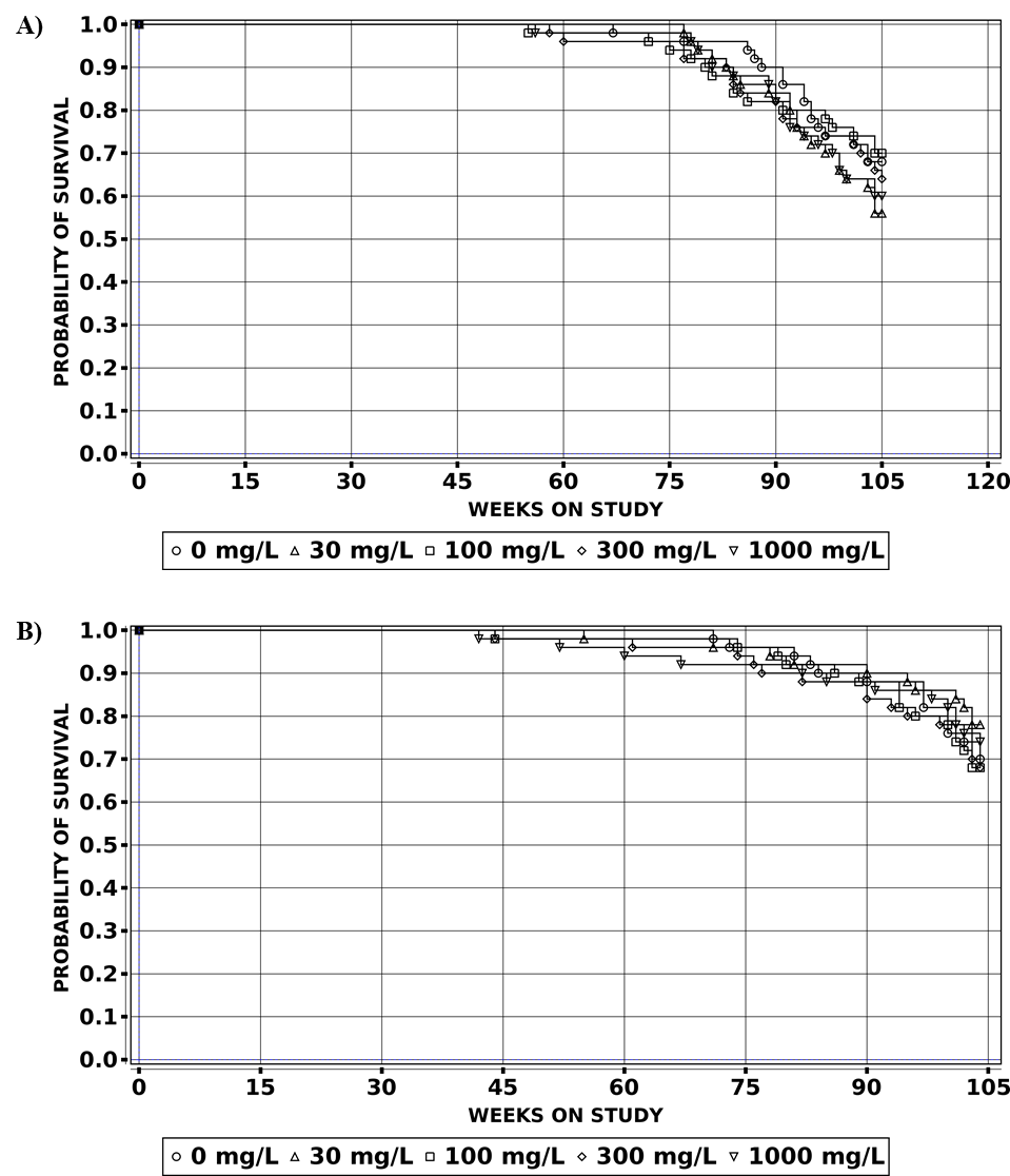
Two-year Study (Three-month Interim Evaluation)
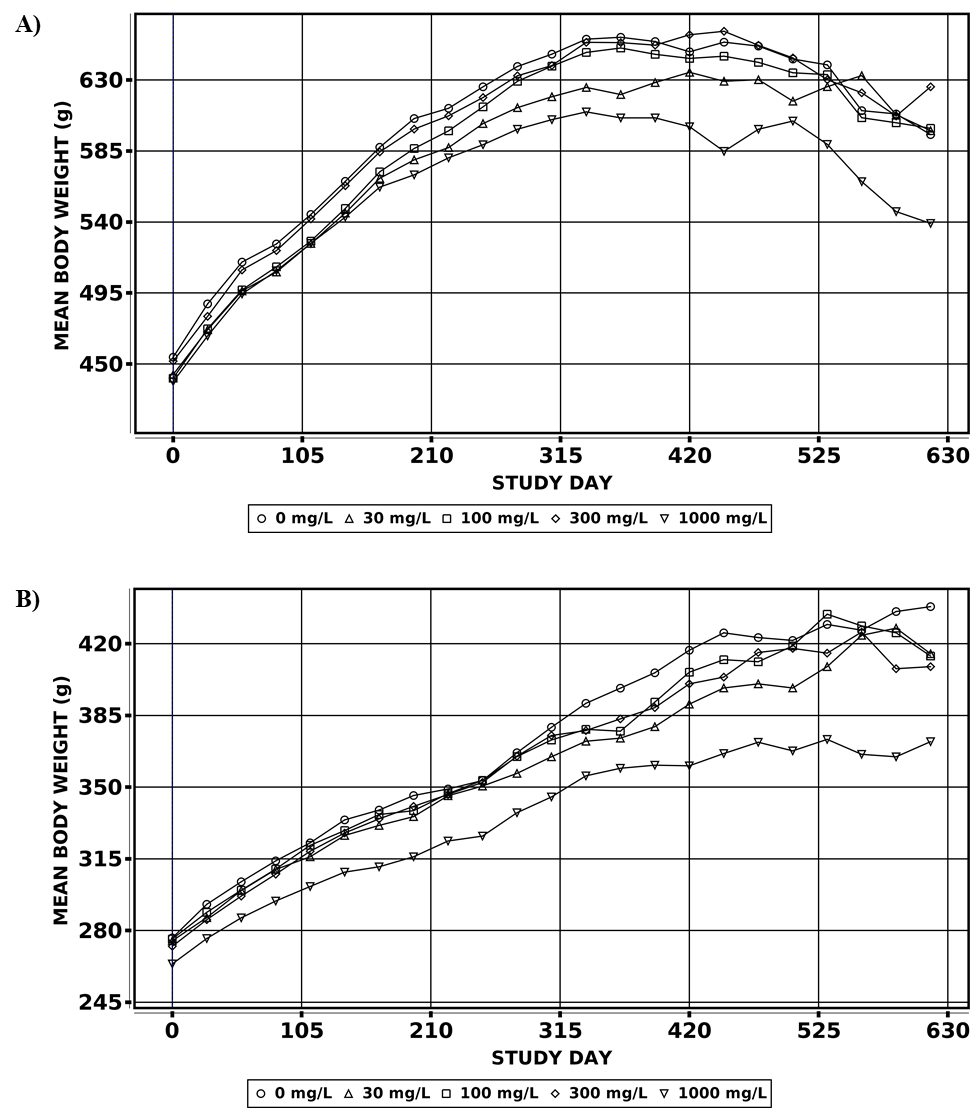
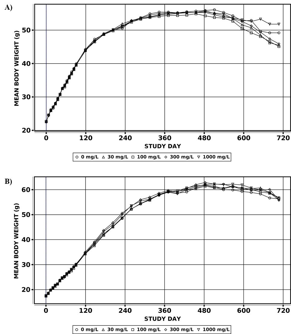
There were no exposure-related effects on survival or on clinical observations in male and female rats at the 3-month interim evaluation (Appendix G). There were significant decreases (6%–9%) in terminal body weights of male rats in the 100, 300, and 1,000 mg/L groups compared to the control group, but these differences did not exceed 10% (Table 9, Table 10; Figure 2). There were no effects on body weights of female rats at the 3-month interim evaluation.
Water consumption by F1 rats postweaning to the interim necropsy (PND 28–119) was unaffected by sulfolane exposure (Table 11). In males, sporadic but significant differences compared to the control group were observed that were not considered exposure related, and no differences were observed in females (Appendix G). Average chemical intake was calculated from study day 28 through study day 119. Generally, chemical intake increased in proportion to exposure concentration; however, female chemical intake was marginally higher (average of approximately 1.3-fold) than that of males.
There were no exposure-related effects on absolute or relative organ weights of males or females; organ weight changes noted in male rats were considered secondary to body weight changes (Appendix G). There were no exposure-related effects observed on clinical chemistry or hematology and no exposure-related gross or microscopic lesions (Appendix G).
Sperm count analysis was not conducted because of clumping during the measurement of sperm parameters. No lesions were observed in the testis or epididymis after microscopic evaluation, and sulfolane exposure did not affect estrous cyclicity parameters (Appendix G).
Plasma sulfolane concentrations were quantified in all exposed groups and were below the limit of detection (LOD) for the control group (Table 12). Average chemical intake (PND 28–119) by males and females in the internal concentration assessment groups was calculated to be 3, 8, 26, and 80 mg/kg/day and 3, 11, 30, and 96 mg/kg/day, respectively (Appendix G). Sulfolane plasma concentrations were generally consistent between male and female rats. The proportional increase in sulfolane plasma concentrations relative to exposure concentration was much higher in the 1,000 mg/L groups, indicating a saturation in metabolism occurs between 300 and 1,000 mg/L.
Two-year Study (Postweaning Phase)
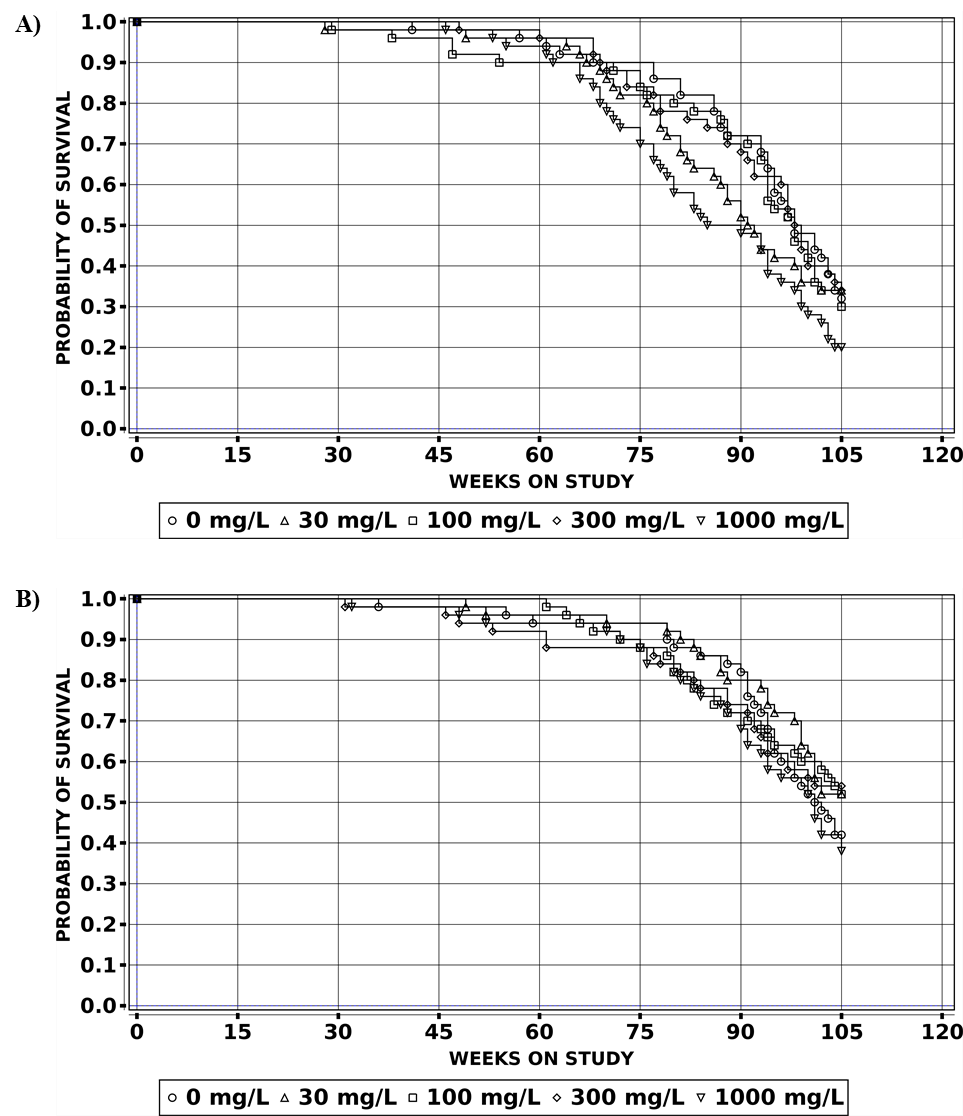
Chronic exposure to sulfolane did not affect the survival of female rats (Table 13; Figure 3), and no clinical observations were attributable to exposure. There was a significant negative trend for survival in male rats; however, no clinical findings or histological lesions explained the lower survival. Survival in the highest exposure group was marginally outside the NTP historical control range of 24%–64% in male Sprague Dawley rats across 10 studies.80 It is not clear if the survival of male rats was affected by sulfolane exposure. In males exposed to 1,000 mg/L, significant decreases (<11%) in body weights compared to the control group were observed. In females exposed to 1,000 mg/L, significant decreases in body weight compared to the control group (5%–16%) were observed throughout the study and were not associated with clinical observations. (Table 14, Table 15; Figure 4).
Water consumption by exposed groups was generally not statistically different from that of the control groups throughout the 2-year period, although water consumption (g/kg/day) by the 1,000 mg/L groups was somewhat higher than that of the control groups. Average chemical intake was calculated over the study day 21–616 interval (corresponding to PND 144–149 through PND 739–744). Generally, chemical intake (mg/kg/day) increased in proportion to exposure concentration. Additionally, intake by males was consistently lower than that of females throughout the 2-year study period (Table 16), which is consistent with findings from other drinking water studies.
Histopathology
This section describes the statistically significant or biologically noteworthy changes in the incidences of neoplasms of the mammary gland.
Mammary gland: In female rats, there were significant increases in the incidences of adenoma and adenoma or carcinoma (combined) in the 100 mg/L group compared to the control group (Table 17). The incidences of these lesions in all other exposed groups were also higher than that in the control group but were not significant following pairwise or trend tests. The incidences of adenoma and adenoma or carcinoma (combined) in the 100 mg/L group exceeded the historical control range for all routes of exposure. Though not significant, the incidence of mammary gland adenoma in the 1,000 mg/L group also exceeded the historical control range for all routes of exposure.
Other lesions: In the lung of male rats, there were slight but significant increases in the incidences of alveolar histiocytic infiltration (1,000 mg/L group) and chronic interstitial inflammation (100 and 300 mg/L groups) compared to the control group (Appendix G). A positive trend was also observed in the incidence of cellular infiltration. There was a slight increase in the severity of cellular infiltration in the 30 and 1,000 mg/L groups compared to the control group, but the increase was negligible. This lesion was characterized by a slight increase in the number of macrophages in the alveoli of the lungs. The chronic inflammation was characterized by mild, multifocal thickening of the alveolar septa associated with an increase in the number of alveolar macrophages and scattered lymphocytes. Because these findings are very common in laboratory rodents, their incidence varies, and the severity was relatively low, the above lesions were not considered to be related to sulfolane exposure.
Mice
Two-year Study (Three-month Interim Evaluation)
There were no exposure-related effects on survival or on clinical observations in male and female mice at the 3-month interim evaluation (Appendix G). Body weights of both male and female mice remained similar to those of the control groups throughout the 3-month study period (Table 18, Table 19; Figure 5).
Water consumption by male and female mice in the interim evaluation was unaffected by exposure (Table 20). Average chemical intake was calculated from study day 0 through study day 91, and chemical intake generally increased in proportion to exposure concentration and was similar between males and females (Table 20).
There were no exposure-related effects observed in hematology or relative organ weights of males or females (Appendix G). In both males and females, there were no exposure-related gross or microscopic lesions.
Sperm count analysis was not conducted because of clumping during the measurement of sperm parameters. No lesions were observed in the testis or epididymis after microscopic evaluation, and sulfolane exposure did not affect estrous cyclicity parameters (Appendix G).
Plasma sulfolane concentrations were quantified in all exposed groups and were below the LOD for the control group (Table 21). Average chemical intake in the internal concentration assessment groups was calculated to be 3, 11, 32, and 103 mg/kg/day for males and 4, 12, 34, and 112 mg/kg/day for females (Appendix G). Sulfolane concentrations were generally consistent between male and female mice in the 30 and 100 mg/L groups, but plasma concentrations were approximately 2-fold and 13-fold higher in females than in males in the 300 and 1,000 mg/L groups, respectively. The proportional increase in sulfolane plasma concentrations relative to exposure concentration was much higher in the female 300 and 1,000 mg/L groups, indicating a saturation in metabolism occurs at exposure concentrations between 100 and 300 mg/L in females.
Two-year Study
Chronic exposure to sulfolane did not affect the survival of male and female mice (Table 22; Figure 6), and no clinical observations were attributable to exposure. There were no significant effects on body weight attributed to sulfolane exposure (Table 23, Table 24; Figure 7).
Water consumption by male and female mice over the 2-year study period was unaffected by exposure (Table 25). Although slight differences in consumption between exposed groups and control groups were noted, these changes were intermittent and likely due to variability in body weights. Average chemical intake was calculated over the study day 0–707 interval. Generally, chemical intake increased in proportion to exposure concentration; additionally, intake by females was consistently lower than that of males throughout the 2-year study period (Table 25).
Histopathology
This section describes the statistically significant or biologically noteworthy changes in the incidences of neoplasms and/or nonneoplastic lesions of all organs (systemic), liver, and ovary.
All organs (systemic): In male mice, the incidence of hemangioma in all organs was higher in the 100 mg/L group relative to the control group; the incidence of hemangiosarcoma in all organs was higher in the 30, 100, and 1,000 mg/L groups relative to the control group; and the incidence of hemangioma or hemangiosarcoma (combined) in all organs was higher in the 30, 100, and 1,000 mg/L groups relative to the control group (Table 26). Only the incidences of hemangiosarcoma alone and hemangioma or hemangiosarcoma (combined) exhibited positive trends and were significantly increased in the 1,000 mg/L group, using pairwise comparisons to the control group. The incidence of hemangioma in all organs exceeded the historical control range for all routes of exposure in the 100 mg/L group. The incidence of hemangiosarcoma exceeded the historical control range for all routes in the 30 and 1,000 mg/L groups, and the incidence of hemangioma or hemangiosarcoma (combined) exceeded the historical control range in the 30, 100, and 1,000 mg/L groups. There were no corresponding increases in lesion incidences observed in sulfolane-exposed female mice.
Hemangiomas and/or hemangiosarcomas were observed in the liver, spleen, bone marrow, heart, mesenteric lymph node, prostate, skeletal muscle, and skin. Significantly increased incidences of hemangiosarcoma, using a pairwise comparison to the control group, were seen in the spleen of males exposed to 30 mg/L and in the liver of males exposed to 1,000 mg/L sulfolane. Positive trends were observed for hemangiosarcoma in the liver and for hemangioma in the spleen. In the liver, the incidence of hemangiosarcoma in the 30 and 1,000 mg/L groups exceeded the historical control range for all routes of exposure. The incidences of hemangioma in the mesenteric lymph node in the 100 mg/L group and in the spleen in the 1,000 mg/L group also exceeded the historical control range for all routes of exposure. The incidences of hemangiosarcoma in the spleen in the 30 mg/L group and in the mesenteric lymph node in the 100 mg/L group exceeded the historical control range for all routes of exposure.
Liver: In female mice, the incidence of hepatocellular carcinoma in the liver was significantly increased in the 1,000 mg/L group, with a positive trend (Table 27). These are relatively common neoplasms in B6C3F1/N mice, and the incidence in all exposure groups, with the exception of the 1,000 mg/L group, was within the historical control range for all routes of exposure. The incidence of hepatocellular adenoma or carcinoma (combined) exhibited a positive trend, but all groups were within the historical control range for all routes of exposure. The incidences of other hepatocellular neoplasms in female mice were not considered exposure related.
In male mice, the incidence of mixed cell focus was significantly increased in the 30, 100, and 300 mg/L groups relative to the control group (Table 27). In female mice, the incidence of clear cell focus exhibited a positive trend, and the incidence of focal fatty change was significantly increased in the 100 and 300 mg/L groups relative to the control group.
Ovary: In female mice, the incidence of follicular cysts was significantly increased in the 30, 300, and 1,000 mg/L groups compared to the control group (Table 28). The toxicological significance of this finding is unknown.
Genetic Toxicology
The genetic toxicity of sulfolane was evaluated in the peripheral blood micronucleus tests in rats and mice after exposure to sulfolane via drinking water. Micronucleated reticulocytes were not increased in male and female rats exposed to sulfolane for 3 months via drinking water (0, 30, 100, 300, 1,000 mg/L) (Table E-1), and there were no increases in micronucleated reticulocytes and micronucleated erythrocytes in male and female mice exposed to sulfolane for 3 months via drinking water (0, 30, 100, 300, 1,000 mg/L) (Table E-2). A positive trend in the percentage of reticulocytes was observed in female rats in the 3-month study, but the increase was within the historical control 95% confidence interval. Data from all NTP genetic toxicity tests with sulfolane are available in the NTP Chemical Effects in Biological Systems database: https://doi.org/10.22427/NTP-DATA-TR-605.79
Discussion
Sulfolane was nominated for study because of exposure concerns for residents surrounding a refinery in North Pole, Alaska. Because of likely long-term exposure and the presence of sulfolane around other similar refineries, these 2-year carcinogenicity studies in rats and mice were conducted. Drinking water was selected as the route of exposure, with 1,000 mg/L as the highest exposure concentration, on the basis of previously published National Toxicology Program (NTP) studies and the literature. The design of this study included several evaluations of potential developmental toxicity in rats and interim evaluations in both rats and mice, including measurements of internal exposure.
Marginal effects were observed following sulfolane exposure during gestation and lactation. Reduced male and female pup weights (up to 11%) in the 1,000 mg/L groups compared to control rats was the only exposure-related finding. This observation is consistent with the OECD 421 study,26,33 as maternal exposures of 200 mg/kg/day showed minimal effects. Sulfolane intake in the current study was 126 mg/kg/day during gestation (gestational day [GD] 6–21) and 206 mg/kg/day during lactation (lactational day [LD] 1–14). Vaginal opening was delayed in the 300 and 1,000 mg/L F1 female rats, with and without adjustment for body weight at weaning. This observation is suggestive of sulfolane inducing a developmental delay in female rats at these exposure concentrations in a developmental endpoint that has not been evaluated in other sulfolane studies.
The interim evaluation in rats and mice at 3 months showed marginal effects in male rats, with reduced body weights that were <10% different from control values. Clinical chemistry (rats only), hematology, and organ weights were unaffected by sulfolane exposure in rats and mice. In female rats, no effects were observed in lymphocyte counts, whereas a significant decrease in lymphocyte counts was previously observed in female rats exposed to >100 mg/L sulfolane in another 3-month drinking water study.28 Although this observation is inconsistent with the Huntingdon Life Sciences (HLS) 2001 drinking water study, marginal to no exposure-related effects on white blood cell counts were observed in other studies within the NTP research program on sulfolane, including in studies that evaluated multiple species after 28 days of exposure to sulfolane4 or in immunotoxicity studies of sulfolane.5 The reason for this is unclear. Additionally, no exposure-related nonneoplastic lesions in immune tissues were observed in these 3-month interim or 2-year studies.
Kidney pathological observations in previous studies on male rats included hyaline droplet accumulation and/or cortical tubular basophilia and granular casts in multiple exposure groups4,28; however, no exposure-related effects in the kidney were observed in this study at the interim or 2-year evaluations. Hyaline droplet accumulation in the NTP 28-day gavage study was significantly increased in all exposed groups (1–800 mg/kg/day), while similar increases were only observed in the higher exposed groups of 400 and 1,600 mg/L (35.0 and 131.7 mg/kg/day, respectively) in the HLS drinking water study. This suggests that the method of administration (e.g., drinking water versus gavage) influences these kidney lesions, likely because of absorption, distribution, metabolism, and exposure (ADME) differences. It is not clear why there was a lack of these findings in the current studies, as chemical intake from weaning to the 3-month interim evaluation (postnatal day [PND] 28–119) averaged 80 mg/kg/day in the 1,000 mg/L male rats, which was within the range of the higher exposure (1,600 mg/L) in the HLS 13-week study. It is possible the difference in findings was due to sensitivity differences between Charles River CD and Hsd Sprague Dawley rats at lower exposure concentrations. Other histopathological findings in the NTP 28-day gavage study that were not observed in the current study were most likely due to the higher doses used in the 28-day study and administration differences (gavage versus drinking water).
In male and female rats, chemical intake (mg/kg/day) increased in proportion to drinking water concentration (mg/L) with consistent sex differences (i.e., increased intake in females in comparison to males) typically found in drinking water studies. However, sulfolane plasma concentrations measured at the 3-month interim timepoint were consistent between the sexes and metabolic saturation occurred at higher exposures. In mice, chemical intake was generally similar between the sexes at 3 months, whereas plasma concentrations were higher in females in the 300 and 1,000 mg/mL groups compared to respective males. Metabolic saturation was evident in females starting at 300 mg/L, but metabolic saturation was not evident in males. When comparing plasma concentrations between this drinking water study and the NTP 28-day toxicity study (gavage),4 concentrations in male and female rats exposed to 30 or 100 mg/kg/day sulfolane were 4–9 times higher in the 28-day study (2 hours after the last dose) compared to the 300 and 1,000 mg/L interim data in the current study. Similarly, concentrations in mice exposed to 30 and 100 mg/kg/day sulfolane were 5–9 times higher at the 2-hour time point in the 28-day toxicity study compared to the 300 and 1,000 mg/L concentrations in this study. An exception was the ratio between the 1,000 mg/L male mice in this study and the 100 mg/kg/day male mice in the 28-day study, which showed a considerably higher difference (134×). Previous studies showed evidence of saturation in both male and female mice,2,4 but there was no evidence of saturation in male mice in this study, which could be due to differences in the method of administration (drinking water versus gavage) for this sex and species.
In the chronic portion of the rat study, survival was unaffected, while body weights were significantly decreased in male and female rats compared to their respective control groups (up to 11% and 16% in males and females, respectively, in the 1,000 mg/L group). In female rats, the incidence of mammary gland adenoma in the exposed groups was higher than the control group incidence and a significant increase was observed in female rats exposed to 100 mg/L sulfolane, which was outside the historical control range (14% versus 0%–9%). Although not significant, the incidence in the 1,000 mg/L group (10%) also exceeded the historical control range. However, there was no corresponding increase in nonneoplastic lesions or adenocarcinoma within the mammary gland. Given that the statistical significance occurred in a mid-exposure group only, and no increase in corresponding nonneoplastic lesions was observed, the increase in the incidence of mammary gland adenoma was considered equivocal evidence of carcinogenic activity. There was no evidence of carcinogenicity in male rats.
In mice, survival and body weights were not affected by sulfolane exposure. In female mice, the incidence of follicle cysts of the ovary was significantly increased in the 30, 300, and 1,000 mg/L groups. However, the toxicological significance of this lesion is unknown, this finding was not observed in rats, and no other findings in male and female reproductive tissues or estrous cyclicity effects were observed. In male mice, there was a positive trend and significant increase in the incidences of hemangiosarcomas of all organs (systemic) and of the liver in the 1,000 mg/L group compared to the concurrent control group, and the incidence rate was outside the historical control range for all organs (24% versus 2%–10%) and for the liver (22% versus 0%–6%). The incidences of hemangiomas of all organs (systemic) or individual organs was not increased in the exposed groups compared to the concurrent control group, aside from a positive trend observed in the spleen. The combined incidence of hemangioma and hemangiosarcoma of all organs, due to the increase in hemangiosarcoma, was also significantly increased in the 1,000 mg/L group, with a positive trend, and was also well outside the historical range (28% versus 3%–12%). Taken together, the increase in hemangiosarcoma supported clear evidence of carcinogenic activity in male mice.
Hemangiosarcomas are more common in mice than in rats and humans.81 Using NTP’s “Organ Sites with Neoplasia” database, 31 chemicals have been found to be positive (“positive,” “clear,” “some”) for hemangiosarcoma or hemangioma and hemangiosarcoma. Of these, 28 were positive in mice, 8 were positive in rats, and 5 were positive in both mice and rats.82 In a review by Cohen et al.,81 genotoxicity and/or nongenotoxic pathways are suggested as possible mechanisms of action. The relevance and mechanism for inducing these tumors varies with genotoxic chemicals inducing hemangiosarcomas in humans (e.g., vinyl chloride, vinyl bromide, and thorotrast), whereas there is more uncertainty with human relevance for the non-genotoxic pathway. In the current study, there was no evidence for genotoxicity by sulfolane exposure. Most of the hemangiosarcomas in the 1,000 mg/L group occurred in the liver, a common site for this neoplasm. 1,3-Butadiene exposure, which can be used as a precursor in sulfolane production, has been found to increase the incidence of hemangiosarcoma within the heart in male and female mice.83 In this study, there was no evidence of 1,3-butadiene contamination in the sulfolane analysis, and no increase in the incidence of hemangiosarcoma in the heart in male or female mice. Although the mechanism is unknown, the finding of an increased incidence of hemangiosarcoma was considered clear evidence of carcinogenic activity in male mice after sulfolane exposure.
A significant increase in the incidence of hepatocellular carcinoma was observed in the highest exposure group in female mice. The incidence (16%) marginally exceeded the historical control range (2%–14%), whereas there was no change in hepatocellular adenoma or preneoplastic lesions relative to control animals. The combined incidence of hepatocellular adenoma and carcinoma was not increased by pairwise comparison to the control group, but there was a positive trend. Taken together, the increase in incidences of hepatocellular carcinoma was considered to be equivocal evidence of carcinogenic activity due to no increase in the incidences of preneoplastic lesions, hepatocellular adenoma, or adenoma or carcinoma (combined) compared to the concurrent control.
Exposure concentrations within this study were higher than has been measured in drinking water wells, which were as high as approximately 100–400 ppb (100–400 µg/L) onsite or offsite the refinery.15,16,84 Previously, a chronic reference dose of 10 µg/kg/day was calculated, which was based on the finding of reduced leukocytes within female rats following a 90-day drinking water exposure28 and for which a drinking water screening level of 365 ppb was derived.85 The data presented here provide context for potential chronic toxicity and carcinogenic activity to support the risk assessment of sulfolane exposures in the general public and for potentially higher worksite exposures.
References
1. National Information Standards Organization (NISO). CRediT (Contributor Roles Taxonomy). Baltimore, MD: National Information Standards Organization; 2024. [Accessed: July 26, 2024]. https://credit.niso.org/
2. Waidyanatha S, Black SR, Blystone CR, Patel PR, Watson SL, Snyder RW, Fennell TR. Disposition and metabolism of sulfolane in Harlan Sprague Dawley rats and B6C3F1/N mice and in vitro in hepatocytes from rats, mice, and humans. Xenobiotica. 2020; 50(4):442-453. DOI: 10.1080/00498254.2019.1630786 PubMed: 31184953
3. Waidyanatha S, Black SR, Fennell TR, Watson SL, Patel PR, Cooper SD, Blake J, Robinson VG, Fernando RA, Blystone CR. Toxicokinetics and bioavailability of sulfolane, a ground water contaminant, following oral and intravenous administration in rodents: A dose, species, and sex comparison. Toxicol Appl Pharmacol. 2019; 379:114690. DOI: 10.1016/j.taap.2019.114690 PubMed: 31344372
4. Shipkowski KA, Cora MC, Cesta MF, Robinson VG, Waidyanatha S, Witt KL, Vallant MK, Fallacara DM, Hejtmancik MR, Masten SA, et al. Comparison of sulfolane effects in Sprague Dawley rats, B6C3F1/N mice, and Hartley guinea pigs after 28 days of exposure via oral gavage. Toxicol Rep. 2021; 8:581-591. DOI: 10.1016/j.toxrep.2021.02.004 PubMed: 33777704
5. Watson ATD, Johnson VJ, Luster MI, Burleson GR, Fallacara DM, Sparrow BR, Cesta MF, Cora MC, Shockley KR, Stout MD, et al. Immunotoxicity studies of sulfolane following developmental exposure in Hsd:Sprague Dawley SD rats and adult exposure in B6C3F1/N mice. J Immunotoxicol. 2021; 18(1):1-12. DOI: 10.1080/1547691X.2020.1869355 PubMed: 34357831
6. National Center for Biotechnology Information (NCBI). PubChem compound summary for CID 31347, sulfolane: Computed properties. Bethesda, MD: U.S. Department of Health and Human Services, National Institutes of Health, National Library of Medicine, National Center for Biotechnology Information; 2022. [Accessed: June 28, 2022]. https://pubchem.ncbi.nlm.nih.gov/compound/sulfolane#section=Computed-Properties
7. Kroschwitz JI, Seidel A. Kirk-Othmer encyclopedia of chemical technology. Hoboken, NJ: Wiley-Interscience; 2001. DOI: 10.1002/0471238961
8. Andersen ME, Jones RA, Kurlansik L, Mehl RG, Jenkins LJ Jr. Sulfolane-induced convulsions in rodents. Res Commun Chem Pathol Pharmacol. 1976; 15(3):571-580. PubMed: 825938
9. Brown VKH, Ferrigan LW, Stevenson DE. Acute toxicity and skin irritant properties of sulfolane. Br J Ind Med. 1966; 23(4):302-304. DOI: 10.1136/oem.23.4.302 PubMed: 5926894
10. National Center for Biotechnology Information (NCBI). PubChem annotation record for sulfolane, source: Hazardous Substances Data Bank (HSDB). Bethesda, MD: U.S. Department of Health and Human Services, National Institutes of Health, National Library of Medicine, National Center for Biotechnology Information; 2011. HSDB No. 122. [Accessed: June 28, 2022]. https://pubchem.ncbi.nlm.nih.gov/source/hsdb/122
11. U.S. Environmental Protection Agency (USEPA). ChemView: CASRN 126-33-0: 2016 CDR data. Washington, DC: U.S. Environmental Protection Agency; 2016. [Accessed: June 27, 2022]. https://chemview.epa.gov/chemview
12. Greene EA, Fedorak PM. Nutrient stimulation of sulfolane biodegradation in a contaminated soil from a sour natural gas plant and in a pristine soil. Environ Technol. 2001; 22(6):619-629. DOI: 10.1080/09593332208618246 PubMed: 11482381
13. Leo T. Uptake and release kinetics of sulfolane by cattail plants. Logan, UT: Utah State University; 2008. DOI: 10.26076/dd9f-d5d0
14. North Pole Utilities. Drinking water quality report. North Pole, AK: City Hall; 2015. [Accessed: June 27, 2022]. https://web.archive.org/web/20150908033354/https://www.northpolealaska.com/sites/default/files/fileattachments/utilities/page/565/water_quality_report_2015.pdf
15. Alaska Department of Environmental Conservation (DEC). Sulfolane sampling of groundwater on North Pole Refinery for November 2010 through January 2011. Juneau, AK: Alaska Department of Environmental Conservation; 2011. [Accessed: January 25, 2023]. https://dec.alaska.gov/media/7283/2011-onsite-mw-jan.pdf
16. Alaska Department of Environmental Conservation (DEC). Sulfolane sampling of groundwater in off site locations for Nov 2010 through January 2011. Juneau, AK: Alaska Department of Environmental Conservation; 2011. [Accessed: January 25, 2023]. https://dec.alaska.gov/media/7285/2011jan-mon-wells-offsite.pdf
17. Canadian Council of Ministers of the Environment (CCME). Canadian environmental quality guidelines for sulfolane: Water and soil. Winnipeg, Manitoba: Canadian Council of Ministers of the Environment; 2006. [Accessed: January 25, 2023]. https://ccme.ca/en/res/2006-sulfolane-ceqg-ssd-1368-en.pdf
18. Oasis Environmental. Sulfolane technical assistance and evaluation report. Juneau, AK: Alaska Department of Environmental Conservation; 2010. [Accessed: January 25, 2023]. https://dec.alaska.gov/media/6169/sulfolane-tech-assist-report.pdf
19. Eberbach J. Threat of chemical contamination fuels neighbors' ire. Detroit, MI: Detroit Free Press; 2016. [Accessed: January 25, 2023]. https://www.freep.com/story/news/local/michigan/2016/06/28/merit-energy-sulfolane-contamination/86460292/
20. Warnica M. Alberta government admits it knew of earlier sulfolane leak. Toronto, ON: Canadian Broadcasting Corporation; 2014. [Accessed: January 25, 2023]. https://www.cbc.ca/news/canada/edmonton/alberta-government-admits-it-knew-of-earlier-sulfolane-leak-1.2633095
21. Zhu ZH, Sun ML, Li ZS, Yang ZC, Zhang TB, Heng ZC, Xiao BL, Li QY, Peng QY, Dong YH. [An investigation of the maximum allowable concentration of sulfolane in surface water]. Hua Xi Yi Ke da Xue Xue Bao. 1987; 18(4):376-380. PubMed: 3449430
22. Agency for Toxic Substances and Disease Registry (ATSDR). Health consultation: Sulfolane. Atlanta, GA: U.S. Department of Health and Human Services, Public Health Service, Agency for Toxic Substances and Disease Registry; 2011. [Accessed: June 27, 2022]. https://ntp.niehs.nih.gov/ntp/noms/support_docs/atsdrsulfolanehc_2011_508.pdf
23. U.S. Environmental Protection Agency (USEPA). Provisional peer-reviewed toxicity values for sulfolane (CASRN 126-33-0). Cincinnati, OH: U.S. Environmental Protection Agency, Office of Research and Development, National Center for Environmental Assessment, Superfund Health Risk Technical Support Center; 2012. EPA Report No. EPA/690/R-12/024F. [Accessed: June 27, 2022]. https://cfpub.epa.gov/ncea/pprtv/documents/Sulfolane.pdf
24. Roberts JJ, Warwick GP. The mode of action of alkylating agents—III: The formation of 3-hydroxytetrahydrothiophene-1:1-dioxide from 1:4-dimethanesulphonyloxybutane (Myleran), S-β-L-alanyltetrahydrothiophenium mesylate, tetrahydro-thiophene and tetrahydrothiophene-1:1-dioxide in the rat, rabbit and mouse. Biochem Pharmacol. 1961; 6(4):217-227. DOI: 10.1016/0006-2952(61)90133-2
25. Andersen ME, Jones RA, Mehl RG, Hill TA, Kurlansik L, Jenkins LJ Jr. The inhalation toxicity of sulfolane (tetrahydrothiophene-1,1-dioxide). Toxicol Appl Pharmacol. 1977; 40(3):463-472. DOI: 10.1016/0041-008X(77)90073-4 PubMed: 407670
26. Organisation for Economic Co-operation and Development (OECD). SIDS initial assessment report for SIAM 19: Tetrahydrothiophene-1,1-dioxide. Paris, France: Organisation for Economic Co-operation and Development; 2004. [Accessed: June 27, 2022]. https://hpvchemicals.oecd.org/UI/handler.axd?id=cbde8e42-6a4f-4112-acf2-09ffcbcf210b
27. Ministry of Health and Welfare (MHW). [Twenty-eight-day repeat dose oral toxicity test of tetrahydrothiophene 1,1-dioxide in rats]. Tokyo, Japan: Ministry of Health and Welfare; 1996. [Accessed: January 25, 2023]. https://dra4.nihs.go.jp/mhlw_data/home/paper/paper126-33-0b.html
28. Huntingdon Life Sciences. Sulfolane toxicity study by oral administration via the drinking water to CD rats for 13 weeks, vol. 1 and 2. Sponsored by Shell Canada; 2001.
29. Ruppert PH, Dyer RS. Acute behavioral toxicity of sulfolane: Influence of hypothermia. Toxicol Lett. 1985; 28(2-3):111-116. DOI: 10.1016/0378-4274(85)90018-9 PubMed: 4071560
30. Dyer RS, Boyes WK, Hetzler BE. Acute sulfolane exposure produces temperature-independent and dependent changes in visual evoked potentials. Neurobehav Toxicol Teratol. 1986; 8(6):687-693. PubMed: 3808184
31. Gordon CJ, Long MD, Dyer RS. Effect of ambient temperature on the hypometabolic and hypothermic effects of sulfolane in rats. Arch Toxicol. 1984; 56(2):123-127. DOI: 10.1007/BF00349084 PubMed: 6532377
32. Gordon CJ, Long MD, Fehlner KS, Dyer RS. Sulfolane-induced hypothermia enhances survivability in mice. Environ Res. 1986; 40(1):92-97. DOI: 10.1016/S0013-9351(86)80084-6 PubMed: 3709502
33. Ministry of Health and Welfare (MHW). [Preliminary reproduction toxicity screening test of tetrahydrothiophene-1,1-dioxide by oral administration in rats]. Tokyo, Japan: Ministry of Health and Welfare; 1999. [Accessed: January 25, 2023]. https://dra4.nihs.go.jp/mhlw_data/home/paper/paper126-33-0c.html
34. National Cancer Institute (NCI). Bioassay of 3-sulfolene for possible carcinogenicity. Bethesda, MD: U.S. Department of Health, Education, and Welfare, Public Health Service, National Institutes of Health, National Cancer Institute; 1978. DHEW Publication No. (NIH) 78-1352. National Cancer Institute Carcinogenesis Technical Report Series No. 102 (NCI-CG-TR-102). [Accessed: June 28, 2022]. https://ntp.niehs.nih.gov/publications/reports/tr/100s/tr102/index.html
35. Sigma-Aldrich. Product 88965 (Fluka), FTIR008848. [Accessed: June 27, 2022]. https://www.sigmaaldrich.com/
36. Sigma-Aldrich. Product 88965 (Fluka), FNMR011407. [Accessed: June 27, 2022]. https://www.sigmaaldrich.com/
37. National Institute of Advanced Industrial Science and Technology (AIST). Spectral Database for Organic Compounds: SDBS-749: Tetrahydrothiophene 1,1-dioxide. Tokyo, Japan: National Institute of Advanced Industrial Science and Technology; 1999. [Accessed: June 28, 2022]. https://sdbs.db.aist.go.jp/CompoundLanding.aspx?sdbsno=749
38. Wiley. Wiley registry: 8th edition/NIST 2005 mass spectral library. Hoboken, NJ: Wiley; 2006.
39. Silinski MAR, Uenoyama T, Cooper SD, Fernando RA, Robinson VG, Waidyanatha S. Development and validation of an analytical method for quantitation of sulfolane in rat and mouse plasma by GC-MS. J Anal Toxicol. 2019; 43(6):477-481. DOI: 10.1093/jat/bkz031 PubMed: 31044244
40. National Toxicology Program (NTP). Specifications for the conduct of studies to evaluate the toxic and carcinogenic potential of chemical, biological and physical agents in laboratory animals for the National Toxicology Program (NTP). Research Triangle Park, NC: U.S. Department of Health and Human Services, Public Health Service, National Institutes of Health, National Toxicology Program; 2011.
41. Sills RC, Cesta MF, Willson CJ, Brix AE, Berridge BR. National Toxicology Program position statement on informed (“nonblinded”) analysis in toxicologic pathology evaluation. Toxicol Pathol. 2019; 47(7):887-890. DOI: 10.1177/0192623319873974 PubMed: 31522628
42. Maronpot RR, Boorman GA. Interpretation of rodent hepatocellular proliferative alterations and hepatocellular tumors in chemical safety assessment. Toxicol Pathol. 1982; 10(2):71-78. DOI: 10.1177/019262338201000210 PubMed: 28094716
43. Boorman GA, Haseman JK, Waters MD, Hardisty JF, Sills RC. Quality review procedures necessary for rodent pathology databases and toxicogenomic studies: The National Toxicology Program experience. Toxicol Pathol. 2002; 30(1):88-92. DOI: 10.1080/01926230252824752 PubMed: 11890481
44. Brix AE, Hardisty JF, McConnell EE. Combining neoplasms for evaluation of rodent carcinogenesis studies. In: Hsu CH, Stedeford T, editors. Cancer Risk Assessment: Chemical Carcinogenesis, Hazard Evaluation, and Risk Quantification. Hoboken, NJ: Wiley; 2010. p. 699-715. DOI: 10.1002/9780470622728.ch28
45. Kaplan EL, Meier P. Nonparametric estimation from incomplete observations. J Am Stat Assoc. 1958; 53(282):457-481. DOI: 10.1080/01621459.1958.10501452
46. Tarone RE. Tests for trend in life table analysis. Biometrika. 1975; 62(3):679-690. DOI: 10.1093/biomet/62.3.679
47. Cox DR. Regression models and life‐tables. J R Stat Soc Series B Methodol. 1972; 34(2):187-202. DOI: 10.1111/j.2517-6161.1972.tb00899.x
48. Bailer AJ, Portier CJ. Effects of treatment-induced mortality and tumor-induced mortality on tests for carcinogenicity in small samples. Biometrics. 1988; 44(2):417-431. DOI: 10.2307/2531856 PubMed: 3390507
49. Piegorsch WW, Bailer AJ. Statistics for environmental biology and toxicology: Section 6.3.2. London, UK: Chapman and Hall; 1997.
50. Portier CJ, Bailer AJ. Testing for increased carcinogenicity using a survival-adjusted quantal response test. Fundam Appl Toxicol. 1989; 12(4):731-737. DOI: 10.1016/0272-0590(89)90004-3 PubMed: 2744275
51. Portier CJ, Hedges JC, Hoel DG. Age-specific models of mortality and tumor onset for historical control animals in the National Toxicology Program’s carcinogenicity experiments. Cancer Res. 1986; 46(9):4372-4378. PubMed: 3731095
52. Bieler GS, Williams RL. Ratio estimates, the delta method, and quantal response tests for increased carcinogenicity. Biometrics. 1993; 49(3):793-801. DOI: 10.2307/2532200 PubMed: 8241374
53. Nam JM. A simple approximation for calculating sample sizes for detecting linear trend in proportions. Biometrics. 1987; 43(3):701-705. DOI: 10.2307/2532006 PubMed: 3663825
54. Rao JNK, Scott AJ. A simple method for the analysis of clustered binary data. Biometrics. 1992; 48(2):577-585. DOI: 10.2307/2532311 PubMed: 1637980
55. Fung KY, Krewski D, Rao JNK, Scott AJ. Tests for trend in developmental toxicity experiments with correlated binary data. Risk Anal. 1994; 14(4):639-648. DOI: 10.1111/j.1539-6924.1994.tb00277.x PubMed: 7972964
56. Gart JJ, Chu KC, Tarone RE. Statistical issues in interpretation of chronic bioassay tests for carcinogenicity. J Natl Cancer Inst. 1979; 62(4):957-974. DOI: 10.1093/jnci/62.4.957 PubMed: 285297
57. Dixon WJ, Massey FJ. Introduction to statistical analysis. 2nd ed. New York, NY: McGraw-Hill; 1957.
58. Tukey JW. Easy summaries--numerical and graphical. In: Exploratory Data Analysis. Reading, MA: Addison-Wesley; 1977. p. 27-56.
59. Dunnett CW. A multiple comparison procedure for comparing several treatments with a control. J Am Stat Assoc. 1955; 50(272):1096-1121. DOI: 10.1080/01621459.1955.10501294
60. Williams DA. A test for differences between treatment means when several dose levels are compared with a zero dose control. Biometrics. 1971; 27(1):103-117. DOI: 10.2307/2528930 PubMed: 5547548
61. Williams DA. The comparison of several dose levels with a zero dose control. Biometrics. 1972; 28(2):519-531. DOI: 10.2307/2556164 PubMed: 5037867
62. Shirley E. A non-parametric equivalent of Williams’ test for contrasting increasing dose levels of a treatment. Biometrics. 1977; 33(2):386-389. DOI: 10.2307/2529789 PubMed: 884197
63. Williams DA. A note on Shirley’s nonparametric test for comparing several dose levels with a zero-dose control. Biometrics. 1986; 42(1):183-186. DOI: 10.2307/2531254 PubMed: 3719054
64. Dunn OJ. Multiple comparisons using rank sums. Technometrics. 1964; 6(3):241-252. DOI: 10.1080/00401706.1964.10490181
65. Jonckheere AR. A distribution-free k-sample test against ordered alternatives. Biometrika. 1954; 41(1-2):133-145. DOI: 10.1093/biomet/41.1-2.133
66. Hsu JC. The factor analytic approach to simultaneous inference in the general linear model. J Comput Graph Stat. 1992; 1(2):151-168. DOI: 10.1080/10618600.1992.10477011
67. Haseman JK. Value of historical controls in the interpretation of rodent tumor data. Drug Inf J. 1992; 26(2):191-200. DOI: 10.1177/009286159202600210
68. Haseman JK, Rao GN. Effects of corn oil, time-related changes, and inter-laboratory variability on tumor occurrence in control Fischer 344 (F344/N) rats. Toxicol Pathol. 1992; 20(1):52-60. DOI: 10.1177/019262339202000107 PubMed: 1411131
69. Haseman JK. Data analysis: Statistical analysis and use of historical control data. Regul Toxicol Pharmacol. 1995; 21(1):52-59. DOI: 10.1006/rtph.1995.1009 PubMed: 7784636
70. U.S. Food and Drug Administration (FDA). 21 CFR Part 58. https://www.ecfr.gov/current/title-21/chapter-I/subchapter-A/part-58
71. Miller JA, Miller EC. Ultimate chemical carcinogens as reactive mutagenic electrophiles. In: Hiatt HH, Watson JD, Winsten JA, editors. Origins of Human Cancer. Cold Spring Harbor, NY: Cold Spring Harbor Laboratory; 1977. p. 605-627.
72. Straus DS. Somatic mutation, cellular differentiation, and cancer causation. J Natl Cancer Inst. 1981; 67(2):233-241. DOI: 10.1093/jnci/67.2.233 PubMed: 7021938
73. Crawford BD. Perspectives on the somatic mutation model of carcinogenesis. In: Flamm WG, Lorentzen RJ, editors. Mechanisms and Toxicity of Chemical Carcinogens and Mutagens. Princeton, NJ: Princeton Scientific Publishing; 1985. p. 13-59.
74. Schmid W. The micronucleus test. Mutat Res. 1975; 31(1):9-15. DOI: 10.1016/0165-1161(75)90058-8 PubMed: 48190
75. Heddle JA, Hite M, Kirkhart B, Mavournin K, MacGregor JT, Newell GW, Salamone MF. The induction of micronuclei as a measure of genotoxicity: A report of the U.S. Environmental Protection Agency Gene-Tox Program. Mutat Res. 1983; 123(1):61-118. DOI: 10.1016/0165-1110(83)90047-7 PubMed: 6888413
76. Shelby MD, Erexson GL, Hook GJ, Tice RR. Evaluation of a three-exposure mouse bone marrow micronucleus protocol: Results with 49 chemicals. Environ Mol Mutagen. 1993; 21(2):160-179. DOI: 10.1002/em.2850210210 PubMed: 8444144
77. Shelby MD, Witt KL. Comparison of results from mouse bone marrow chromosome aberration and micronucleus tests. Environ Mol Mutagen. 1995; 25(4):302-313. DOI: 10.1002/em.2850250407 PubMed: 7607185
78. Witt KL, Knapton A, Wehr CM, Hook GJ, Mirsalis J, Shelby MD, MacGregor JT. Micronucleated erythrocyte frequency in peripheral blood of B6C3F(1) mice from short-term, prechronic, and chronic studies of the NTP carcinogenesis bioassay program. Environ Mol Mutagen. 2000; 36(3):163-194. DOI: 10.1002/1098-2280(2000)36:3%3C163::AID-EM1%3E3.0.CO;2-P PubMed: 11044899
79. National Toxicology Program (NTP). TR-605: Pathology tables, survival and growth curves from NTP long term studies. Research Triangle Park, NC: U.S. Department of Health and Human Services, Public Health Service, National Toxicology Program; 2026. DOI: 10.22427/NTP-DATA-TR-605
80. National Toxicology Program (NTP). NTP historical controls report: All routes and vehicles: Harlan Sprague-Dawley rats. Research Triangle Park, NC: U.S. Department of Health and Human Services, Public Health Service, National Toxicology Program; 2022. [Accessed: August 11, 2025]. https://ntp.niehs.nih.gov/sites/default/files/2023-06/r_hcrpt_allrte20221000.pdf
81. Cohen SM, Storer RD, Criswell KA, Doerrer NG, Dellarco VL, Pegg DG, Wojcinski ZW, Malarkey DE, Jacobs AC, Klaunig JE, et al. Hemangiosarcoma in rodents: Mode-of-action evaluation and human relevance. Toxicol Sci. 2009; 111(1):4-18. DOI: 10.1093/toxsci/kfp131 PubMed: 19525443
82. National Toxicology Program (NTP). Organ sites with neoplasia. Research Triangle Park, NC: U.S. Department of Health and Human Services, Public Health Service, National Toxicology Program; 2023. [Accessed: May 24, 2023]. https://cebs.niehs.nih.gov/organsites/
83. National Toxicology Program (NTP). NTP technical report on the toxicology and carcinogenesis studies of 1,3-butadiene (CAS No. 106-99-0) in B6C3F1 mice (inhalation studies). Research Triangle Park, NC: U.S. Department of Health and Human Services, Public Health Service, National Institutes of Health, National Toxicology Program; 1993. NTP Technical Report No. 434. NIH Publication No. 93-3165. [Accessed: January 25, 2023]. https://ntp.niehs.nih.gov/go/tr434abs
84. Parshley L. The pollution plumes of North Pole. Bozeman, MT: Environmental Health News; 2021. [Accessed: January 25, 2023]. https://www.ehn.org/north-pole-alaska-pollution-2651333766.html
85. Thompson CM, Gaylor DW, Tachovsky JA, Perry C, Carakostas MC, Haws LC. Development of a chronic noncancer oral reference dose and drinking water screening level for sulfolane using benchmark dose modeling. J Appl Toxicol. 2013; 33(12):1395-1406. DOI: 10.1002/jat.2799 PubMed: 22936336
86. Witt KL, Livanos E, Kissling GE, Torous DK, Caspary W, Tice RR, Recio L. Comparison of flow cytometry- and microscopy-based methods for measuring micronucleated reticulocyte frequencies in rodents treated with nongenotoxic and genotoxic chemicals. Mutat Res. 2008; 649(1-2):101-113. DOI: 10.1016/j.mrgentox.2007.08.004 PubMed: 17869571
87. Dertinger SD, Camphausen K, MacGregor JT, Bishop ME, Torous DK, Avlasevich S, Cairns S, Tometsko CR, Menard C, Muanza T, et al. Three‐color labeling method for flow cytometric measurement of cytogenetic damage in rodent and human blood. Environ Mol Mutagen. 2004; 44(5):427-435. DOI: 10.1002/em.20075 PubMed: 15517570
88. Kissling GE, Dertinger SD, Hayashi M, MacGregor JT. Sensitivity of the erythrocyte micronucleus assay: Dependence on number of cells scored and inter-animal variability. Mutat Res. 2007; 634(1-2):235-240. DOI: 10.1016/j.mrgentox.2007.07.010 PubMed: 17851117
Conclusions
Under the conditions of these 2-year drinking water studies, there was no evidence of carcinogenic activity of sulfolane in male Hsd:Sprague Dawley SD rats at exposure concentrations of 30, 100, 300, or 1,000 mg/L. There was equivocal evidence of carcinogenic activity of sulfolane in female Hsd:Sprague Dawley SD rats based on the increased incidence of mammary gland adenoma.
There was clear evidence of carcinogenic activity of sulfolane in male B6C3F1/N mice based on the increased incidence of hemangiosarcoma (all organs), which predominately occurred in the liver. There was equivocal evidence of carcinogenic activity of sulfolane in female B6C3F1/N mice based on the increased incidence of hepatocellular carcinoma.
Exposure to sulfolane resulted in increased incidences of nonneoplastic lesions of the liver in male and female mice and the ovary in female mice. Sulfolane exposure also induced a delay in vaginal opening in female rats.
Appendices
Appendix A.
Chemical Characterization and Dose Formulation Studies
A.1.
Procurement and Characterization of Sulfolane
Sulfolane was obtained from Sigma-Aldrich (Milwaukee, WI) in a single lot (lot MKBN9784V). Identity, purity, and stability analyses were conducted by the analytical chemistry laboratory at RTI International (Research Triangle Park, NC) and the study laboratory at Battelle (Columbus, OH). Reports on analyses performed in support of the sulfolane studies are on file at the National Institute of Environmental Health Sciences (NIEHS).
Lot MKBN9784V was a solid to a gel at room temperature, formed a colorless liquid upon warming, and was consistent with the supplier’s Certificate of Analysis. The identity of the test lot was evaluated using infrared (IR) spectroscopy, 1H nuclear magnetic resonance (NMR) spectroscopy, 13C NMR spectroscopy, and ultra-high performance liquid chromatography (UHPLC) with high resolution mass spectrometry (MS). The IR, 1H NMR, and 13C NMR spectra (Figure A-1, Figure A-2, and Figure A-3, respectively) cohered with the structure of sulfolane and were consistent with reference spectra from the Sigma-Aldrich library (IR and 1H NMR)35,36 and the Spectral Database for Organic Compounds (13C NMR).37 UHPLC with high resolution MS further confirmed the identity of the test lot as sulfolane, as the observed molecular mass (121.0321 m/z) was within 2.5 m/z of the calculated molecular mass (121.0318 m/z). Elemental analyses were performed by Galbraith Laboratory (Knoxville, TN) to aid in identification. The relative amounts of carbon (39.51%), hydrogen (7.08%), and sulfur (26.34%) were within 6% of the theoretical values (39.98%, 6.71%, and 26.68%, respectively). An accurate determination of oxygen could not be made because of interference from the high elemental sulfur content. The boiling point (285°C) and density (1.31 g/cm3 at 21.9°C) measured by Galbraith Laboratory were consistent with literature values from the Hazardous Substance Data Bank (285°C and 1.26 g/cm3 at 30°C, respectively).10
Purity evaluation was conducted using gas chromatography (GC) with flame ionization detection (FID) (Table A-1, System A) and GC with MS detection (Table A-1, System B). The GC/FID chromatogram showed one major peak accounting for 99.9% of total peak area; no impurity peaks with areas ≥0.1% of the total integrated peak area were detected. The GC/MS spectrum for the major component was consistent with the library spectrum for sulfolane.38 Karl Fischer titration yielded a water content of 0.22%. The overall purity of the test lot was determined to be >99%. Because butadiene is a precursor of sulfolane synthesis, additional GC/MS analysis was conducted to determine whether butadiene was present at trace levels (Table A-1, System C). Butadiene was not present at ≥0.05% of the total ion chromatogram, with an analytical system limit of detection of butadiene at 12 ng/mL.
An accelerated stability study was previously conducted using a different lot (MKBH1265V, Sigma-Aldrich [Milwaukee, WI]) and was not repeated using lot MKBN9784V. The stability of lot MKBH1265V was confirmed for 14 days at frozen (−20°C), refrigerated (5°C), room (25°C), and elevated (60°C) temperatures when stored in amber glass vials sealed with Teflon-lined caps.
One 30-gallon drum of lot MKBN9784V was warmed for 2.5 hours to liquify the contents. Once the chemical was liquified, the drum was rolled for approximately 5 minutes to homogenize the contents. A spigot was screwed into the drum, and it was placed on a drum cradle. The chemical was transferred to thirty 80-ounce amber glass bottles sealed with a nitrogen headspace for bulk storage at room temperature. Reanalysis of the bulk chemical was performed by the study laboratory prior to and after the 2-year study; no degradation was detected by GC/FID (Table A-1, System D).
A.2.
Preparation and Analysis of Dose Formulations
Dose formulations of sulfolane (lot MKBN9784V) in tap water (West Jefferson, OH municipal supply) were prepared approximately monthly at the study laboratory following the protocols outlined in Table A-2. Formulations were prepared at 0, 30, 100, 300, and 1,000 mg/L for both rats and mice (29 batches; May 2015–April 2017). For the rat study, two additional batches were prepared in May and June 2017. The formulations were stored refrigerated (5°C) in amber glass bottles sealed with Teflon-lined lids and were used within 42 days of preparation.
The stability of sulfolane formulations in tap water was determined by the analytical laboratory at RTI International using GC/FID (Table A-1, System E). Samples were prepared at 30 mg/L and stored at refrigerated (5°C) or room (25°C) temperatures. Stability was confirmed for 42 days at both refrigerated and room temperatures. An additional study of the 30 mg/L formulation under dosing conditions was conducted. A single test solution was stored in a clear glass bottle in an animal cage in a fume hood exposed to normal laboratory lighting. Stability was confirmed for 8 days under these simulated dosing conditions.
Periodic analyses of the preadministration and postadministration dose formulations of sulfolane were conducted every 2 to 3 months by the Battelle analytical laboratory using GC/FID (Table A-1, System F). Postadministration samples were collected from the animal rooms and formulation carboys. A 1,000 mg/L formulation was prepared on August 12, 2015, to replace the original preparation from July 23, 2015, which did not meet acceptance criteria and was deemed not suitable for use. With that exception, all other preadministration dose formulations were within 10% of the target concentrations (Table A-3). All postadministration samples were within 10% of the target concentrations with the following exceptions: The carboy samples of the 30 and 1,000 mg/L formulations prepared on May 4, 2015, were 10.9% and 10.2% below the target concentrations, respectively. The carboy samples of the 300 and 1,000 mg/L formulations prepared on September 21, 2015, were 10.2% and 13.0% below the target concentrations, respectively. The rat animal room sample of the 30 mg/L formulation prepared on July 23, 2015, was 10.5% below the target concentration, and the mouse animal room sample of the 300 mg/L formulation prepared on May 4, 2015, was 12.4% below the target concentration.
Table A-1. Gas Chromatography Systems Used in the Two-year Drinking Water Studies of Sulfolane
| Detection System | Column | Carrier Gas | Oven Temperature Program |
|---|---|---|---|
| System A | |||
| Flame ionization (260°C) | J&W DB-WAX (15 m × 0.32 mm ID, 0.25 µm film thickness) | Helium at 1.5 mL/minute | 80°C for 1 minute, then 20°C/minute to 250°C, then held for 7 minutes at 250°C |
| System B | |||
| Mass spectrometry with electron impact ionization | J&W DB-5MS (30 m × 0.25 mm ID, 0.25 µm film thickness) | Helium at 1.0 mL/minute | 60°C for 3 minutes, then 30°C/minute to 270°C, then held for 2 minutes at 270°C |
| System C | |||
| Mass spectrometry | J&W DB-624 (30 m × 0.32 mm ID, 1.80 µm film thickness) | Helium at 1.5 mL/minute | 35°C for 3 minutes, then 30°C/minute to 245°C, then held for 5 minutes at 245°C |
| System D | |||
| Flame ionization (260°C) | J&W DB-WAX (15 m × 0.32 mm ID, 0.25 µm film thickness) | Helium at 1.5 mL/minute | 80°C for 1 minute, then 20°C/minute to 200°C, then held for 5 minutes at 200°C |
| System E | |||
| Flame ionization (260°C) | J&W DB-WAX (15 m × 0.32 mm ID, 0.25 µm film thickness) | Helium at 1.5 mL/minute | 80°C for 1 minute, then 20°C/minute to 200°C, then held for 3 minutes at 200°C |
| System F | |||
| Flame ionization (260°C) | J&W DB-WAX (15 m × 0.32 mm ID, 0.25 µm film thickness) | Helium at 1.5 mL/minute | 80°C for 1 minute, then 20°C/minute to 200°C, then held for 7 minutes at 200°C |
Table A-2. Preparation and Storage of Dose Formulations Administered to Male and Female Rats and Mice in the Two-year Drinking Water Studies of Sulfolane
| Preparation |
|---|
| For the 0 mg/L formulation, blank tap water vehicle was used. For remaining formulations, a target amount of test article was weighed into individually tared weighing containers. The weighed test article was transferred to the appropriate tared mixing drum containing ~80% of the target final volume of vehicle. The weighing container was rinsed three times with vehicle, and the rinses were added to the mixing drum. The vehicle was slowly added to the mixing drum to achieve the target final total weight. The mixing drum contents were mixed with a drum stirrer for ~10 minutes or until the test article was visibly in solution. |
| Chemical lot number |
| MKBN9784V (Sigma-Aldrich, Milwaukee, WI) |
| Maximum storage time |
| 42 days |
| Storage conditions |
| Stored in amber glass bottles sealed with Teflon-lined lids at 5°C (refrigerated). |
| Analytical laboratory |
| Battelle (Columbus, OH) |
| Study laboratory |
| Battelle (West Jefferson, OH) |
Table A-3. Results of Analyses of Dose Formulations Administered to Male and Female Rats and Mice in the Two-year Drinking Water Studies of Sulfolane
| Date Prepared | Date Analyzed | Target Concentration (mg/L) | Determined Concentration (mg/L)a | Difference from Target (%) |
|---|---|---|---|---|
| May 4, 2015 | May 5, 2015 | 0 | BLOQ | NA |
| 30 | 29.7 ± 0.2 | −1.1 | ||
| 100 | 92.3 ± 0.3 | −7.7 | ||
| 300 | 276 ± 10 | −8.1 | ||
| 1,000 | 940 ± 48 | −6.0 | ||
| July 23, 2015 | July 27, 2015 | 0 | BLOQ | NA |
| 30 | 30.5 ± 0.3 | 1.7 | ||
| 30 | 29.0 ± 0.3 | −3.4 | ||
| 100 | 90.8 ± 5.0 | −9.2 | ||
| 100 | 98.7 ± 2.0 | −1.3 | ||
| July 23, 2015 | July 30, 2015 | 300 | 305 ± 8 | 1.7 |
| 300 | 299 ± 7 | −0.3 | ||
| 1,000 | 933 ± 59 | −6.7 | ||
| 1,000b,c | 980 ± 33 | −2.0 | ||
| September 21, 2015 | September 21, 2015 | 0 | BLOQ | NA |
| 30 | 29.1 ± 0.3 | −2.9 | ||
| 30 | 28.3 ± 0.8 | −5.6 | ||
| 100 | 91.7 ± 0.8 | −8.3 | ||
| 100 | 96.8 ± 1.0 | −3.2 | ||
| 300 | 284 ± 10 | −5.3 | ||
| 300 | 279 ± 10 | −6.9 | ||
| 1,000 | 905 ± NAd | −9.5 | ||
| 1,000 | 938 ± 64 | −6.2 | ||
| December 8, 2015 | December 9, 2015 | 0 | BLOQ | NA |
| 30 | 30.1 ± 0.6 | 0.2 | ||
| 30 | 30.4 ± 0.4 | 1.5 | ||
| 100 | 97.5 ± 1.0 | −2.5 | ||
| 100 | 103 ± 2 | 2.7 | ||
| 300 | 295 ± 5 | −1.8 | ||
| 300 | 286 ± 8 | −4.8 | ||
| 1,000 | 1,010 ± 30 | 0.5 | ||
| 1,000 | 980 ± 26 | −2.0 | ||
| February 2, 2016 | February 4, 2016 | 0 | BLOQ | NA |
| 30 | 31.3 ± 0.4 | 4.2 | ||
| 30 | 31.2 ± 0.8 | 3.9 | ||
| 100 | 97.8 ± 1.0 | −2.2 | ||
| 100 | 101 ± 2 | 1.3 | ||
| 300 | 309 ± 6 | 3.0 | ||
| 300 | 306 ± 13 | 2.1 | ||
| 1,000 | 973 ± 8 | −2.7 | ||
| 1,000 | 987 ± 20 | −1.3 | ||
| April 26, 2016 | April 29, 2016 | 0 | BLOQ | NA |
| 30 | 29.2 ± 0.5 | −2.8 | ||
| 30 | 29.7 ± 0.5 | −1.0 | ||
| 100 | 97.8 ± 0.8 | −2.2 | ||
| 100 | 97.3 ± 0.3 | −2.7 | ||
| 300 | 298 ± 5 | −0.8 | ||
| 300 | 301 ± 1 | 0.3 | ||
| 1,000 | 988 ± 19 | −1.2 | ||
| 1,000 | 977 ± 8 | −2.3 | ||
| June 21, 2016 | June 23, 2016 | 0 | BLOQ | NA |
| 30 | 29.5 ± 0.1 | −1.8 | ||
| 30 | 30.2 ± 0.4 | 0.8 | ||
| 100 | 97.5 ± 0.9 | −2.5 | ||
| 100 | 98.5 ± 0.0 | −1.5 | ||
| 300 | 305 ± 1 | 1.8 | ||
| 300 | 301 ± 5 | 0.5 | ||
| 1,000 | 998 ± 10 | −0.2 | ||
| 1,000 | 992 ± 8 | −0.8 | ||
| September 13, 2016 | September 14, 2016 | 0 | BLOQ | NA |
| 30 | 29.4 ± 0.2 | −1.9 | ||
| 30 | 29.3 ± 0.2 | −2.2 | ||
| 100 | 98.0 ± 0.5 | −2.0 | ||
| 100 | 96.3 ± 0.3 | −3.7 | ||
| 300 | 296 ± 3 | −1.2 | ||
| 300 | 290 ± 4 | −3.5 | ||
| 1,000 | 977 ± 8 | −2.3 | ||
| 1,000 | 972 ± 15 | −2.8 | ||
| November 8, 2016 | November 11, 2016 | 0 | BLOQ | NA |
| 30 | 29.2 ± 0.6 | −2.8 | ||
| 30 | 28.5 ± 0.9 | −5.1 | ||
| 100 | 94.7 ± 2.3 | −5.3 | ||
| 100 | 93.0 ± 0.5 | −7.0 | ||
| 300 | 293 ± 7 | −2.5 | ||
| 300 | 290 ± 2 | −3.4 | ||
| 1,000 | 967 ± 20 | −3.3 | ||
| 1,000 | 968 ± 14 | −3.2 | ||
| February 2, 2017 | February 2, 2017 | 0 | BLOQ | NA |
| 30 | 29.6 ± 0.7 | −1.3 | ||
| 30 | 29.6 ± 0.5 | −1.3 | ||
| 100 | 101 ± 1 | 0.5 | ||
| 100 | 102 ± 1 | 1.7 | ||
| 300 | 306 ± 2 | 1.9 | ||
| 300 | 299 ± 6 | −0.4 | ||
| 1,000 | 1,010 ± 10 | 0.5 | ||
| 1,000 | 977 ± 18 | −2.3 | ||
| March 28, 2017 | March 30, 2017 | 0 | BLOQ | NA |
| 30 | 29.2 ± 0.2 | −2.7 | ||
| 30 | 28.8 ± 0.3 | −3.9 | ||
| 100 | 95.0 ± 1.3 | −5.0 | ||
| 100 | 97.3 ± 1.0 | −2.7 | ||
| 300 | 288 ± 6 | −4.1 | ||
| 300 | 290 ± 1 | −3.2 | ||
| 1,000 | 972 ± 15 | −2.8 | ||
| 1,000 | 995 ± 15 | −0.5 | ||
| June 20, 2017e | June 26, 2017 | 0 | BLOQ | NA |
| 30 | 30.7 ± 0.6 | 2.3 | ||
| 100 | 101 ± 1 | 1.3 | ||
| 300 | 292 ± 5 | −2.6 | ||
| 1,000 | 918 ± 8 | −8.2 | ||
| Carboy samples | ||||
| May 4, 2015 | June 15, 2015 | 0 | BLOQ | NA |
| 30 | 26.7 ± 0.2 | −10.9 | ||
| 100 | 96.2 ± 0.3 | −3.8 | ||
| 300 | 295 ± 3 | −1.8 | ||
| 1,000 | 898 ± 6 | −10.2 | ||
| July 23, 2015 | September 3, 2015 | 0 | BLOQ | NA |
| 30 | 27.7 ± 0.7 | −7.7 | ||
| 100 | 93.8 ± 1.4 | −6.2 | ||
| 300 | 276 ± 2 | −8.1 | ||
| 1,000b | 987 ± 20 | −1.3 | ||
| September 21, 2015 | October 28, 2015 | 0 | BLOQ | NA |
| 30 | 27.8 ± 0.2 | −7.5 | ||
| 100 | 91.5 ± 1.3 | −8.5 | ||
| 300 | 269 ± 4 | −10.2 | ||
| 1,000 | 870 ± 10 | −13.0 | ||
| December 8, 2015 | January 20, 2016 | 0 | BLOQ | NA |
| 30 | 29.4 ± 0.3 | −2.0 | ||
| 100 | 95.8 ± 0.8 | −4.2 | ||
| 300 | 316 ± 8 | 5.3 | ||
| 1,000 | 973 ± 10 | −2.7 | ||
| June 21, 2016 | August 3, 2016 | 0 | BLOQ | NA |
| 30 | 29.7 ± 1.4 | −1.0 | ||
| 100 | 103 ± 2 | 3.3 | ||
| 300 | 310 ± 3 | 3.5 | ||
| 1,000 | 1,050 ± 10 | 4.7 | ||
| February 2, 2017 | March 16, 2017 | 0 | BLOQ | NA |
| 30 | 28.7 ± 0.3 | −4.3 | ||
| 100 | 95.3 ± 0.8 | −4.7 | ||
| 300f | 287 ± 6 | −4.4 | ||
| 1,000f | 952 ± 8 | −4.8 | ||
| Animal room samples (rats) | ||||
| May 4, 2015 | June 15, 2015 | 0 | BLOQ | NA |
| 30 | 29.8 ± 0.5 | −0.6 | ||
| 100 | 97.5 ± 0.0 | −2.5 | ||
| 300 | 283 ± 8 | −5.6 | ||
| 1,000 | 935 ± 26 | −6.5 | ||
| July 23, 2015 | September 3, 2015 | 0 | BLOQ | NA |
| 30 | 26.9 ± 0.6 | −10.5 | ||
| 100 | 93.8 ± 0.3 | −6.2 | ||
| 300 | 278 ± 1 | −7.4 | ||
| 1,000b | 972 ± 13 | −2.8 | ||
| September 21, 2015 | October 28, 2015 | 0 | BLOQ | NA |
| 30 | 27.5 ± 0.2 | −8.3 | ||
| 100 | 96.2 ± 0.3 | −3.8 | ||
| 300 | 270 ± 4 | −10.0 | ||
| 1,000 | 912 ± 6 | −8.8 | ||
| December 8, 2015 | January 20, 2016 | 0 | BLOQ | NA |
| 30 | 28.8 ± 0.3 | −3.9 | ||
| 100 | 92.8 ± 2.5 | −7.2 | ||
| 300 | 296 ± 2 | −1.2 | ||
| 1,000 | 955 ± 13 | −4.5 | ||
| June 21, 2016 | August 3, 2016 | 0 | BLOQ | NA |
| 30 | 31.0 ± 1.8 | 3.2 | ||
| 100 | 101 ± 2 | 1.2 | ||
| 300 | 306 ± 3 | 1.9 | ||
| 1,000 | 1,050 ± 20 | 4.7 | ||
| February 2, 2017 | March 16, 2017 | 0 | BLOQ | NA |
| 30 | 28.9 ± 0.2 | −3.7 | ||
| 100 | 96.0 ± 1.3 | −4.0 | ||
| 300f | 286 ± 6 | −4.7 | ||
| 1,000f | 970 ± 13 | −3.0 | ||
| Animal room samples (mice) | ||||
| May 4, 2015 | June 15, 2015 | 0 | BLOQ | NA |
| 30 | 30.1 ± 0.2 | 0.3 | ||
| 100 | 99.2 ± 0.3 | −0.8 | ||
| 300 | 263 ± 10 | −12.4 | ||
| 1,000 | 927 ± 8 | −7.3 | ||
| July 23, 2015 | September 3, 2015 | 0 | BLOQ | NA |
| 30 | 27.6 ± 1.0 | −8.1 | ||
| 100 | 91.5 ± 2.8 | −8.5 | ||
| 300 | 276 ± 5 | −7.9 | ||
| 1,000b | 982 ± 8 | −1.8 | ||
| September 21, 2015 | October 28, 2015 | 0 | BLOQ | NA |
| 30 | 27.7 ± 0.1 | −7.7 | ||
| 100 | 91.2 ± 0.8 | −8.8 | ||
| 300 | 279 ± 8 | −7.0 | ||
| 1,000 | 922 ± 25 | −7.8 | ||
| December 8, 2015 | January 20, 2016 | 0 | BLOQ | NA |
| 30 | 29.3 ± 0.3 | −2.3 | ||
| 100 | 97.2 ± 0.6 | −2.8 | ||
| 300 | 289 ± 9 | −3.6 | ||
| 1,000 | 930 ± 33 | −7.0 | ||
| June 21, 2016 | August 3, 2016 | 0 | BLOQ | NA |
| 30 | 30.6 ± 0.4 | 2.0 | ||
| 100 | 103 ± 1 | 3.0 | ||
| 300 | 275 ± 9 | −8.4 | ||
| 1,000 | 1,060 ± 10 | 6.0 | ||
| February 2, 2017 | March 16, 2017 | 0 | BLOQ | NA |
| 30 | 29.1 ± 0.2 | −3.0 | ||
| 100 | 96.7 ± 0.8 | −3.3 | ||
| 300f | 279 ± 5 | −6.9 | ||
| 1,000f | 960 ± 13 | −4.0 | ||
Figure A-1. Infrared Absorption Spectrum of Sulfolane
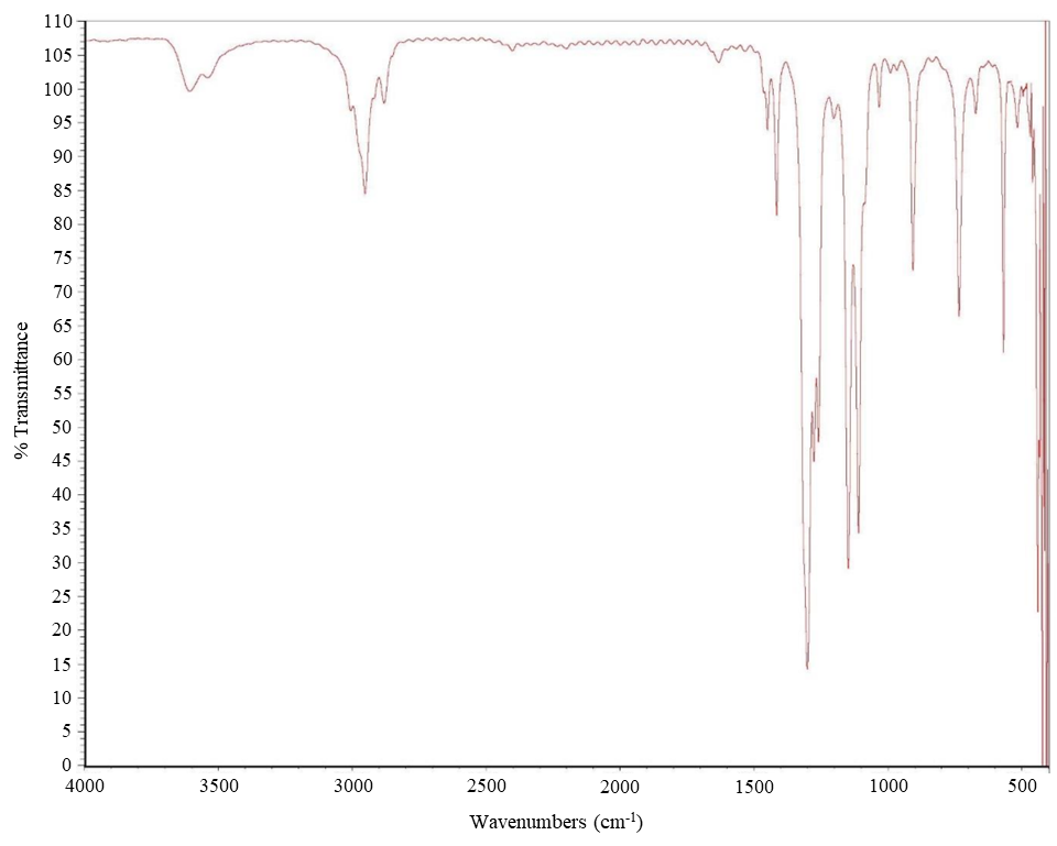
Figure A-2. 1H Nuclear Magnetic Resonance Spectrum of Sulfolane
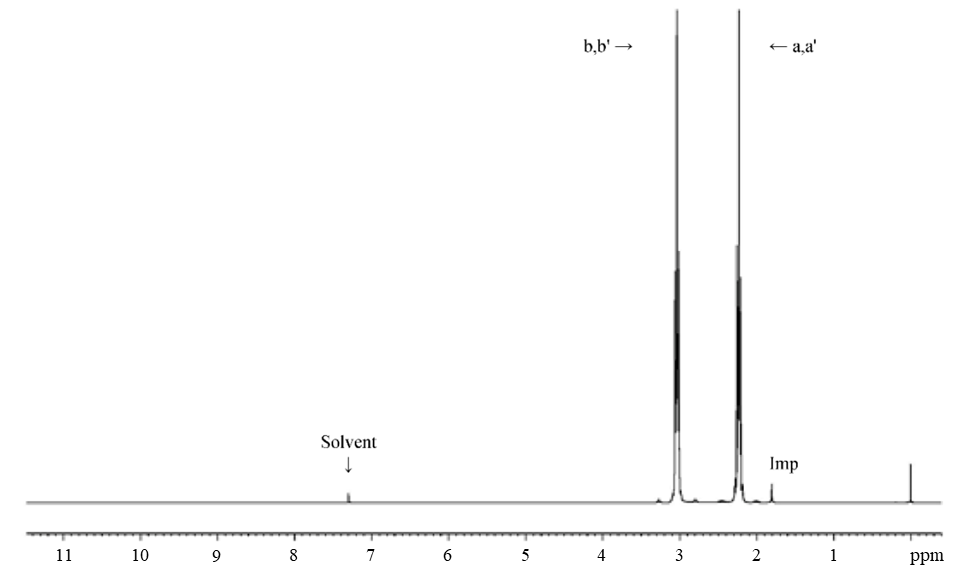
Figure A-3. 13C Nuclear Magnetic Resonance Spectrum of Sulfolane
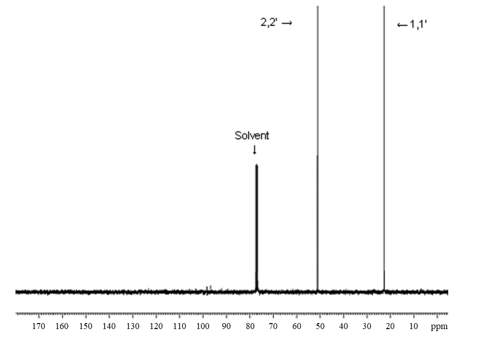
Appendix B.
Ingredients, Nutrient Composition, and Contaminant Levels in NIH-07 and NTP-2000 Rat and Mouse Rations
B.1.
NIH-07 Feed
Table B-1. Ingredients of NIH-07 Rat Ration
| Ingredients | Percent by Weight |
|---|---|
| Ground hard winter wheat | 23.00 |
| Ground #2 yellow shelled corn | 24.25 |
| Wheat middlings | 10.0 |
| Oat hulls | 0.0 |
| Alfalfa meal (dehydrated, 17% protein) | 4.0 |
| Purified cellulose | 0.0 |
| Soybean meal (47% protein) | 12.0 |
| Fish meal (62% protein) | 10.0 |
| Corn oil (without preservatives) | 0.0 |
| Soy oil (without preservatives) | 2.5 |
| Dried brewer’s yeast | 2.0 |
| Calcium carbonate (USP) | 0.5 |
| Vitamin premixa | 0.25 |
| Mineral premixb | 0.15 |
| Calcium phosphate, dibasic (USP) | 1.25 |
| Sodium chloride | 0.5 |
| Choline chloride (70% choline) | 0.10 |
| Dried skim milk | 5.0 |
| Dried molasses | 1.50 |
| Corn gluten meal (60% protein) | 3.00 |
| Methionine | 0.0 |
Table B-2. Vitamins and Minerals in NIH-07 Rat Ration
| Vitamins and Minerals | Amounta | Source |
|---|---|---|
| Vitamins | ||
| Vitamin A | 6,062 IU | Stabilized vitamin A palmitate or acetate |
| Vitamin D | 5,070 IU | D-activated animal sterol |
| Vitamin K | 3.1 mg | Menadione sodium bisulfite complex |
| Vitamin E | 22 IU | α-Tocopheryl Acetate |
| Niacin | 33 mg | – |
| Folic acid | 2.4 mg | – |
| d-Pantothenic Acid | 19.8 mg | d-Calcium pantothenate |
| Riboflavin | 3.8 mg | – |
| Thiamine | 11 mg | Thiamine mononitrate |
| B12 | 50 µg | – |
| Pyridoxine | 6.5 mg | Pyridoxine hydrochloride |
| Biotin | 0.15 mg | d-Biotin |
| Minerals | ||
| Iron | 132 mg | Iron sulfate |
| Zinc | 18 mg | Zinc oxide |
| Manganese | 66 mg | Manganese oxide |
| Copper | 4.4 mg | Copper sulfate |
| Iodine | 2.0 mg | Calcium iodate |
| Cobalt | 0.44 mg | Cobalt carbonate |
Table B-3. Nutrient Composition of NIH-07 Rat Ration
| Nutrient | Mean ± Standard Deviation | Range | Number of Samples |
|---|---|---|---|
| Protein (% by weight) | 22.7 ± 0.566 | 22.3–23.1 | 2 |
| Crude fat (% by weight) | 5.2 ± 0.283 | 5.0–5.4 | 2 |
| Crude fiber (% by weight) | 3.6 ± 0.198 | 3.46–3.74 | 2 |
| Ash (% by weight) | 6.23 ± 0.537 | 5.85–5.61 | 2 |
| Amino acids (% of total diet) | |||
| Arginine | 1.278 ± 0.343 | 0.258–1.49 | 11 |
| Cystine | 0.307 ± 0.059 | 0.153–0.372 | 11 |
| Glycine | 1.065 ± 0.289 | 0.217–1.31 | 11 |
| Histidine | 0.482 ± 0.121 | 0.125–0.553 | 11 |
| Isoleucine | 0.914 ± 0.233 | 0.214–1.03 | 11 |
| Leucine | 1.873 ± 0.485 | 0.423–2.13 | 11 |
| Lysine | 1.140 ± 0.345 | 0.111–1.32 | 11 |
| Methionine | 0.453 ± 0.117 | 0.102–0.515 | 11 |
| Phenylalanine | 1.023 ± 0.245 | 0.286–1.12 | 11 |
| Threonine | 0.850 ± 0.228 | 0.168–0.961 | 11 |
| Tryptophan | 0.259 ± 0.064 | 0.076–0.326 | 11 |
| Tyrosine | 0.801 ± 0.200 | 0.209–0.894 | 11 |
| Valine | 1.055 ± 0.264 | 0.262–1.17 | 11 |
| Essential fatty acids (% of total diet) | |||
| Linoleic | 2.436 ± 0.489 | 0.199–3.77 | 11 |
| Linolenic | 0.367 ± 0.397 | 0.214–1.56 | 11 |
| Vitamins | |||
| Vitamin A (IU/kg) | 5,080 ± 42.26 | 4,780–5,380 | 2 |
| α-Tocopherol (ppm) | 6,097 ± 20,067 | 31.36–66,600 | 11 |
| Thiamine (ppm)a | 11.05 ± 0.495 | 10.7–11.4 | 2 |
| Riboflavin (ppm) | 13.54 ± 4.438 | 4.2–19.8 | 11 |
| Niacin (ppm) | 95.02 ± 16.30 | 51.9–112.0 | 11 |
| Pantothenic aacid (ppm) | 40.69 ± 12.76 | 3.8–51.1 | 11 |
| Pyridoxine (ppm)a | 11.74 ± 4.81 | 0.42–19.7 | 11 |
| Folic acid (ppm) | 2.38 ± 0.571 | 1.37–3.09 | 11 |
| Biotin (ppm) | 0.300 ± 0.187 | 0.0–0.638 | 11 |
| B12 (ppb) | 45.27 ± 15.14 | 4.0–61.6 | 11 |
| Choline (as chloride) (ppm) | 1,719.0 ± 386.0 | 700.0–2,200.0 | 11 |
| Minerals | |||
| Calcium (%) | 1.18 ± 0.113 | 1.1–1.26 | 2 |
| Phosphorus (%) | 5.62 ± 6.62 | 0.939–10.3 | 2 |
| Potassium (%) | 0.762 ± 0.226 | 0.088–0.88 | 11 |
| Chloride (%) | 0.656 ± 0.102 | 0.411–0.8 | 11 |
| Sodium (%) | 0.409 ± 0.112 | 0.318–0.721 | 11 |
| Magnesium (%) | 0.171 ± 0.053 | 0.0162–0.218 | 11 |
| Iron (ppm) | 353.3 ± 117.5 | 35.7–469.0 | 11 |
| Manganese (ppm) | 82.88 ± 27.28 | 3.53–104.0 | 11 |
| Zinc (ppm) | 58.75 ± 20.3 | 4.74–89.2 | 11 |
| Copper (ppm) | 12.91 ± 4.73 | 0.683–21.1 | 11 |
| Iodine (ppm) | 1.647 ± 1.088 | 0.0–3.45 | 11 |
| Chromium (ppm) | 3.95 ± 0.035 | 3.89–4.0 | 9 |
| Cobalt (ppm) | 0.470 ± 0.296 | 0.01–0.963 | 11 |
Table B-4. Contaminant Levels in NIH-07 Rat Ration
| Contaminants and Pesticides | Mean ± Standard Deviation | Range | Number of Samples |
|---|---|---|---|
| Contaminants | |||
| Arsenic (ppm) | 0.408 ± 0.022 | 0.392–0.423 | 2 |
| Cadmium (ppm) | 0.087 ± 0.009 | 0.08–0.093 | 2 |
| Lead (ppm) | 0.160 ± 0.025 | 0.142–0.177 | 2 |
| Mercury (ppm) | 0.01 | 0.01–0.01 | 2 |
| Selenium (ppm) | 0.314 ± 0.030 | 0.292–0.335 | 2 |
| Aflatoxins (ppb)a | <5.0 | – | 2 |
| Nitrate nitrogen (ppm)b | 9.25 ± 2.06 | 7.79–10.7 | 2 |
| Nitrite nitrogen (ppm)a,b | <0.61 | – | 2 |
| BHA (ppm)a,c | <1.0 | – | 2 |
| BHT (ppm)a,c | <1.0 | – | 2 |
| Aerobic plate count (CFU/g)d | <10.0 | – | 2 |
| Coliform (MPN/g)d | <3.0 | – | 2 |
| Escherichia coli (MPN/g)d | <3.0 | – | 2 |
| Salmonella sp. (MPN/g) | Negative | – | 2 |
| Total nitrosamines (ppb)e | 3.35 ± 4.74 | 0.0–6.7 | 2 |
| N-Nitrosodimethylamine (ppb)e | 1.4 ± 1.98 | 0.0–2.8 | 2 |
| N-Nitrosopyrrolidine (ppb)e | 1.95 ± 2.76 | 0.0–3.69 | 2 |
| Pesticides (ppm) | |||
| α-BHCa | <0.01 | – | 2 |
| β-BHCa | <0.02 | – | 2 |
| γ-BHCa | <0.01 | – | 2 |
| δ-BHCa | <0.01 | – | 2 |
| Heptachlora | <0.01 | – | 2 |
| Aldrina | <0.01 | – | 2 |
| Heptachlor epoxidea | <0.01 | – | 2 |
| DDEa | <0.01 | – | 2 |
| DDDa | <0.01 | – | 2 |
| DDTa | <0.01 | – | 2 |
| HCBa | <0.01 | – | 2 |
| Mirexa | <0.01 | – | 2 |
| Methoxychlora | <0.05 | – | 2 |
| Dieldrina | <0.01 | – | 2 |
| Endrina | <0.01 | – | 2 |
| Telodrina | <0.01 | – | 2 |
| Chlordanea | <0.05 | – | 2 |
| Toxaphenea | <0.10 | – | 2 |
| Estimated PCBsa | <0.20 | – | 2 |
| Ronnela | <0.01 | – | 2 |
| Ethiona | <0.02 | – | 2 |
| Trithiona | <0.05 | – | 2 |
| Diazinona | <0.10 | – | 2 |
| Methyl chlorpyrifos | 0.047 ± 0.018 | 0.034–0.060 | 2 |
| Methyl parathiona | <0.02 | – | 2 |
| Ethyl parathiona | <0.02 | – | 2 |
| Malathion | 0.0405 ± 0.012 | 0.032–0.049 | 2 |
| Endosulfan Ia | <0.01 | – | 2 |
| Endosulfan IIa | <0.01 | – | 2 |
| Endosulfane sulfatea | <0.03 | – | 2 |
B.2.
NTP-2000 Feed
Table B-5. Ingredients of NTP-2000 Rat and Mouse Ration
| Ingredients | Percent by Weight |
|---|---|
| Ground hard winter wheat | 23.00 |
| Ground #2 yellow shelled corn | 22.44 |
| Wheat middlings | 15.0 |
| Oat hulls | 8.5 |
| Alfalfa meal (dehydrated, 17% protein) | 7.5 |
| Purified cellulose | 5.5 |
| Soybean meal (49% protein) | 4.0 |
| Fish meal (60% protein) | 4.0 |
| Corn oil (without preservatives) | 3.0 |
| Soy oil (without preservatives) | 3.0 |
| Dried brewer’s yeast | 1.0 |
| Calcium carbonate (USP) | 0.9 |
| Vitamin premixa | 0.5 |
| Mineral premixb | 0.5 |
| Calcium phosphate, dibasic (USP) | 0.4 |
| Sodium chloride | 0.3 |
| Choline chloride (70% choline) | 0.26 |
| Dried skim milk | 0.2 |
Table B-6. Vitamins and Minerals in NTP-2000 Rat and Mouse Ration
| Vitamins and Minerals | Amounta | Source |
|---|---|---|
| Vitamins | ||
| Vitamin A | 4,000 IU | Stabilized vitamin A palmitate or acetate |
| Vitamin D | 1,000 IU | D-activated animal sterol |
| Vitamin K | 1.0 mg | Menadione sodium bisulfite complex |
| α-Tocopheryl acetate | 100 IU | – |
| Niacin | 23 mg | – |
| Folic acid | 1.1 mg | – |
| d-Pantothenic acid | 10 mg | d-Calcium pantothenate |
| Riboflavin | 3.3 mg | – |
| Thiamine | 4 mg | Thiamine mononitrate |
| B12 | 52 µg | – |
| Pyridoxine | 6.3 mg | Pyridoxine hydrochloride |
| Biotin | 0.2 mg | d-Biotin |
| Minerals | ||
| Magnesium | 514 mg | Magnesium oxide |
| Iron | 35 mg | Iron sulfate |
| Zinc | 12 mg | Zinc oxide |
| Manganese | 10 mg | Magnesium oxide |
| Copper | 2.0 mg | Copper sulfate |
| Iodine | 0.2 mg | Calcium iodate |
| Chromium | 0.2 mg | Chromium acetate |
Table B-7. Nutrient Composition of NTP-2000 Rat and Mouse Ration
| Nutrient | Mean ± Standard Deviation | Range | Number of Samples |
|---|---|---|---|
| Protein (% by weight) | 14.7 ± 0.495 | 13.9–15.7 | 25 |
| Crude fat (% by weight) | 8.08 ± 0.654 | 5.1–8.6 | 25 |
| Crude fiber (% by weight) | 9.56 ± 0.523 | 7.7–10.3 | 25 |
| Ash (% by weight) | 4.95 ± 0.177 | 4.41–5.22 | 25 |
| Amino acids (% of total diet) | |||
| Arginine | 0.808 ± 0.073 | 0.67–0.97 | 31 |
| Cystine | 0.220 ± 0.021 | 0.15–0.25 | 31 |
| Glycine | 0.703 ± 0.037 | 0.62–0.8 | 31 |
| Histidine | 0.341 ± 0.068 | 0.27–0.68 | 31 |
| Isoleucine | 0.548 ± 0.039 | 0.43–0.66 | 31 |
| Leucine | 1.096 ± 0.061 | 0.96–1.24 | 31 |
| Lysine | 0.070 ± 0.101 | 0.31–0.86 | 31 |
| Methionine | 0.409 ± 0.040 | 0.26–0.49 | 31 |
| Phenylalanine | 0.623 ± 0.045 | 0.471–0.72 | 31 |
| Threonine | 0.513 ± 0.040 | 0.43–0.61 | 31 |
| Tryptophan | 0.156 ± 0.026 | 0.11–0.2 | 31 |
| Tyrosine | 0.425 ± 0.064 | 0.28–0.54 | 31 |
| Valine | 0.666 ± 0.038 | 0.55–0.73 | 31 |
| Essential fatty acids (% of total diet) | |||
| Linoleic | 3.936 ± 0.229 | 3.49–4.55 | 31 |
| Linolenic | 0.306 ± 0.030 | 0.21–0.368 | 31 |
| Vitamins | |||
| Vitamin A (IU/kg) | 3,493 ± 155.0 | 2,080–8,920 | 25 |
| α-Tocopherol (ppm) | 2,376 ± 12,602 | 13.6–69,100 | 30 |
| Thiamine (ppm)a | 7.74 ± 1.143 | 6.8–12.5 | 25 |
| Riboflavin (ppm) | 8.32 ± 2.868 | 4.2–17.5 | 31 |
| Niacin (ppm) | 79.78 ± 8.978 | 66.4–98.2 | 31 |
| Pantothenic acid (ppm) | 26.28 ± 10.69 | 17.4–81.0 | 31 |
| Pyridoxine (ppm)a | 9.832 ± 2.080 | 6.44–14.3 | 31 |
| Folic acid (ppm) | 1.61 ± 0.434 | 1.15–3.27 | 31 |
| Biotin (ppm) | 0.319 ± 0.113 | 0.0–0.704 | 31 |
| B12 (ppb) | 49.82 ± 33.79 | 18.3–174.0 | 31 |
| Choline (as chloride) (ppm) | 2,553 ± 632 | 1,160–3,790 | 31 |
| Minerals | |||
| Calcium (%) | 0.902 ± 0.049 | 0.802–0.992 | 25 |
| Phosphorus (%) | 0.571 ± 0.027 | 0.522–0.639 | 25 |
| Potassium (%) | 0.663 ± 0.035 | 0.563–0.733 | 31 |
| Chloride (%) | 0.389 ± 0.044 | 0.3–0.517 | 31 |
| Sodium (%) | 0.194 ± 0.027 | 0.153–0.283 | 31 |
| Magnesium (%) | 0.216 ± 0.052 | 0.185–0.49 | 31 |
| Iron (ppm) | 190.0 ± 35.69 | 135–311 | 31 |
| Manganese (ppm) | 49.87 ± 9.15 | 21.0–73.1 | 31 |
| Zinc (ppm) | 56.53 ± 24.87 | 42.5–184 | 31 |
| Copper (ppm) | 7.64 ± 2.42 | 3.21–16.3 | 31 |
| Iodine (ppm) | 0.50 ± 0.232 | 0–0.972 | 31 |
| Chromium (ppm) | 1.164 ± 1.16 | 0.33–3.97 | 30 |
| Cobalt (ppm) | 0.217 ± 0.148 | 0.086–0.864 | 29 |
Table B-8. Contaminant Levels in NTP-2000 Rat and Mouse Ration
| Contaminants and Pesticides | Mean ± Standard Deviation | Range | Number of Samples |
|---|---|---|---|
| Contaminants | |||
| Arsenic (ppm) | 0.242 ± 0.036 | 0.187–0.314 | 25 |
| Cadmium (ppm) | 0.052 ± 0.004 | 0.045–0.061 | 25 |
| Lead (ppm) | 0.117 ± 0.136 | 0.058–0.621 | 25 |
| Mercury (ppm) | 0.009 ± 0.001 | 0.005–0.011 | 25 |
| Selenium (ppm) | 0.166 ± 0.027 | 0.127–0.251 | 25 |
| Aflatoxins (ppb)a | <5.0 | – | 25 |
| Nitrate nitrogen (ppm)b | 10.73 ± 3.06 | 5.05–17.7 | 25 |
| Nitrite nitrogen (ppm)a,b | <0.61 | – | 25 |
| BHA (ppm)c | 1.067 ± 0.255 | 1.0–2.24 | 25 |
| BHT (ppm)a,c | <1.0 | – | 25 |
| Aerobic plate count (CFU/g) | 16.8 ± 34.0 | 10.0–180.0 | 25 |
| Coliform (MPN/g)d | <3.0 | – | 25 |
| Escherichia coli (MPN/g)d | <3.0 | – | 25 |
| Salmonella sp. (MPN/g) | Negative | – | 25 |
| Total nitrosamines (ppb)e | 8.3 ± 3.7 | 2.4–17.1 | 25 |
| N-Nitrosodimethylamine (ppb)e | 2.7 ± 2.2 | 0.0–7.9 | 25 |
| N-Nitrosopyrrolidine (ppb)e | 5.6 ± 2.5 | 1.2–12.0 | 25 |
| Pesticides (ppm) | |||
| α-BHCa | <0.01 | – | 25 |
| β-BHCa | <0.02 | – | 25 |
| γ-BHCa | <0.01 | – | 25 |
| δ-BHCa | <0.01 | – | 25 |
| Heptachlora | <0.01 | – | 25 |
| Aldrina | <0.01 | – | 25 |
| Heptachlor epoxidea | <0.01 | – | 25 |
| DDEa | <0.01 | – | 25 |
| DDDa | <0.01 | – | 25 |
| DDTa | <0.01 | – | 25 |
| HCBa | <0.01 | – | 25 |
| Mirexa | <0.01 | – | 25 |
| Methoxychlora | <0.05 | – | 25 |
| Dieldrina | <0.01 | – | 25 |
| Endrina | <0.01 | – | 25 |
| Telodrina | <0.01 | – | 25 |
| Chlordanea | <0.05 | – | 25 |
| Toxaphenea | <0.01 | – | 25 |
| Estimated PCBsa | <0.20 | – | 25 |
| Ronnela | <0.01 | – | 25 |
| Ethiona | <0.02 | – | 25 |
| Trithiona | <0.05 | – | 25 |
| Diazinona | <0.10 | – | 25 |
| Methyl chlorpyrifos | 0.119 ± 0.082 | 0.029–0.36 | 25 |
| Ethyl chlorpyrifos | <0.025 | – | 25 |
| Methyl pirimiphos | 0.027 ± 0.008 | 0.025–0.05 | 25 |
| Methyl parathiona | <0.02 | 25 | |
| Ethyl parathiona | <0.02 | 25 | |
| Malathion | 0.115 ± 0.134 | 0.02–0.54 | 25 |
| Endosulfan Ia | <0.01 | – | 25 |
| Endosulfan IIa | <0.01 | – | 25 |
| Endosulfane sulfatea | <0.03 | – | 25 |
Appendix C.
Sentinel Animal Program
C.1.
Methods
Rodents used in the National Toxicology Program are produced in optimally clean facilities to eliminate potential pathogens that might affect study results. The Sentinel Animal Program is part of the periodic monitoring of animal health that occurs during the toxicological evaluation of test compounds. Under this program, the disease state of the rodents is monitored via sera or feces from extra (sentinel) or exposed animals in the study rooms. The sentinel animals and the study animals are subject to identical environmental conditions. Furthermore, the sentinel animals come from the same production source and weanling groups as the animals used for the studies of test compounds.
In these toxicology and carcinogenesis studies, blood samples were collected from each sentinel animal and allowed to clot, and the serum was separated. Additionally, fecal samples were collected and tested for endoparasites and Helicobacter species. All samples were processed appropriately with serology and Helicobacter testing performed by IDEXX BioResearch (formerly Rodent Animal Diagnostic Laboratory [RADIL], University of Missouri), Columbia, MO, for determination of the presence of pathogens. Evaluation for endo- and ectoparasites was performed in-house by the testing laboratory.
The laboratory methods and agents for which testing was performed are tabulated below; the times at which samples were collected during the studies are also listed (Table C-1, Table C-2).
C.2.
Results
Rats: All test results were negative.
Mice: All test results were negative.
Table C-1. Methods and Results for Sentinel Animal Testing in Male and Female Rats
| Two-year Study | |||||||||
|---|---|---|---|---|---|---|---|---|---|
| Collection Time Points | Quarantinea | Immunotoxicityb | 4 Weeksc | 6 Months | 12 Months | 14 Months | 18 Months | Study Termination | |
| Number examined (males/females) | 0/10 | 0/10 | 5/5 | 5/5 | 5/5 | 1/0 | 3/5 | 5/5 | |
| Method/test | |||||||||
| Multiplex fluorescent immunoassay (MFI) | |||||||||
| Kilham rat virus (KRV) | – | – | – | – | – | – | – | – | |
| Mycoplasma pulmonis | – | – | – | – | – | – | – | – | |
| Pneumocystis carinii | NT | NT | NT | – | NT | NT | NT | NT | |
| Pneumonia virus of mice (PVM) | – | – | – | – | – | – | – | – | |
| Rat coronavirus/sialodacryoadenitis virus (RCV/SDA) | – | – | – | – | – | – | – | – | |
| Rat minute virus (RMV) | – | – | – | – | – | – | – | – | |
| Rat parvo virus (RPV) | – | – | – | – | – | – | – | – | |
| Rat theilovirus (RTV) | – | – | – | – | – | – | – | – | |
| Sendai | – | – | – | – | – | – | – | – | |
| Theiler's murine encephalomyelitis virus (TMEV) | – | – | – | – | – | – | – | – | |
| Toolan's H-1 | – | – | – | – | – | – | – | – | |
| Immunofluorescence assay (IFA) | |||||||||
| Rat coronavirus/sialodacryoadenitis virus (RCV/SDA) | NT | – | NT | NT | NT | NT | NT | – | |
| In-house evaluation | |||||||||
| Endoparasites (evaluation of cecal content) | – | – | – | – | –d | – | – | NT | |
| Ectoparasites (evaluation of perianal surface) | – | – | – | –e | –d | – | – | NT | |
Table C-2. Methods and Results for Sentinel Animal Testing in Male and Female Mice
| Two-year Study | ||||||
|---|---|---|---|---|---|---|
| Collection Time Points | Quarantine | 4 Weeks | 6 Months | 12 Months | 18 Months | Study Termination |
| Number examined (males/females) | 5/5 | 5/5 | 5/5 | 5/5 | 4/5 | 5/5 |
| Method/test | ||||||
| Multiplex fluorescent immunoassay (MFI) | ||||||
| Ectromelia virus | – | – | – | – | – | – |
| Epizootic diarrhea of infant mice (EDIM) | – | – | – | – | – | – |
| Lymphocytic choriomeningitis virus (LCMV) | – | – | – | – | – | – |
| Mycoplasma pulmonis | – | – | – | – | – | – |
| Mouse hepatitis virus (MHV) | – | – | – | – | – | – |
| Mouse norovirus (MNV) | – | – | – | – | – | – |
| Mouse parvovirus (MPV) | – | – | – | – | – | – |
| Minute virus of mice (MVM) | – | – | – | – | – | – |
| Pneumonia virus of mice (PVM) | – | – | – | – | – | – |
| Reovirus (REO3) | – | – | – | – | – | – |
| Sendai | – | – | – | – | – | – |
| Theiler's murine encephalomyelitis virus (TMEV) GDVII | – | – | – | – | – | – |
| Immunofluorescence assay (IFA) | ||||||
| Mouse norovirus (MNV) | NT | NT | NT | NT | – | NT |
| Polymerase chain reaction (PCR) | ||||||
| Helicobacter species | NT | NT | NT | NT | –a | NT |
| In-house evaluation | ||||||
| Endoparasites (evaluation of cecal content) | – | – | – | – | –a | NT |
| Ectoparasites (evaluation of perianal surface) | – | – | – | – | –a | NT |
Appendix D.
Sulfolane Concentration Determination
D.1.
Sample Collection
At the 3-month interim evaluation, five F1 rats/sex/exposure group and five mice/sex/exposure group were anesthetized with a CO2/O2 mixture, and blood was collected from the retroorbital plexus (rats) or by cardiac puncture (mice). The blood samples were collected within 2 hours after lights were turned on (approximately 6 a.m.), placed into tubes containing tripotassium ethylenediaminetetraacetic acid (K3 EDTA), and centrifuged to separate the plasma. Plasma samples were frozen at approximately −70°C and shipped to the analytical laboratory (RTI International, Research Triangle Park, NC).
D.2.
Sample Analysis
Sulfolane concentrations in samples were quantified using validated analytical methods.39 Briefly, a 100 µL aliquot of plasma was transferred to a screw-cap microcentrifuge tube, and 10 µL of deionized water was added and mixed. To each sample, 10 µL of internal standard solution (2 µg/mL sulfolane-d8) and a 50 µL aliquot of 1 N NaOH solution were added and briefly mixed. A 500 µL aliquot of ethyl acetate was added and mixed by vortex for 5 minutes. The extracts were centrifuged for 5 minutes at 14,000 rpm at 4°C. To each supernatant, 50 µL of isopropanol was added, and the volume was reduced to 50 µL under nitrogen at room temperature. The concentrated extract was mixed and transferred to an autosampler vial for analysis.
Matrix calibration standards (20 to 1,000 ng sulfolane/mL) were prepared in male Sprague Dawley rat plasma similar to study samples except that 10 µL of sulfolane spiking solution was added in place of water. Matrix blanks (no sulfolane added) and quality control (QC) standards (50 and 500 ng sulfolane/mL) were prepared in corresponding study matrices. All samples were analyzed for sulfolane concentration via gas chromatography with mass spectrometry (GC/MS) using an Agilent 7890A (Agilent, Santa Clara, CA) GC/MS with an Agilent DB-5 column (30 m × 0.25 mm internal diameter, 0.25 µm film thickness). Study samples with responses greater than the highest calibration standard were diluted 5-fold with matrix or diluted up to 50-fold using both the matrix and the internal standard blank matrix extract.
D.2.1.
Instrumentation and Quantitation
A linear regression with 1/x2 weighting was used to relate the peak area ratio of sulfolane to the internal standard and sulfolane concentration. The concentration of sulfolane in each study sample was calculated from the peak area ratio, the linear regression equation, and a dilution factor, if necessary. The concentrations were reported as ng sulfolane/mL plasma.
The performance of the calibration curve was evaluated before the analysis of each sample set. A successful calibration was indicated by the following: correlation coefficient (r) ≥0.99; relative standard deviation (RSD) ≤15% (except at the limit of quantitation [LOQ] where RSD was ≤20%); relative error (RE) ≤15% (except at LOQ where RE was ≤20%). All calibration curves met acceptance criteria.
Data from study samples were considered valid if they were bracketed by valid QC sets. In general, each sample set, method blank, and control were bracketed by two QC sets, which consisted of a calibration blank and two concentrations of calibration standards (QC low and QC high). A QC set passed when the measured concentration for QC standards was within 15% of its nominal value. If the QC standard failed, it was necessary to reanalyze the bracketed samples. All QC standards were within 15% of nominal concentrations.
Appendix E.
Genetic Toxicology
E.1.
Evaluation Protocol
National Toxicology Program (NTP) reports consider biological as well as statistical factors to determine an overall assay result. For an individual assay, the statistical procedures for data analysis are described in the following protocols. There have been instances, however, in which multiple samples of a chemical were tested in the same assay, and different results were obtained among these samples and/or among laboratories. In such cases, all the data are critically evaluated with attention given to possible protocol variations in determining the weight of evidence for an overall conclusion of chemical activity in an assay. The summary table in the abstract of this technical report presents the Division of Translational Toxicology’s (DTT’s) scientific judgment regarding the overall evidence for activity of the chemical in an assay.
E.2.
Micronucleus Assay
E.2.1.
Peripheral Blood Micronucleus Test Protocol
Peripheral blood samples were analyzed by Integrated Laboratory Systems, LLC (ILS; Research Triangle Park, NC) for determination of erythrocyte micronucleus frequencies. At the 3-month interim evaluation of sulfolane, blood samples (approximately 200 μL) were collected from male and female rats and mice, placed in tripotassium ethylenediaminetetraacetic acid (K3 EDTA)-coated tubes, and shipped overnight to the testing laboratory. Upon arrival, blood samples were fixed in ultracold methanol using a MicroFlowPLUS Kit (Litron Laboratories, Rochester, NY) according to the manufacturer’s instructions. Fixed samples were stored in a −80°C freezer until analysis. Thawed blood samples were analyzed for frequency of micronucleated immature erythrocytes (i.e., reticulocytes or polychromatic erythrocytes [PCEs]) and mature erythrocytes (i.e., normochromatic erythrocytes [NCEs]) using a flow cytometer86; both the mature and immature erythrocyte populations can be analyzed separately by employing special cell surface markers to differentiate the two cell types. Because the very young reticulocyte subpopulation (CD71+ cells) can be targeted using this technique, rat blood samples can be analyzed for damage that occurred in the bone marrow within the past 24–48 hours, before the rat spleen appreciably alters the percentage of reticulocytes in circulation.87 In mice, both the mature and immature erythrocyte populations can be evaluated for micronucleus frequency because the mouse spleen does not sequester and eliminate damaged erythrocytes. Damaged erythrocytes achieve steady state in the peripheral blood of mice after 4 weeks of continuous exposure. Approximately 20,000 reticulocytes and 1 × 10⁶ erythrocytes were analyzed per animal for frequency of micronucleated cells, and the percentage of immature erythrocytes (% PCE) was calculated as a measure of bone marrow toxicity resulting from chemical exposure.
Prior experience with the large number of cells scored using flow cytometric scoring techniques88 suggests it is reasonable to assume that the proportion of micronucleated reticulocytes is approximately normally distributed. The statistical tests selected for trend and for pairwise comparisons with the control group depend on whether the variances among the groups are equal. The Levene test at α = 0.05 is used to test for equal variances. In the case of equal variances, linear regression is used to test for a linear trend with exposure concentration, and the Williams test is used to test for pairwise differences between each exposed group and the control group. In the case of unequal variances, the Jonckheere test is used to test for linear trend, and the Dunn test is used for pairwise comparisons of each exposed group with the control group. To correct for multiple pairwise comparisons, the p value for each comparison with the control group is multiplied by the number of comparisons made. In the event that this product is >1.00, it is replaced with 1.00. Trend tests and pairwise comparisons with the control group are considered statistically significant at p ≤ 0.025.
In the micronucleus test, it is preferable to base a positive result on the presence of both a positive trend as well as at least one significantly elevated exposed group compared with the corresponding control group. In addition, historical control data are used to evaluate the biological significance of any observed response. Both statistical significance and biological significance are considered when arriving at a call. The presence of either a positive trend or a single significant exposed group generally results in an equivocal call. The absence of both a trend and any significant differences between exposed groups and the control group results in a negative call. Ultimately, the scientific staff determines the final call after considering the results of statistical analyses, reproducibility of any effects observed (in acute studies), and the magnitudes of those effects.
E.2.2.
Results
The genetic toxicity of sulfolane was evaluated in the peripheral blood micronucleus tests in rats and mice after exposure to sulfolane via drinking water. Micronucleated reticulocytes were not increased in male and female rats exposed to sulfolane for 3 months via drinking water (0, 30, 100, 300, 1,000 mg/L) (Table E-1), and there were no increases in micronucleated reticulocytes and micronucleated erythrocytes in male and female mice exposed to sulfolane for 3 months via drinking water (0, 30, 100, 300, 1,000 mg/L) (Table E-2). A positive trend in the percentage of reticulocytes was observed for female rats in the 3-month study, but the increase was within the historical 95% confidence interval. Data from all NTP genetic toxicity tests with sulfolane are available in the NTP Chemical Effects in Biological Systems database: https://doi.org/10.22427/NTP-DATA-TR-605.79
Table E-1. Frequency of Micronuclei in Peripheral Blood Erythrocytes of Male and Female Rats in the Three-month Interim Evaluation during the Perinatal and Two-year Drinking Water Study of Sulfolane
| Number of Rats with Erythrocytes Scored | Micronucleated PCEs/1,000 PCEsa | P Valueb | Micronucleated NCEs/1,000 NCEsa | P Valueb | PCEs (%)a | P Valueb | |
|---|---|---|---|---|---|---|---|
| Male | |||||||
| Exposure concentration (mg/L) | |||||||
| 0 | 6 | 0.783 ± 0.139 | 0.164 ± 0.028 | 1.214 ± 0.177 | |||
| 30 | 5 | 0.850 ± 0.208 | 0.6413 | 0.124 ± 0.043 | 0.7729 | 1.128 ± 0.105 | 0.7672 |
| 100 | 5 | 0.677 ± 0.122 | 0.7334 | 0.101 ± 0.014 | 0.8536 | 1.077 ± 0.053 | 0.8860 |
| 300 | 5 | 0.640 ± 0.080 | 0.7713 | 0.161 ± 0.035 | 0.8830 | 1.176 ± 0.075 | 0.9251 |
| 1,000 | 5 | 0.600 ± 0.091 | 0.7920 | 0.098 ± 0.030 | 0.8975 | 1.145 ± 0.076 | 0.9432 |
| Trendc | p = 0.8790 | p = 0.8350 | p = 0.9011 | ||||
| Female | |||||||
| Exposure concentration (mg/L) | |||||||
| 0 | 5 | 0.580 ± 0.132 | 0.080 ± 0.023 | 1.156 ± 0.082 | |||
| 30 | 5 | 0.760 ± 0.116 | 0.4516 | 0.109 ± 0.031 | 0.2964 | 1.041 ± 0.129 | 1.0000 |
| 100 | 5 | 0.530 ± 0.098 | 0.5318 | 0.090 ± 0.023 | 0.3542 | 1.206 ± 0.036 | 1.0000 |
| 300 | 5 | 0.540 ± 0.073 | 0.5647 | 0.141 ± 0.029 | 0.2756 | 1.289 ± 0.139 | 1.0000 |
| 1,000 | 5 | 0.560 ± 0.081 | 0.5820 | 0.077 ± 0.015 | 0.2867 | 1.511 ± 0.142 | 0.2347 |
| Trend | p = 0.7247 | p = 0.6948 | p = 0.0191 | ||||
Table E-2. Frequency of Micronuclei in Peripheral Blood Erythrocytes of Male and Female Mice in the Three-month Interim Evaluation during the Two-year Drinking Water Study of Sulfolane
| Number of Mice with Erythrocytes Scored | Micronucleated PCEs/1,000 PCEsa | P Valueb | Micronucleated NCEs/1,000 NCEsa | P Valueb | PCEs (%)a | P Valueb | |
|---|---|---|---|---|---|---|---|
| Male | |||||||
| Exposure concentration (mg/L) | |||||||
| 0 | 5 | 2.630 ± 0.203 | 1.544 ± 0.025 | 1.538 ± 0.079 | |||
| 30 | 5 | 2.600 ± 0.156 | 0.5209 | 1.585 ± 0.052 | 0.4767 | 1.513 ± 0.092 | 1.0000 |
| 100 | 5 | 2.670 ± 0.143 | 0.5230 | 1.510 ± 0.042 | 0.5592 | 1.516 ± 0.063 | 1.0000 |
| 300 | 5 | 2.920 ± 0.241 | 0.3417 | 1.581 ± 0.028 | 0.5040 | 1.585 ± 0.096 | 0.8652 |
| 1,000 | 5 | 2.700 ± 0.239 | 0.3535 | 1.537 ± 0.051 | 0.5213 | 1.611 ± 0.053 | 0.6563 |
| Trendc | p = 0.3697 | p = 0.6003 | p = 0.2922 | ||||
| Female | |||||||
| Exposure concentration (mg/L) | |||||||
| 0 | 5 | 2.870 ± 0.244 | 1.383 ± 0.302 | 1.960 ± 0.379 | |||
| 30 | 5 | 2.060 ± 0.206 | 0.9866 | 1.006 ± 0.020 | 1.0000 | 1.798 ± 0.220 | 1.0000 |
| 100 | 5 | 2.020 ± 0.101 | 0.9960 | 0.978 ± 0.039 | 1.0000 | 1.799 ± 0.107 | 1.0000 |
| 300 | 5 | 2.400 ± 0.174 | 0.9975 | 1.077 ± 0.054 | 1.0000 | 1.830 ± 0.104 | 1.0000 |
| 1,000 | 5 | 2.020 ± 0.115 | 0.9984 | 1.007 ± 0.043 | 1.0000 | 1.794 ± 0.128 | 1.0000 |
| Trend | p = 0.9091 | p = 0.8642 | p = 0.9238 | ||||
Appendix F.
Peer Review Comments
F.1.
Peer Reviewers
External peer review of the draft NTP Technical Report on the Toxicology and Carcinogenesis Studies of Sulfolane (CASRN 126-33-0) Administered in Drinking Water to Sprague Dawley (Hsd:Sprague Dawley SD) Rats and B6C3F1/N Mice was conducted by four subject matter experts who individually provided comments via letter. Individuals outside the federal government were invited to serve as peer reviewers because of their expertise and then vetted, following established National Toxicology Program (NTP) practices, to ensure no conflicts of interest. For peer review of this draft NTP report, the peer reviewers had expertise in chemical toxicology, in-vitro biology, pharmacology, regulatory toxicology, reproductive toxicology, toxicologic pathology, and vascular carcinogenicity.
The peer reviewers were charged to peer review the draft NTP report and provide recommended revisions that would strengthen the scientific analyses or conclusions.
The report preparation team carefully considered the peer reviewers’ comments in revising the report. The peer reviewers’ anonymized comments, presented in random order, are provided verbatim in this appendix, other than for the correction of minor grammatical or typographical errors.
Reviewers:
Charles A. Easley, Ph.D.
Associate Professor, Department of Environmental Health Science
University of Georgia
Athens, Georgia, USA
Elana R. Elkin, Ph.D., M.P.H.
Assistant Professor, Division of Environmental Health
San Diego State University
San Diego, California, USA
Debie J. Hoivik, Ph.D.
Vice President, Early Development
Akebia Therapeutics
Cambridge, Massachusetts, USA
Kevin A. Keane, D.V.M., Ph.D.
Senior Director of Pathology
Blueprint Medicines
Cambridge, Massachusetts, USA
F.2.
Peer Review Charge and Instructions
Charge:
Peer review the draft NTP Technical Report on the Toxicology and Carcinogenesis Studies of Sulfolane (CASRN 126-33-0) Administered in Drinking Water to Sprague Dawley (Hsd:Sprague Dawley SD) Rats and B6C3F1/N Mice.
Instructions:
For the numbered charge questions, provide any recommended revisions necessary for strengthening the scientific analyses or conclusions of the draft NTP Technical Report. As available, please provide the page and line number(s), table number, and/or figure number to which the specific comments refer.
F.3.
Peer Review Comments
F.3.1.
Reviewer 1
1. Information presentation:
a. Please comment on whether the information presented in the draft NTP Technical Report, including presentation of data in any tables and figures, is technically correct, clearly stated, and objectively presented.
Reviewer Comments: The Technical Report is very well-written. The data are clearly stated, the methodologies are well-described and detailed, the statistical tests are very detailed and well-justified, and the overall presentation of the data is excellent. I particularly like the background provided and the commentary on the current literature.
b. Please suggest any improvements to the information presented and provide your rationale or scientific support for proposed improvements where applicable.
Reviewer Comments: I recommend adding more details on why the doses were chosen especially since they differ from the 28-day report and are substantially higher than levels reported in well water in Alaska.
Especially with the rat studies, a number of animals died during the 2-year study, I recommend the authors comment on that in more detail and discuss whether these death rates are common in the historical studies. I imagine that they are, but I believe that adding more information will provide better context. Considering that the number of animals that died was higher in the 1000 mg/L dose, it might be difficult to say that there isn’t a sulfolane-mediated effect.
The 28-day study observed a number of non-neoplastic lesions, it appears these weren’t observed in the 2-year study in the same strain of rats, can the authors add more information as to why they did not see the same lesions in this Report?
Especially in the rat studies, can the authors comment more about whether intake by males being less than females impacted the overall results (specifically in the section on page 43)?
For the mice studies, do female animals retain more sulfolane or more slowly metabolize it compared to males? If so, can the authors add more detail into how that might impact the results?
The follicular cyst data in mice are really interesting, the authors mention that the toxicological significance of this finding is unknown, but I would recommend expanding on this notion. This knowledge could be useful for physicians in regions with potential exposure to sulfolane to treat their patients, especially if there is a potential impact on fertility long-term.
In two different places in the Discussion section, the authors comment: “It is possible the difference in findings was due to sensitivity differences between rat stocks” when discussing previously published results, but other previous studies, including the 28-day Technical Report suggest that there is not species sensitivity to sulfolane. I would recommend editing these statements or proposing more precise explanations for why there are differences in this study. The Technical Report here is incredibly rigorous, so the presented data is likely more accurate.
c. Please identify any information that should be added or deleted and provide your rationale behind the additions or deletions.
Reviewer Comments: In Appendix D, I would either include Tables 12 and 21 in this section or at least reference them in the Appendix.
On page 7, there is the following error: Table A 1Error! Reference source not found. Please add back the reference source
In the “Preparation and Analysis of Dose Formulations,” only one dose appears to be tested for stability. What is the rationale for this? Why just the lowest dose vs the highest dose?
In this same section, the authors importantly note differences in target concentrations. Did these differences affect the results in any way? Presumably not, but the authors should add a statement addressing whether this impacted the results or not.
While the authors note that sperm counts were not assessed due to sample clumping, were any histology assessments conducted on testis tissue? If so, discussing those results (even if there were no changes in seminiferous tubule morphology, etc.) would give more credence to the idea that the concentrations tested did not impact male fertility
In Table 11, were the Chemical Intake Calculations validated by internal plasma concentration values per animal?
In the Conclusion section, I recommend adding the follicular cyst data.
2. Study design, conduct, and findings:
a. Please comment on the adequacy of the experimental design and methods used for execution of the studies. Provide your rationale or scientific support for proposed changes where the approach is deemed insufficient.
Reviewer Comments: The experimental design and methods are rigorous and robust and give lots of credence to the results. This is a very well-designed, well-executed study with meaningful results.
b. Please comment on whether the statistical analyses have been appropriately applied and scientifically justified. Provide your rationale or scientific support for any proposed changes to the statistical analyses.
Reviewer Comments: The statistical tests and analyses are very well described, are detailed well, and are easily justified. My only recommendation is that in the methods, significance is less than 0.05, but in some of the tables and graphs, significance less than 0.01 is noted. I would include this in the methodology section as well.
c. Please comment on whether the scientific interpretations of the data are objectively presented and scientifically justified. Please specify any study conclusions that are not justified and provide alternate interpretation of the data.
Reviewer Comments: All the interpretations of the data are justified and scientifically sound. I have no objections as to how the data were interpreted.
3. Levels of Evidence of Carcinogenic Activity categories:
Information on the NTP Levels of Evidence of Carcinogenic Activity is available at https://ntp.niehs.nih.gov/sites/default/files/ntp/test_info/cartox_loe_508.pdf and in the draft NTP Technical Report.
a. Indicate your agreement or disagreement with each draft NTP Level of Evidence conclusion, taking into consideration the strength of the toxicology and carcinogenicity results in Hsd:Sprague Dawley SD rats and B6C3F1/N mice exposed to sulfolane:
Male Hsd:Sprague Dawley SD rats:
No evidence of carcinogenic activity
☒ Agree
☐ Agree in principle with the exceptions listed below:
☐ Do not agree with interpretations because:
Female Hsd:Sprague Dawley SD rats:
Equivocal evidence of carcinogenic activity
Increased incidence of mammary gland adenoma
☒ Agree
☐ Agree in principle with the exceptions listed below:
☐ Do not agree with interpretations because:
Male B6C3F1/N mice:
Clear evidence of carcinogenic activity
Increased incidence of hemangiosarcoma (all organs), predominantly occurring in the liver
☒ Agree
☐ Agree in principle with the exceptions listed below:
☐ Do not agree with interpretations because:
Female B6C3F1/N mice:
Equivocal evidence of carcinogenic activity
Increased incidence of hepatocellular carcinoma
☒ Agree
☐ Agree in principle with the exceptions listed below:
☐ Do not agree with interpretations because:
4. Please provide any additional comments, suggestions, or recommendations for improving the report and provide rationale or scientific support for proposed improvements where applicable.
Reviewer Comments: At the end of the Discussion section, the authors write: “Previously, a chronic reference dose of 10 µg/kg/day was calculated, which was based on the finding of reduced leukocytes within female rats following a 90-day drinking water exposure27 and for which a drinking water screening level of 365 ppb was derived.83 The data presented here may influence these values.” I would consider re-wording this statement because it might lead policymakers to assume that higher levels of sulfolane in drinking water are safe.
The authors accurately state that hemangiosarcomas are more prevalent in mice compared to rats and humans, and as presented, this statement draws concerns about the mouse model for these types of studies. I would add additional text to highlight the limitations (as they have done) but also talk about the strengths and how the observed data still indicate clear evidence for carcinogenicity.
F.3.2.
Reviewer 2
1. Information presentation:
a. Please comment on whether the information presented in the draft NTP Technical Report, including presentation of data in any tables and figures, is technically correct, clearly stated, and objectively presented.
Reviewer Comments: I have no issues with the presentation of the data. No changes proposed.
b. Please suggest any improvements to the information presented and provide your rationale or scientific support for proposed improvements where applicable.
Reviewer Comments: See my comments in part 1C.
c. Please identify any information that should be added or deleted and provide your rationale behind the additions or deletions.
Reviewer Comments: The section titled “Exposure Concentration Selection Rationale” gives a brief rationale for the doses used in the rat and mice experiments outlined in the technical report. This section is very important. In the early years of toxicology studies, very high doses of toxicants were given to animals with little regard to the relevance of probable human exposures levels. Now, that idea has drastically changed. Human relevance and justification of the doses is one of the most important aspects of the study for translatability and extrapolation. Although the study gives some justification for doses, I believe they are inadequate. Many sentences in the paragraph refer the reader to other sources/references that the reader must look up on their own to further understand the dosing justifications. Because of the importance of dosing justification in the context of acceptable toxicology studies done in this field today, these justifications should be more explicitly stated and cited properly so that the reader does not have to do more work to understand the justification for doses. I think this section needs to be reviewed by the authors carefully and make sure they are making it as easy for the reader as possible to understand why they are using doses as high as 1000 mg/L. Page 8 line 38-39 and page 9 line 1-6.
2. Study design, conduct, and findings:
a. Please comment on the adequacy of the experimental design and methods used for execution of the studies. Provide your rationale or scientific support for proposed changes where the approach is deemed insufficient.
Reviewer Comments:
What is the rationale for using “teflon-lined” lids to seal the sulfolane dose preparations? There would a slight concern for PFAS-family chemical contamination depending on what chemicals were used to manufacture the “teflon-lined” lids and whether there is any risk of sulfolane touching/mixing with “teflon-lined.” If this specific lid serves a specific purpose, I would suggest stating that purpose (ie to prevent evaporation etc.). Otherwise, it seems like a pointless fact to add to the methods. Page 8, Lines 5-6.
Why were more female F1 rats held in a single cage compared to males? Again may want to explain why. Page 10, Line 16.
b. Please comment on whether the statistical analyses have been appropriately applied and scientifically justified. Provide your rationale or scientific support for any proposed changes to the statistical analyses.
Reviewer Comments: No changes proposed.
c. Please comment on whether the scientific interpretations of the data are objectively presented and scientifically justified. Please specify any study conclusions that are not justified and provide alternate interpretation of the data.
Reviewer Comments: Please see my level of evidence comments.
3. Levels of Evidence of Carcinogenic Activity categories:
Information on the NTP Levels of Evidence of Carcinogenic Activity is available at https://ntp.niehs.nih.gov/sites/default/files/ntp/test_info/cartox_loe_508.pdf and in the draft NTP Technical Report.
a. Indicate your agreement or disagreement with each draft NTP Level of Evidence conclusion, taking into consideration the strength of the toxicology and carcinogenicity results in Hsd:Sprague Dawley SD rats and B6C3F1/N mice exposed to sulfolane:
Male Hsd:Sprague Dawley SD rats:
No evidence of carcinogenic activity
☒ Agree
☐ Agree in principle with the exceptions listed below:
☐ Do not agree with interpretations because:
Female Hsd:Sprague Dawley SD rats:
Equivocal evidence of carcinogenic activity
Increased incidence of mammary gland adenoma
☐ Agree
☐ Agree in principle with the exceptions listed below:
☒ Do not agree with interpretations because:
I don’t believe there is enough evidence to make the overall conclusion of equivocal evidence of carcinogenic activity based only “increased incidence” of mammary gland adenoma in one treatment group. In Table 17, it shows only one dose, the middle dose of 100 mg/L increasing the incidence over the other treatment groups with a p-value 0.036. Although this p-value is technically significant based on a p<0.05 definition, it is still not particularly convincing. Looking at the full context of the results for the female rats, there was no dose-response relationship, only one group showing higher incidence in one type of tumor out of two listed. I don’t think this evidence rises to the level of any carcinogenic activity. I would change this conclusion to no evidence of carcinogenic activity.
Male B6C3F1/N mice:
Clear evidence of carcinogenic activity
Increased incidence of hemangiosarcoma (all organs), predominantly occurring in the liver
☒ Agree
☐ Agree in principle with the exceptions listed below:
☒ Do not agree with interpretations because:
I agree that the data at the 1000 mg/L are consistent with increased incidence of carcinogenesis, however, the concentration is so high that I’m not sure how relevant is it to actual human exposures.
Female B6C3F1/N mice:
Equivocal evidence of carcinogenic activity
Increased incidence of hepatocellular carcinoma
☒ Agree
☐ Agree in principle with the exceptions listed below:
☐ Do not agree with interpretations because:
4. Please provide any additional comments, suggestions, or recommendations for improving the report and provide rationale or scientific support for proposed improvements where applicable.
Reviewer Comments: [No comments]
F.3.3.
Reviewer 3
1. Information presentation:
a. Please comment on whether the information presented in the draft NTP Technical Report, including presentation of data in any tables and figures, is technically correct, clearly stated, and objectively presented.
Reviewer Comments: The integrated and summarized word document was presented in a standard and easily understood format (e.g., intro, M&M, results, etc.). The pertinent tables were expertly prepared and inserted into this document and this made it quite easy to review. Additionally, the folders with background reports were useful. There was a minor issue in getting the individual data tables (due to some PDF watermarking format) but this was readily resolved by the NTP staff, and these data were subsequently received in a timely manner.
b. Please suggest any improvements to the information presented and provide your rationale or scientific support for proposed improvements where applicable.
Reviewer Comments: It would be ideal if there could be links in the integrated document [that] could take one to the ancillary file (e.g. individual data) of interest rather than the reviewer having to hunt for this document in the review packet. The vast number of files associated with this technical report makes searching for an individual data file a cumbersome process.
c. Please identify any information that should be added or deleted and provide your rationale behind the additions or deletions.
Reviewer Comments: Given that there were minor findings in the associated immunotoxicity studies (e.g., reduced NK cells), it could be more clearly stated if there were any non-neoplastic immune tissue findings that were observed that maybe associated with the neoplastic findings, especially in the same tissues as those identified below.
2. Study design, conduct, and findings:
a. Please comment on the adequacy of the experimental design and methods used for execution of the studies. Provide your rationale or scientific support for proposed changes where the approach is deemed insufficient.
Reviewer Comments: The unusual study design which includes gestational exposure may be something that warrants further discussion and justification. I am not inherently opposed to this study design but consider that a non-neoplastic developmental issue may skew the results of the assessment of neoplasia. I admit that I did not find any evidence even with careful review of the developmental data provided. Thus, I admit this concern is more hypothetical than real based on the data herein.
b. Please comment on whether the statistical analyses have been appropriately applied and scientifically justified. Provide your rationale or scientific support for any proposed changes to the statistical analyses.
Reviewer Comments: The statistical analyses were complete and appropriately applied.
c. Please comment on whether the scientific interpretations of the data are objectively presented and scientifically justified. Please specify any study conclusions that are not justified and provide alternate interpretation of the data.
Reviewer Comments: I believe your conclusions below are conservative (albeit justified) in the two cases below for the reasons stated.
3. Levels of Evidence of Carcinogenic Activity categories:
Information on the NTP Levels of Evidence of Carcinogenic Activity is available at https://ntp.niehs.nih.gov/sites/default/files/ntp/test_info/cartox_loe_508.pdf and in the draft NTP Technical Report.
a. Indicate your agreement or disagreement with each draft NTP Level of Evidence conclusion, taking into consideration the strength of the toxicology and carcinogenicity results in Hsd:Sprague Dawley SD rats and B6C3F1/N mice exposed to sulfolane:
Male Hsd:Sprague Dawley SD rats:
No evidence of carcinogenic activity
☒ Agree
☐ Agree in principle with the exceptions listed below:
☐ Do not agree with interpretations because:
Female Hsd:Sprague Dawley SD rats:
Equivocal evidence of carcinogenic activity
Increased incidence of mammary gland adenoma
☐ Agree
☒ Agree in principle with the exceptions listed below:
The non-dose response of this common finding (presence in the 100 mg/L group) gives considerable concern that this is a spurious background finding.
☐ Do not agree with interpretations because:
Male B6C3F1/N mice:
Clear evidence of carcinogenic activity
Increased incidence of hemangiosarcoma (all organs), predominantly occurring in the liver
☒ Agree
☐ Agree in principle with the exceptions listed below:
☐ Do not agree with interpretations because:
Female B6C3F1/N mice:
Equivocal evidence of carcinogenic activity
Increased incidence of hepatocellular carcinoma
☐ Agree
☒ Agree in principle with the exceptions listed below:
The high incidence of these findings in the control group (combined 11/50 or 22%) gives concern that this is a spurious background finding in all dose groups.
☐ Do not agree with interpretations because:
4. Please provide any additional comments, suggestions, or recommendations for improving the report and provide rationale or scientific support for proposed improvements where applicable.
Reviewer Comments: Accessing the report in the ECB system is quite cumbersome. It is far easier to work in the Kiteworks system.
F.3.4.
Reviewer 4
1. Information presentation:
a. Please comment on whether the information presented in the draft NTP Technical Report, including presentation of data in any tables and figures, is technically correct, clearly stated, and objectively presented.
Reviewer Comments: Information was clearly presented.
b. Please suggest any improvements to the information presented and provide your rationale or scientific support for proposed improvements where applicable.
Reviewer Comments: None
c. Please identify any information that should be added or deleted and provide your rationale behind the additions or deletions.
Reviewer Comments: None
2. Study design, conduct, and findings:
a. Please comment on the adequacy of the experimental design and methods used for execution of the studies. Provide your rationale or scientific support for proposed changes where the approach is deemed insufficient.
Reviewer Comments: Experimental design adequate.
b. Please comment on whether the statistical analyses have been appropriately applied and scientifically justified. Provide your rationale or scientific support for any proposed changes to the statistical analyses.
Reviewer Comments: No comment
c. Please comment on whether the scientific interpretations of the data are objectively presented and scientifically justified. Please specify any study conclusions that are not justified and provide alternate interpretation of the data.
Reviewer Comments: Data is presented in a balanced, objective manner.
3. Levels of Evidence of Carcinogenic Activity categories:
Information on the NTP Levels of Evidence of Carcinogenic Activity is available at https://ntp.niehs.nih.gov/sites/default/files/ntp/test_info/cartox_loe_508.pdf and in the draft NTP Technical Report.
a. Indicate your agreement or disagreement with each draft NTP Level of Evidence conclusion, taking into consideration the strength of the toxicology and carcinogenicity results in Hsd:Sprague Dawley SD rats and B6C3F1/N mice exposed to sulfolane:
Male Hsd:Sprague Dawley SD rats:
No evidence of carcinogenic activity
☒ Agree
☐ Agree in principle with the exceptions listed below:
☐ Do not agree with interpretations because:
Female Hsd:Sprague Dawley SD rats:
Equivocal evidence of carcinogenic activity
Increased incidence of mammary gland adenoma
☒ Agree
☐ Agree in principle with the exceptions listed below:
☐ Do not agree with interpretations because:
Male B6C3F1/N mice:
Clear evidence of carcinogenic activity
Increased incidence of hemangiosarcoma (all organs), predominantly occurring in the liver
☒ Agree
☐ Agree in principle with the exceptions listed below:
☐ Do not agree with interpretations because:
Female B6C3F1/N mice:
Equivocal evidence of carcinogenic activity
Increased incidence of hepatocellular carcinoma
☒ Agree
☐ Agree in principle with the exceptions listed below:
☐ Do not agree with interpretations because:
4. Please provide any additional comments, suggestions, or recommendations for improving the report and provide rationale or scientific support for proposed improvements where applicable.
Reviewer Comments: Studies and report are well done.
Appendix G.
Supplemental Data
Tables with supplemental data can be found here: https://doi.org/10.22427/NTP-DATA-TR-605.
About This Report
Foreword
The National Toxicology Program (NTP), established in 1978, is an interagency program within the Public Health Service of the U.S. Department of Health and Human Services. Its activities are executed through a partnership of the National Institute for Occupational Safety and Health (NIOSH, part of the Centers for Disease Control and Prevention), the Food and Drug Administration (FDA, primarily at the National Center for Toxicological Research), and the National Institute of Environmental Health Sciences (NIEHS, part of the National Institutes of Health), where the program is administratively located. NTP offers a unique venue for the testing, research, and analysis of agents of concern to identify toxic and biological effects, provide information that strengthens the science base, and inform decisions by health regulatory and research agencies to safeguard public health. NTP also works to develop and apply new and improved methods and approaches that advance toxicology and better assess health effects from environmental exposures.
The Technical Report series began in 1976 with carcinogenesis studies conducted by the National Cancer Institute. In 1981, this bioassay program was transferred to NTP. The studies described in the NTP Technical Report series are designed and conducted to characterize and evaluate the toxicological potential, including carcinogenic activity, of selected substances in laboratory animals (usually two species, rats and mice). Substances (e.g., chemicals, physical agents, and mixtures) selected for NTP toxicity and carcinogenicity studies are chosen primarily on the basis of human exposure, level of commercial production, and chemical structure. The interpretive conclusions presented in NTP Technical Reports are derived solely from the results of these NTP studies and should not be misconstrued to represent an official policy of the individual agencies that participate in the NTP partnership (NIEHS, NIOSH, or FDA). Extrapolation of the results to other species, including characterization of hazards and risks to humans, requires analyses beyond the intent of these reports. Selection for study per se is not an indicator of a substance’s carcinogenic potential.
NTP conducts its studies in compliance with its laboratory health and safety guidelines and the FDA Good Laboratory Practice Regulations and meets or exceeds all applicable federal, state, and local health and safety regulations. Animal care and use are in accordance with the Public Health Service Policy on Humane Care and Use of Laboratory Animals. Studies are subjected to retrospective quality assurance audits before they are presented for public review. Draft reports undergo external peer review before they are finalized and published.
NTP Technical Reports are available free of charge on the NTP website and cataloged in PubMed, a free resource developed and maintained by the National Library of Medicine (part of the National Institutes of Health). Data for these studies are included in NTP’s Chemical Effects in Biological Systems database.
For questions about the reports and studies, please email NTP or call 984-287-3211.
Collaborators and Contributors
Collaborators
Division of Translational Toxicology, National Institute of Environmental Health Sciences, Research Triangle Park, North Carolina, USA
Chad R. Blystone, Ph.D., Lead Toxicologist1,9,10
Mark F. Cesta, D.V.M., Ph.D., Lead Pathologist1,5,9,10
Helen C. Cunny, Ph.D.1,5,10
Michelle J. Hooth, Ph.D.1,10
Angela P. King-Herbert, D.V.M.5,9,10
Barry S. McIntyre, Ph.D. (Retired)1,9,10
Georgia K. Roberts, Ph.D.5,10
Kelly A. Shipkowski, Ph.D.5,10
Keith R. Shockley, Ph.D.2,5,10
Stephanie L. Smith-Roe, Ph.D.5,9,10
Matthew D. Stout, Ph.D.1,5,10
Suramya Waidyanatha, Ph.D.1,5,9,10
Nigel J. Walker, Ph.D.1,10
Social & Scientific Systems, a DLH Holdings Corp Company, Research Triangle Park, North Carolina, USA
Contracts GS-00F-173CA/75N96022F00055 and HHSN273201600011C
Shawn F. Harris, M.S.2,10
ICF, Reston, Virginia, USA
Contracts 75N96025C00003 and GS00Q14OADU417 (Order No. HHSN273201600015U)
Lisa M. Prince, Ph.D.9,10
Contributors
Division of Translational Toxicology, National Institute of Environmental Health Sciences, Research Triangle Park, North Carolina, USA
Danica Andrews, B.S.5
Mamta Behl, Ph.D. (currently at Neurocrine Biosciences, Inc.)1
Milene L. Brownlow, Ph.D. (currently at NIH Center for Scientific Review)12
Michelle C. Cora, D.V.M.1,9
Michael J. DeVito, Ph.D. (currently at U.S. Environmental Protection Agency)1
Jennifer M. Fostel, Ph.D.2,5
Paul M. Foster, Ph.D. (Retired)1
Dori R. Germolec, Ph.D.1
Ronald A. Herbert, D.V.M., Ph.D.5
Grace E. Kissling, Ph.D. (Retired)1
Scott A. Masten, Ph.D.1
Veronica G. Robinson, M.S.1,5
Kristen R. Ryan, Ph.D.1
Brian C. Sayers, Ph.D. (currently at Incyte)1
Jason P. Stanko, Ph.D.5
Molly K. Vallant, M.T. (Retired)1
Shannah K. Witchey, Ph.D. (currently at Inotiv)1,9
Kristine L. Witt, M.S. (Retired)1
Mary S. Wolfe, Ph.D. (Retired)12
Pathology Working Group on Two-year Mice (August 3, 2022), National Institute of Environmental Health Sciences, Triangle Park, North Carolina, USA
Mark F. Cesta, D.V.M., Ph.D., National Institute of Environmental Health Sciences3
Ronald A. Herbert, D.V.M., Ph.D., National Institute of Environmental Health Sciences3
Contract HHSN273201500013C
Georgette Hill, D.V.M., Ph.D., Inotiv3
Rebecca R. Moore, D.V.M., Inotiv3
Cynthia J. Willson, D.V.M., Ph.D., Inotiv3
Battelle, Columbus, Ohio, USA
Contract HHSN273201400015C
Milton R. Hejtmancik, Ph.D., Principal Investigator5,6
Barney R. Sparrow, Ph.D., Principal Investigator5,6
Matthew A. Buccellato, D.V.M., Ph.D.3
Brian L. Burback, Ph.D.3
P. Scott Clemons, Ph.D.3
Dawn M. Fallacara, Ph.D.3
Sam J. Harbo, D.V.M.3
Kathleen H. Mellinger, M.S.3
Eve Mylchreest, Ph.D.3
Michael J. Ryan, D.V.M., Ph.D.3
Jamie S. Richey, M.S.3
Kemla Siddoway, B.S.3
RTI International, Research Triangle Park, North Carolina, USA
Contract HHSN273201400022C
Reshan A. Fernando, Ph.D., Principal Investigator5,6
James C. Blake, B.A., Deputy Principal Investigator5,6
Donna B. Browning, B.S.3
Stephen D. Cooper, M.S.3
Jennifer Gilliam, B.S.3
Charles River Laboratories, Inc., Durham, North Carolina, USA
Contract HHSN273201500012C
Vivian S. Chen, D.V.M., Ph.D.3
Crystal L. Johnson, D.V.M.3
Ashley M. Talley, D.V.M.3
Erin M. Quist, D.V.M., Ph.D.5
Taconic BioSciences, Inc., Germantown, New York, USA
Contract HHSN273201600020C
Jeffrey Lohmiller, D.V.M., Principal Investigator5,6
Emily Hackett3
Jami Southard, A.G.S.3
Instem, Staffordshire, United Kingdom
Contract HHSN273201300004C
Mark Handley, Computing H.N.C., Program Manager2,5,6
Pam Reese, B.S.2
Martin Tyszka, M.S.2
ASRC Federal Data Solutions, Beltsville, Maryland, USA
Contracts 75N96023A00001 and HHSN316201200054W
Julie Berke, B.S.2
Phyllis Brown, B.S.2
Erik Diffin, B.S.2
Karen Gilbert, B.S.2
Marcus A. Jackson, B.S.2
Christina Myers, M.S.2
Tony Silver, B.S.2
Longlong Yang, Ph.D.2
Integrated Laboratory Systems, LLC, an Inotiv Company, Research Triangle Park, North Carolina, USA
Contract HHSN273201300009C
Leslie Recio, Ph.D., Principal Investigator5,6
Cheryl A. Hobbs, Ph.D.3
Experimental Pathology Laboratories, Inc., Research Triangle Park, North Carolina, USA
Contract HHSN273201800006C
Emily Singletary, B.S.,Manager3,5,6
Michael J. Carden3
Leslie C. Couch, B.S.3
Lorri N. Ezedin, B.S.3
Jami L. Heller, B.S.3
CSS Corporation, Research Triangle Park, North Carolina, USA
Contract HHSN273201500006C
Steven Brecher, Ph.D., Principal Investigator5,6,11
Sudha Iyer, B.S.11
Varghese S. Tharakan, D.V.M.11
Social & Scientific Systems, a DLH Holdings Corp Company, Research Triangle Park, North Carolina, USA
Contract GS-00F-173CA/75N96022F00055 and HHSN273201600011C
Sandra J. McBride, Ph.D., Principal Investigator2,5,6
Laura J. Betz, M.S.2
Guanhua Xie, Ph.D.2
ICF, Reston, Virginia, USA
Contracts 75N96025C00003 and GS00Q14OADU417 (Order No. HHSN273201600015U)
David Burch, M.E.M., Principal Investigator5,6
Tamara Dawson12
Katherine S. Duke, Ph.D.10
Hannah J. Eglinton, B.A.10
Tara Hamilton, M.S.10
Anthony K. Hannani, M.P.H.10
Cary E. Haver, M.P.H.5
Aishwarya Javali, M.S.12
Kevin T. O’Donovan, B.A.10
Jennifer I. Powers, M.A.P.10
Nkoli Ukpabi, M.S.10
Jessica A. Wignall, M.S.P.H.5,6
Collaborator and Contributor Roles and Definitionsa
| No. | Role | Definition |
|---|---|---|
| 1 | Conceptualization | Ideas; formulation or evolution of overarching research goals and aims |
| 2 | Data Curation, Formal Analysis, and Software | Management activities to annotate (produce metadata), scrub, and maintain research data (including software code, when it is necessary for interpreting the data) for initial use and later reuse or Application of statistical, mathematical, computational, or other formal techniques to analyze or synthesize study data or Programming and software development; design of computer programs; implementation of computer code and supporting algorithms; testing of existing code components |
| 3 | Investigation | Conduct of the research/investigation process, specifically the performance of experiments or the collection of data/evidence |
| 4 | Methodology | Development or design of methodology; creation of models |
| 5 | Project Administration | Management and coordination responsibility for research planning and execution |
| 6 | Resources for Study Conduct | Provision of study materials, reagents, patients, laboratory samples, animals, instrumentation, computing resources, or other analysis tools |
| 7 | Validation | Verification, whether as a part of the activity or separately, of the overall replication/reproducibility of results/experiments and other research outputs |
| 8 | Visualization | Preparation, creation, and/or presentation of the published work, specifically visualization/data presentation |
| 9 | Writing: Original | Preparation, creation, and/or presentation of the published work, specifically the writing of the initial draft (including substantive translation) |
| 10 | Writing: Review and Editing | Preparation, creation, and/or presentation of the published work by those from the original research group, specifically provision of substantive critical review, commentary, or revision—including pre- or post-publication stages |
| 11 | Quality Assessment | Conduct of independent assessments of accuracy, consistency, and completeness of various aspects of research products and their components, including data; identification of areas in the conduct and documentation of studies that merit correction or improvement of the description of methodologies |
| 12 | Peer Review and Production | Coordination and management of external peer review and publication, including identification of experts, conflict-of-interest screening, correspondence with reviewers, preparation of review documents, and publication activities |
Explanation of Levels of Evidence of Carcinogenic Activity
The National Toxicology Program (NTP) describes the results of individual experiments on a chemical agent and notes the strength of the evidence for conclusions regarding each study. Negative results, in which the study animals do not have a greater incidence of neoplasia than control animals, do not necessarily mean that a chemical is not a carcinogen, in as much as the experiments are conducted under a limited set of conditions. Positive results demonstrate that a chemical is carcinogenic for laboratory animals under the conditions of the study and indicate that exposure to the chemical has the potential for hazard to humans. Other organizations, such as the International Agency for Research on Cancer, assign a strength of evidence for conclusions based on an examination of all available evidence, including animal studies such as those conducted by NTP, epidemiologic studies, and estimates of exposure. Thus, the actual determination of risk to humans from chemicals found to be carcinogenic in laboratory animals requires a wider analysis that extends beyond the purview of these studies.
Five categories of evidence of carcinogenic activity are used in the Technical Report series to summarize the strength of evidence observed in each experiment: two categories for positive results (clear evidence and some evidence); one category for uncertain findings (equivocal evidence); one category for no observable effects (no evidence); and one category for experiments that cannot be evaluated because of major flaws (inadequate study). These categories of interpretative conclusions were first adopted in June 1983 and then revised in March 1986 for use in the Technical Report series to incorporate more specifically the concept of actual weight of evidence of carcinogenic activity. For each separate experiment (male rats, female rats, male mice, female mice), one of the following five categories is selected to describe the findings. These categories refer to the strength of the experimental evidence and not to potency or mechanism.
Clear evidence of carcinogenic activity is demonstrated by studies that are interpreted as showing a dose-related (i) increase of malignant neoplasms, (ii) increase of a combination of malignant and benign neoplasms, or (iii) marked increase of benign neoplasms if there is an indication from this or other studies of the ability of such tumors to progress to malignancy.
Some evidence of carcinogenic activity is demonstrated by studies that are interpreted as showing a chemical-related increased incidence of neoplasms (malignant, benign, or combined) in which the strength of the response is less than that required for clear evidence.
Equivocal evidence of carcinogenic activity is demonstrated by studies that are interpreted as showing a marginal increase of neoplasms that may be chemical related.
No evidence of carcinogenic activity is demonstrated by studies that are interpreted as showing no chemical-related increases in malignant or benign neoplasms.
Inadequate study of carcinogenic activity is demonstrated by studies that, because of major qualitative or quantitative limitations, cannot be interpreted as valid for showing either the presence or absence of carcinogenic activity.
For studies showing multiple chemical-related neoplastic effects that if considered individually would be assigned to different levels of evidence categories, the following convention has been adopted to convey completely the study results. In a study with clear evidence of carcinogenic activity at some tissue sites, other responses that alone might be deemed some evidence are indicated as “were also related” to chemical exposure. In studies with clear or some evidence of carcinogenic activity, other responses that alone might be termed equivocal evidence are indicated as “may have been” related to chemical exposure.
When a conclusion statement for a particular experiment is selected, consideration must be given to key factors that would extend the actual boundary of an individual category of evidence. Such consideration should allow for incorporation of scientific experience and current understanding of long-term carcinogenesis studies in laboratory animals, especially for those evaluations that may be on the borderline between two adjacent levels. These considerations should include:
adequacy of the experimental design and conduct;
occurrence of common versus uncommon neoplasia;
progression (or lack thereof) from benign to malignant neoplasia as well as from preneoplastic to neoplastic lesions;
some benign neoplasms have the capacity to regress but others (of the same morphologic type) progress. At present, it is impossible to identify the difference. Therefore, where progression is known to be a possibility, the most prudent course is to assume that benign neoplasms of those types have the potential to become malignant;
combining benign and malignant tumor incidence known or thought to represent stages of progression in the same organ or tissue;
latency in tumor induction;
multiplicity in site-specific neoplasia;
metastases;
supporting information from proliferative lesions (hyperplasia) in the same site of neoplasia or other experiments (same lesion in another sex or species);
presence or absence of dose relationships;
statistical significance of the observed tumor increase;
concurrent control tumor incidence as well as the historical control rate and variability for a specific neoplasm;
survival-adjusted analyses and false positive or false negative concerns;
structure-activity correlations; and
in some cases, genetic toxicology.
Peer Review
The National Toxicology Program (NTP) conducted a peer review of the draft NTP Technical Report on the Toxicology and Carcinogenesis Studies of Sulfolane (CASRN 126-33-0) Administered in Drinking Water to Sprague Dawley (Hsd:Sprague Dawley SD) Rats and B6C3F1/N Mice by letter in June 2025 by the experts listed below. Reviewer selection and document review followed established NTP practices. The reviewers were charged to:
Peer review the draft NTP Technical Report on the Toxicology and Carcinogenesis Studies of Sulfolane (CASRN 126-33-0) Administered in Drinking Water to Sprague Dawley (Hsd:Sprague Dawley SD) Rats and B6C3F1/N Mice.
NTP carefully considered reviewer comments in finalizing this report.
Peer Reviewers
Charles A. Easley, Ph.D.
Associate Professor, Department of Environmental Health Science
University of Georgia
Athens, Georgia, USA
Elana R. Elkin, Ph.D., M.P.H.
Assistant Professor, Division of Environmental Health
San Diego State University
San Diego, California, USA
Debie J. Hoivik, Ph.D.
Vice President, Early Development
Akebia Therapeutics
Cambridge, Massachusetts, USA
Kevin A. Keane, D.V.M., Ph.D.
Senior Director of Pathology
Blueprint Medicines
Cambridge, Massachusetts, USA
Publication Details
Publisher: National Toxicology Program
Publishing Location: Research Triangle Park, NC
ISSN: 2378-8925
DOI: https://doi.org/10.22427/NTP-TR-605
Report Series: NTP Technical Report Series
Report Series Number: 605
Official citation: National Toxicology Program (NTP). 2026. NTP technical report on the toxicology and carcinogenesis studies of sulfolane (CASRN 126-33-0) administered in drinking water to Sprague Dawley (Hsd:Sprague Dawley SD) rats and B6C3F1/N mice. Research Triangle Park, NC: National Toxicology Program. Technical Report 605.
Overview
Sulfolane was nominated to the National Toxicology Program (NTP) because of its contamination of drinking water supplies near a natural gas and petroleum refinery site. As part of the broader NTP sulfolane research program, the metabolism of sulfolane was evaluated in rats, mice, and humans (in vitro),2 and the kinetics of sulfolane exposure were evaluated in rats and mice.3 In addition, because of questions of species sensitivity, 28-day toxicity studies were conducted to determine whether guinea pigs were more sensitive to sulfolane exposure than rats and mice.4 Immunotoxicity studies were also conducted in rats and mice5 to further explore identified immune toxicity in previous studies in the literature. The 2-year carcinogenicity studies of sulfolane in rats and mice are presented in this Technical Report.
Acknowledgments
This work was supported by the Intramural Research Program (ES103375, ES103376, ES103377, and ES103380) at the National Institute of Environmental Health Sciences, National Institutes of Health and performed for the National Toxicology Program, Public Health Service, U.S. Department of Health and Human Services under contracts 75N96025C00003, GS-00F-173CA/75N96022F00055, 75N96023A00001, HHSN273201800006C, GS00Q14OADU417 (Order No. HHSN273201600015U), HHSN273201600011C, HHSN273201600020C, HHSN273201500013C, HHSN273201500012C, HHSN273201500006C, HHSN273201400015C, HHSN273201400022C, HHSN273201300009C, HHSN273201300004C, and HHSN316201200054W.