Note on Accessibility: Persons using mobile devices may find some tables are not fully accessible. Note that you can view tables on a larger screen or in the PDF version of the report/monograph. If you need additional assistance, email us or use our contact form and identify the tables for which access is required. We will assist you in accessing the content. NIH has helpful information on accessibility.
Technical Report 606
NTP Technical Report on the Toxicology and Carcinogenesis Studies of α-Pinene (CASRN 80-56-8) Administered by Inhalation to Sprague Dawley (Hsd:Sprague Dawley SD) Rats, B6C3F1/N Mice, and CD-1 Mice

α-Pinene is a naturally occurring monoterpene produced by some plants (e.g., pine trees, cannabis, rosemary) and is used as a flavor and fragrance ingredient in addition to being the main constituent in turpentine. Exposure concentrations of α-pinene can reach 27 ppm (α-pinene) or 99 ppm (total terpenes, primarily α-pinene and Δ3-carene) in certain occupational settings, such as softwood lumber processing. Data are insufficient on the long-term effects associated with exposure to α-pinene or turpentine, and occupational exposures are likely to be chronic. Therefore, National Toxicology Program (NTP) carcinogenicity studies were conducted in Sprague Dawley (Hsd:Sprague Dawley SD) rats and B6C3F1/N mice to characterize hazard following chronic exposure to α-pinene. An inhalation route of exposure was selected to mimic the primary route of human exposure in occupational settings.
Based on findings in the urinary bladder, kidney, and male reproductive system in previous 3-month α-pinene inhalation studies in Fischer 344 (F344/N) rats and B6C3F1/N mice,2 3-month evaluations were added for male Sprague Dawley rats, B6C3F1/N mice, and CD-1 mice to compare the responses in target tissues across species and strains and to evaluate reproductive function.
Early mortalities in the chronic study, attributed to mammary masses or nodules, prompted the addition of a follow-up 3-month investigative study in male and female Sprague Dawley rats to evaluate early biomarkers of carcinogenicity in mammary gland and collect definitive internal concentration data for α-pinene and α-pinene oxide in blood and mammary gland using headspace gas chromatography-mass spectrometry (HS-GC-MS) detection for α-pinene and GC-MS for α-pinene oxide. Mammary gland was collected for future analysis, and vaginal cytology and sperm parameters were also evaluated.
Three-month Study in Rats
Male Sprague Dawley rats (n = 25/exposure group) were exposed to 0, 100, 200, or 400 ppm α-pinene via whole-body inhalation (6 hours plus the theoretical value for the time to achieve 90% of the target concentration after the beginning of vapor generation [T90] per day for 5 days per week) for 3 months for the reproductive assessment (see below) and analysis of limited tissues (kidney, urinary bladder). No exposure-related histopathological changes were noted in the kidney or urinary bladder of male rats in the 3-month study.
Two-year Study in Rats
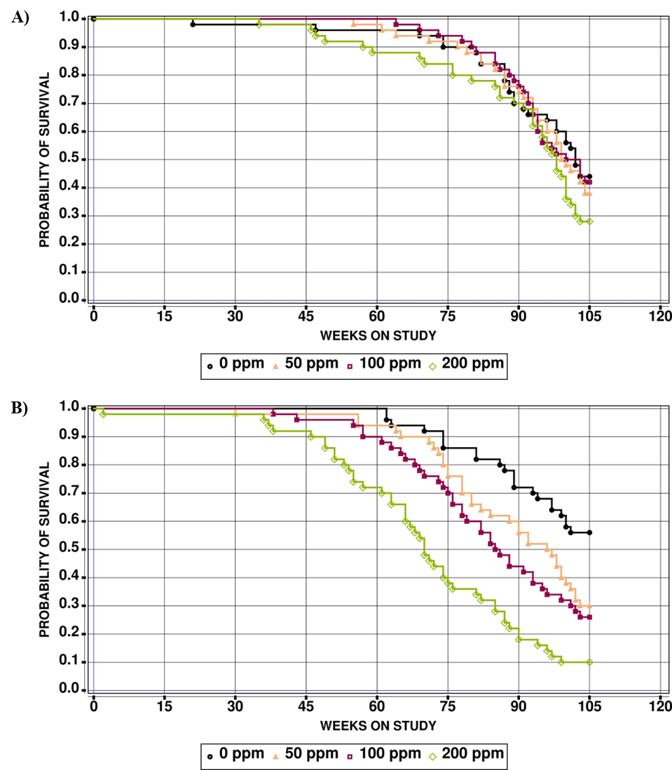
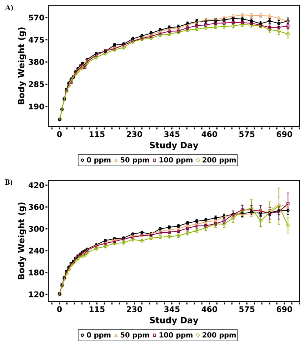
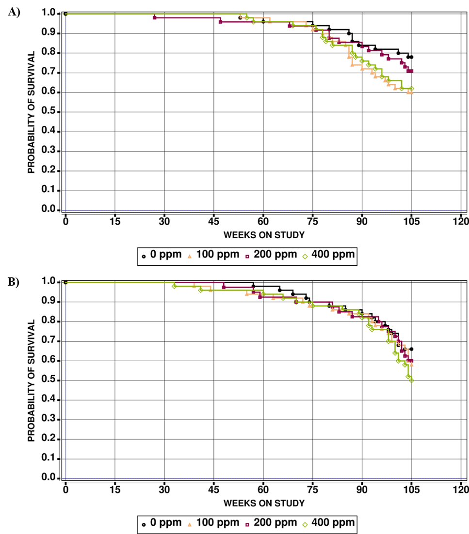
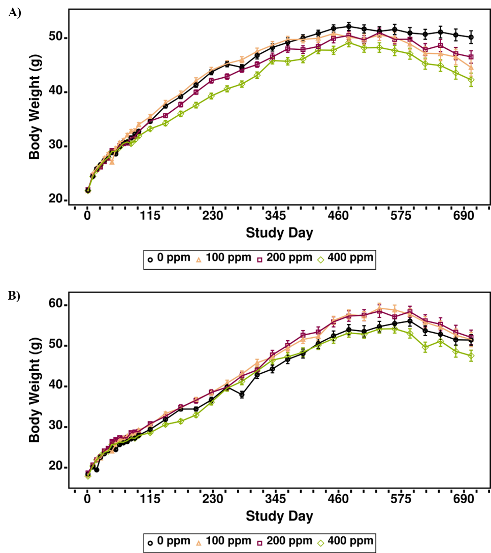
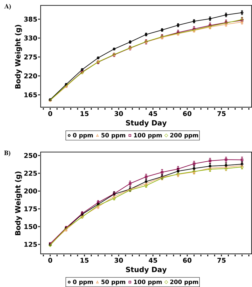
Male and female Sprague Dawley rats (n = 50/sex/exposure group) were exposed to 0, 50, 100, or 200 ppm α-pinene via whole-body inhalation (6 hours plus T90 per day for 5 days per week) for 2 years to investigate chronic toxicity and carcinogenicity. Survival of male rats in all exposed groups was comparable to that of control rats, whereas survival of exposed female rats was significantly decreased compared to control rats. Although there were significant decreases (≤10%) in body weights in male and female rats in the 100 and 200 ppm groups at different time points throughout the study, they were inconsistent throughout the study period and did not indicate overt toxicity. There were no clinical observations associated with α-pinene exposure in male rats, but female rats displayed an exposure-related higher incidence of vaginal discharge in all exposed groups.
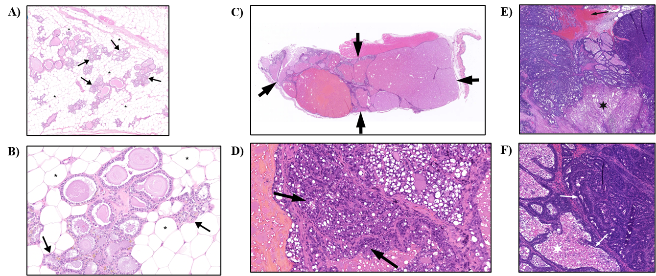
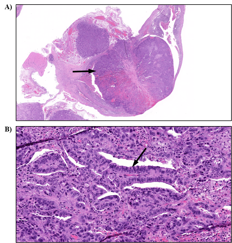
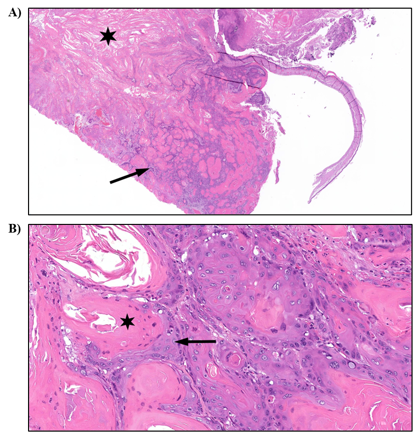
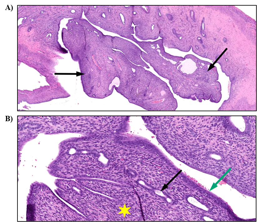
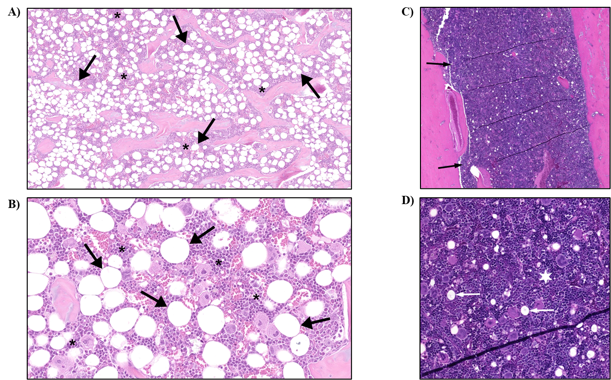
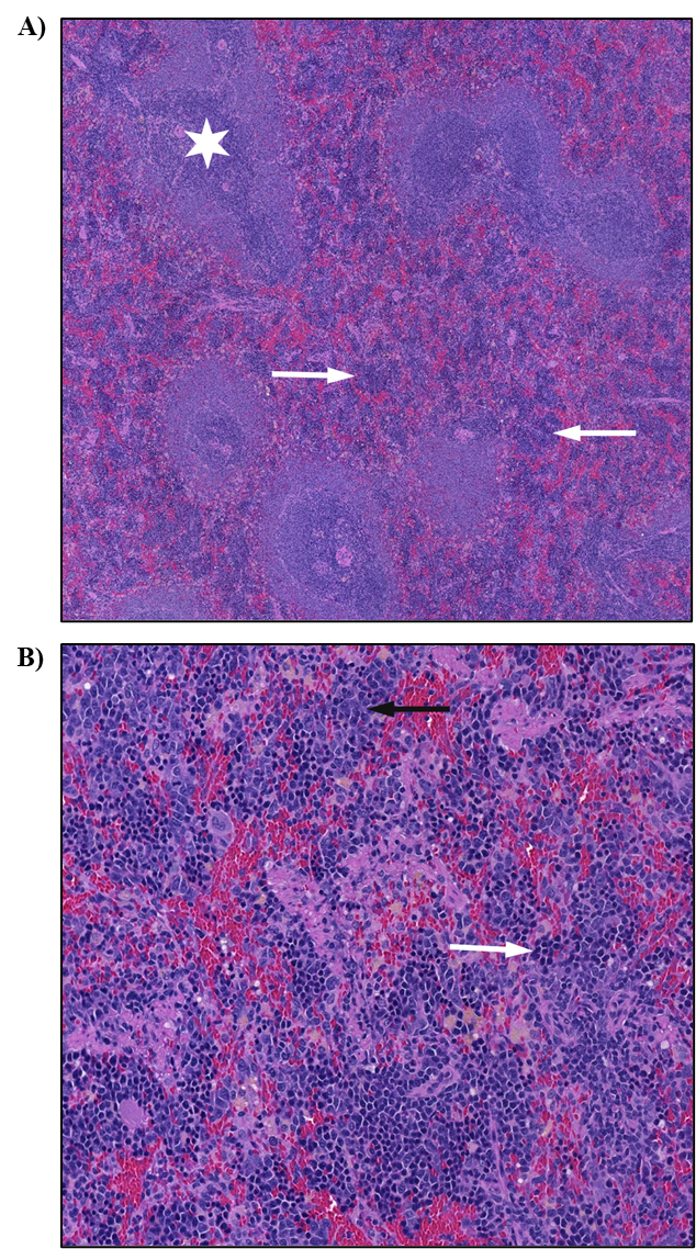
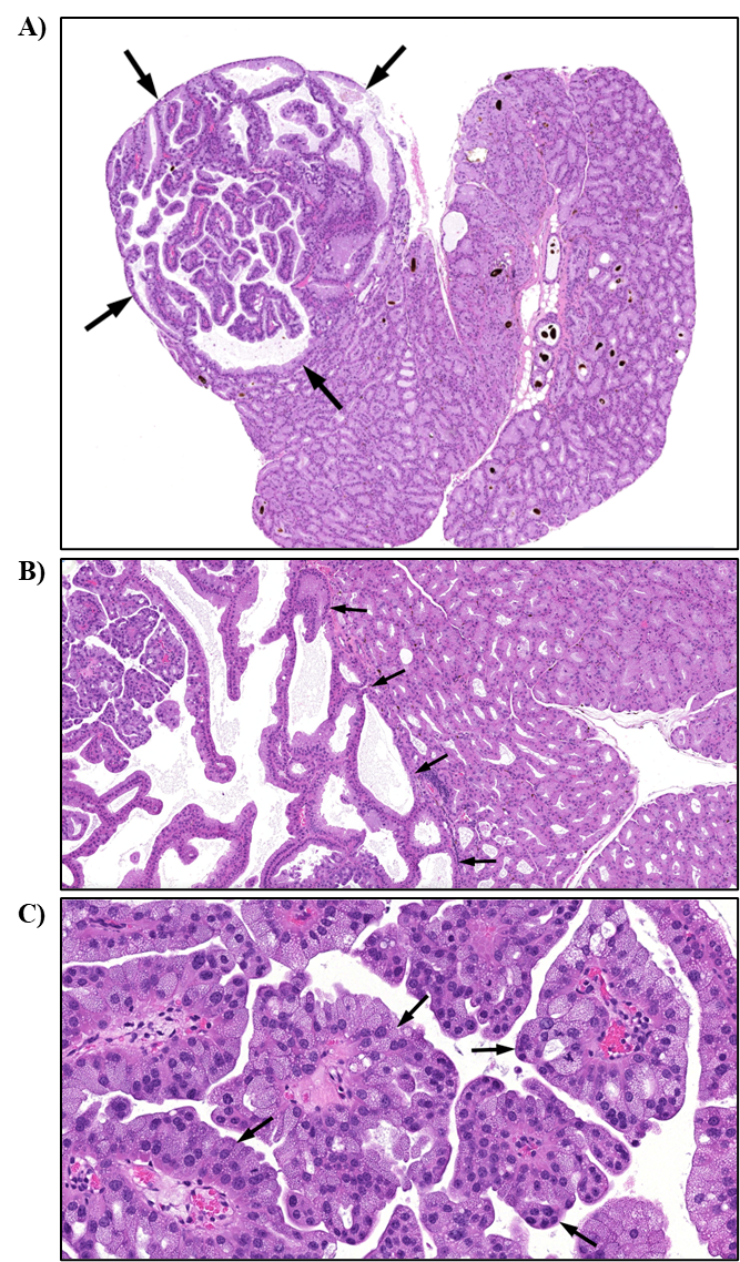
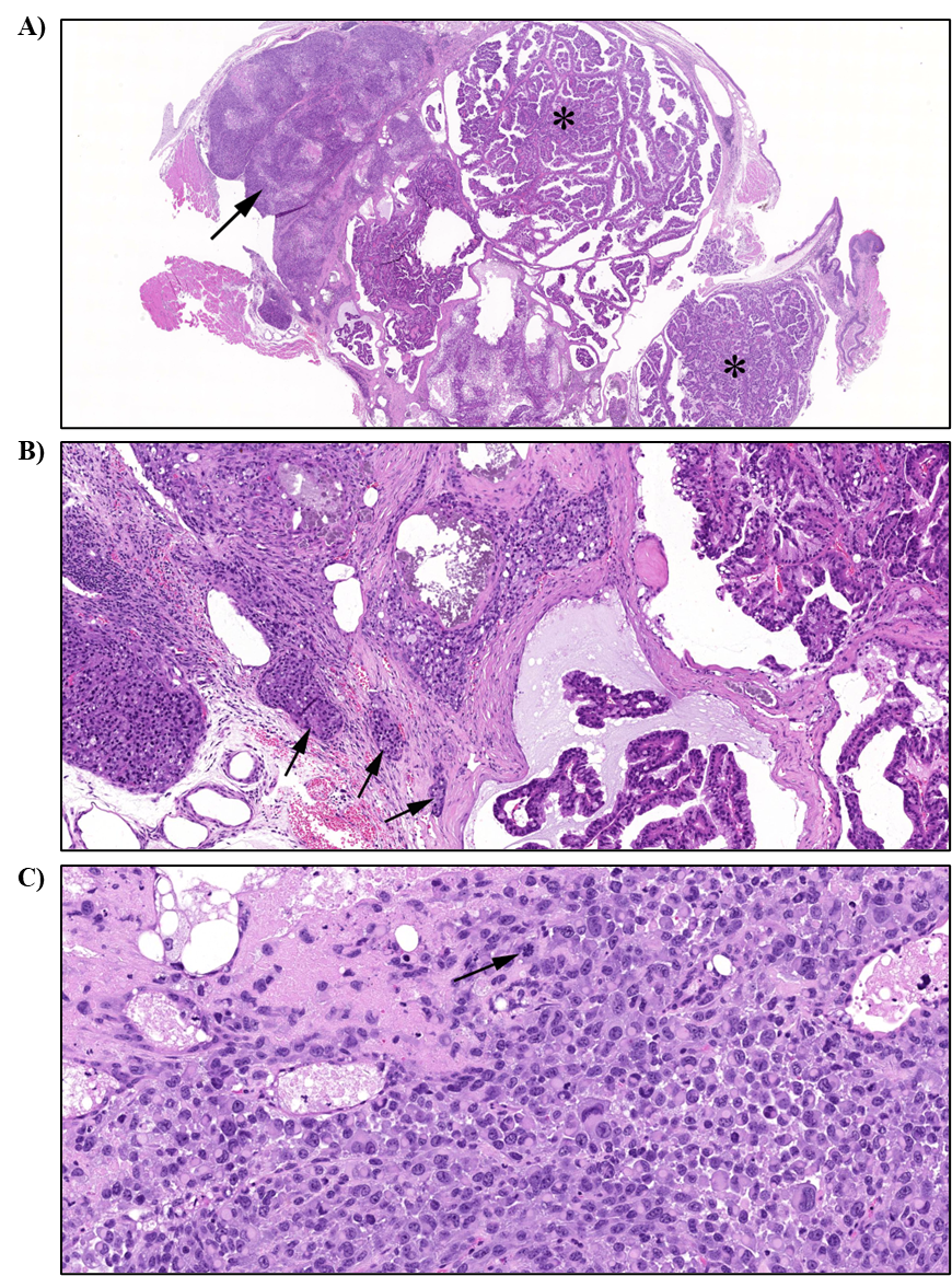
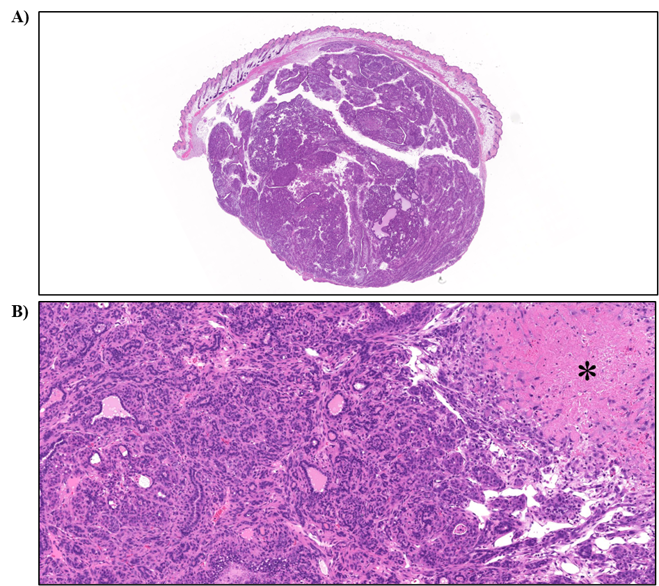
In female rats, the incidences of mammary gland adenocarcinoma and adenoma or adenocarcinoma (combined) were significantly increased in the 100 and 200 ppm groups. The incidences of adenocarcinoma; squamous cell carcinoma; squamous cell papilloma, squamous cell carcinoma, or adenoma or adenocarcinoma (combined); and stromal polyps in the uterus were significantly increased in the 200 ppm group. There were significant increases in the incidences of nonneoplastic lesions in the uterus, including atypical hyperplasia and stromal endometrium hyperplasia. Exposure-related significant increases in the incidences of hypercellularity in the bone marrow and increased extramedullary hematopoiesis in the spleen were also observed in female rats.
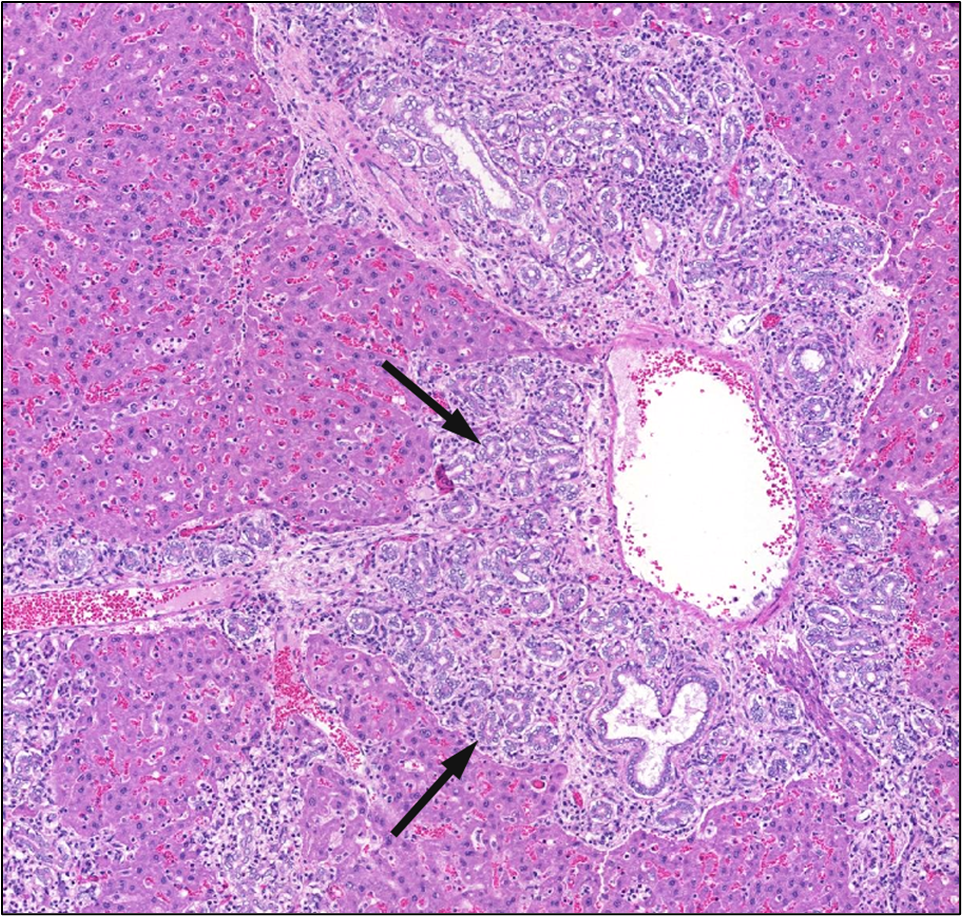
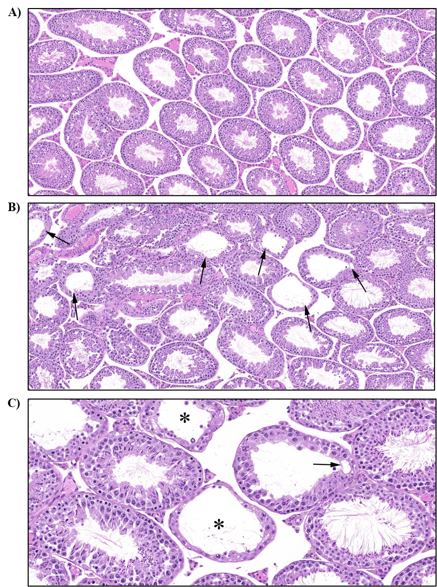
In male rats, there was a positive trend, and the incidence of papilloma in the urinary bladder was higher in the 200 ppm group relative to the control group. Increased incidences of nonneoplastic lesions in male rats included hypospermia in the epididymis, degeneration and degeneration or atrophy (combined) in the germinal epithelium of the testis, focal hyperplasia in the adrenal gland medulla, and bile duct hyperplasia in the liver.
α-Pinene and α-pinene oxide were present in blood and mammary gland.
Three-month Studies in Mice
Male CD-1 mice (n = 25/exposure group) and B6C3F1/N mice (n = 10/exposure group) were exposed to 0, 100, 200, or 400 ppm α-pinene via whole-body inhalation (6 hours plus T90 per day for 5 days per week) for 3 months for the reproductive assessment (see below) and analysis of the urinary bladder. Male B6C3F1/N mice displayed exposure-related urinary bladder nonneoplastic lesions, but there were no findings in CD-1 mice.
Two-year Study in Mice
Male and female B6C3F1/N mice (n = 50/sex/exposure group) were exposed to 0, 100, 200, or 400 ppm α-pinene via whole-body inhalation (6 hours plus T90 per day for 5 days per week) for 2 years to investigate chronic toxicity and carcinogenicity. α-Pinene exposure did not significantly affect survival of male or female mice. Male B6C3F1/N mice in the 400 ppm group had significantly decreased body weights beginning on approximately study day 80 and persisting until study termination, with a decrease of 16% compared to the control group on study day 703. Female B6C3F1/N mice did not display consistently different weights from control animals. Clinical observations were sporadic and not attributed to α-pinene exposure in male or female mice.
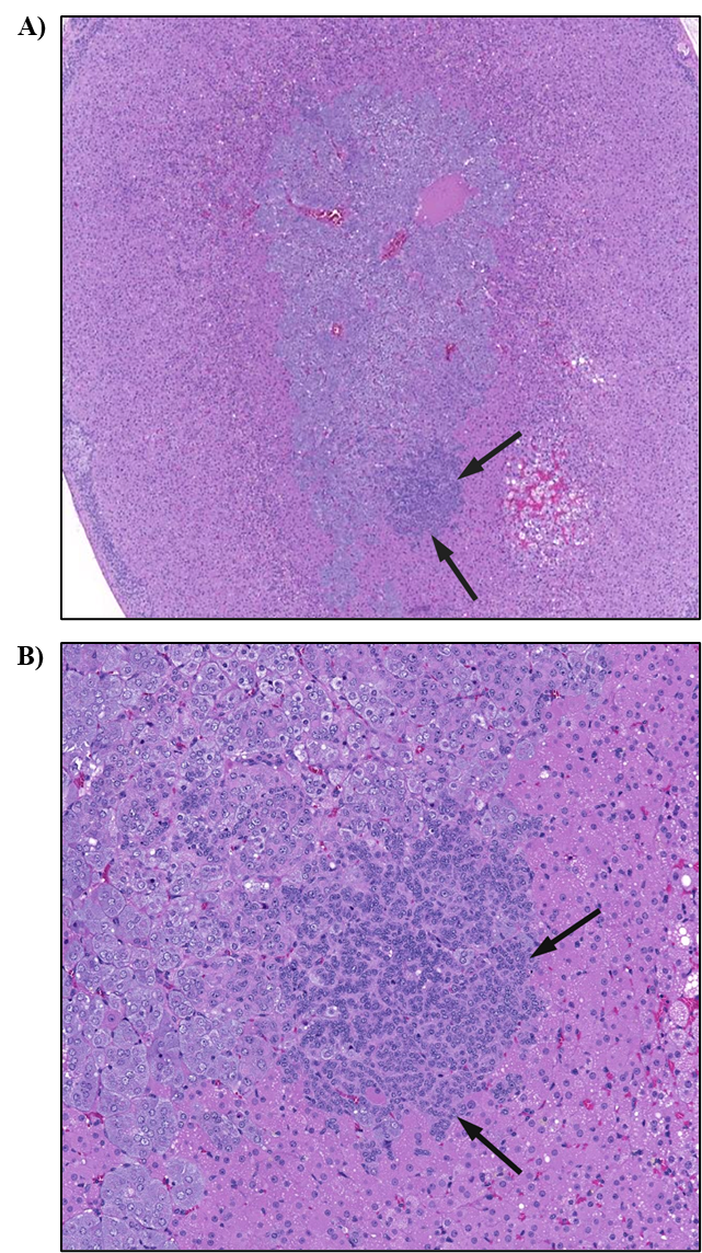
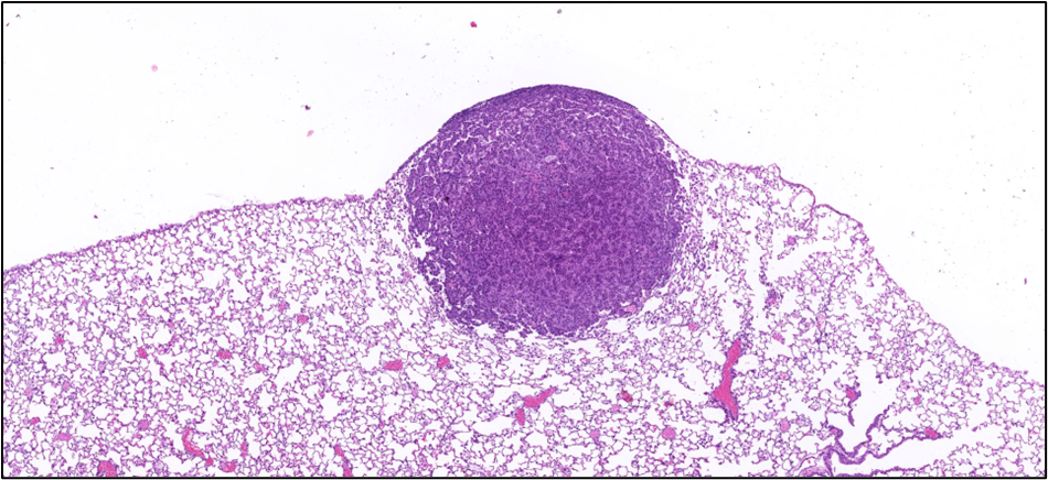
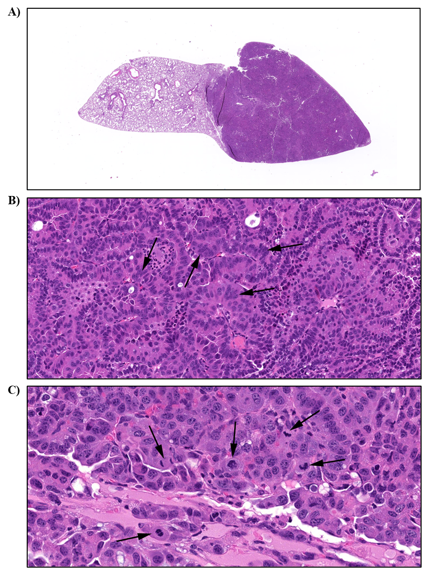
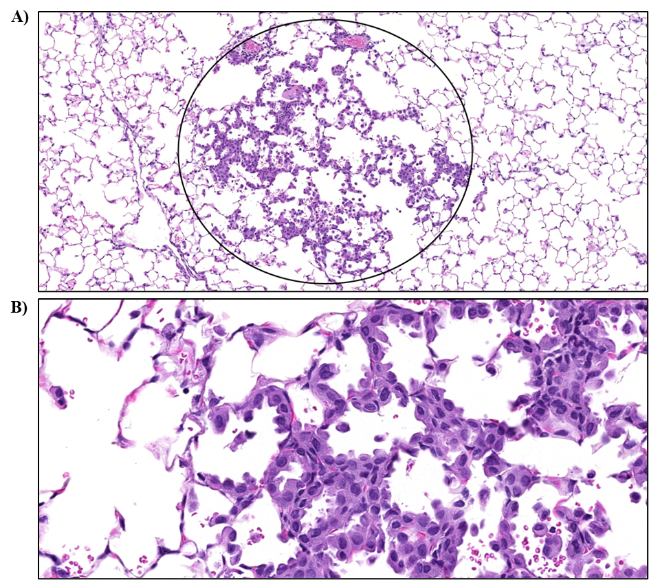
Male and female B6C3F1/N mice exhibited similar patterns of carcinogenicity in the Harderian gland, liver, and lung following exposure to α-pinene. The incidences of Harderian gland adenoma, adenocarcinoma, and adenoma or adenocarcinoma (combined) exhibited positive trends and were significantly increased in the 400, 400, and ≥200 ppm groups, respectively, in male mice, whereas the incidences of Harderian gland adenoma and adenoma or adenocarcinoma (combined) exhibited positive trends and were significantly increased at concentrations ≥200 ppm in female mice. In male and female mice, the incidences of hepatocellular adenoma, carcinoma, and adenoma or carcinoma (combined) exhibited positive trends and were significantly increased at concentrations of ≥200, 400, and ≥100 ppm α-pinene, respectively, in male mice, and ≥100, 400, and ≥100 ppm, respectively, in female mice. These neoplastic lesions in the liver were accompanied by an exposure-related significant increase in multinucleated hepatocyte, basophilic focus, and necrosis in male mice. There were positive trends in the incidences of alveolar/bronchiolar adenoma and adenoma or carcinoma (combined) in the lungs of male and female mice and alveolar/bronchiolar carcinoma in female mice. The incidences of alveolar/bronchiolar adenoma and adenoma or carcinoma (combined) were significantly increased at concentrations of 400 and ≥200 ppm α-pinene, respectively, in male mice, and the incidences of alveolar/bronchiolar adenoma, carcinoma, and adenoma or carcinoma (combined) were significantly increased at concentrations of ≥100, ≥200, and ≥100 ppm, respectively, in female mice. Significant increases in the incidence of alveolar/bronchiolar epithelium hyperplasia were observed in all exposed groups in both male and female mice.
In female mice, there were significant increases in the incidences of mammary gland adenocarcinoma and adenoma or adenocarcinoma (combined) in the 400 ppm group. The incidences of granulosa cell tumor, benign or malignant (combined) and tubulostromal adenoma in the ovary in the 400 ppm group were also significantly increased. An increased incidence in tubulostromal hyperplasia accompanied the neoplastic lesions in the ovary.
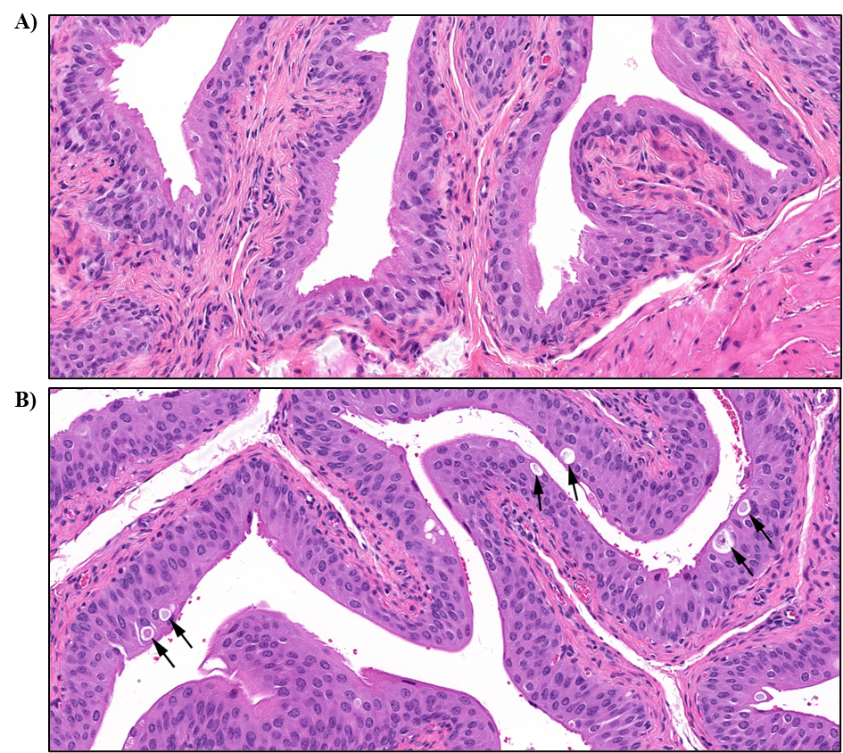
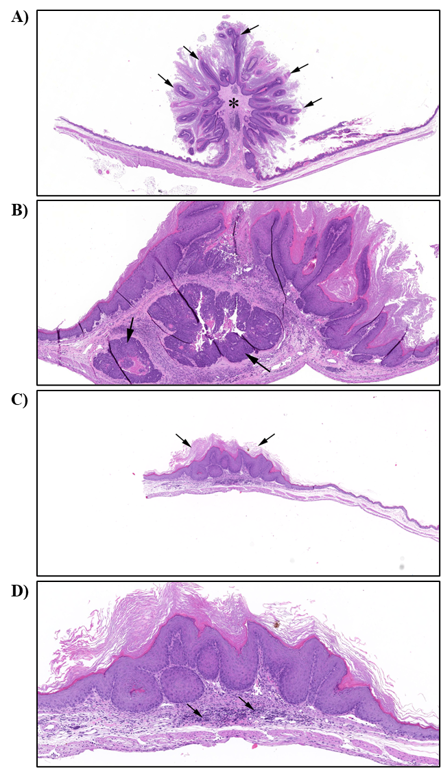
There was a positive trend and a higher incidence of urinary bladder papilloma in the 400 ppm group of male mice. Significant increases in the incidences of urinary bladder nonneoplastic lesions were observed, including urothelium hyperplasia in both male and female mice and lymphocytic cellular infiltration and suppurative inflammation in male mice. In male mice, there was a positive trend and higher incidence of forestomach papilloma in the 200 and 400 ppm groups. This finding was not noted in females, although there was a positive trend and higher incidence of squamous cell carcinoma and a significant increase in the incidence of hyperplasia of the forestomach epithelium in the 400 ppm group.
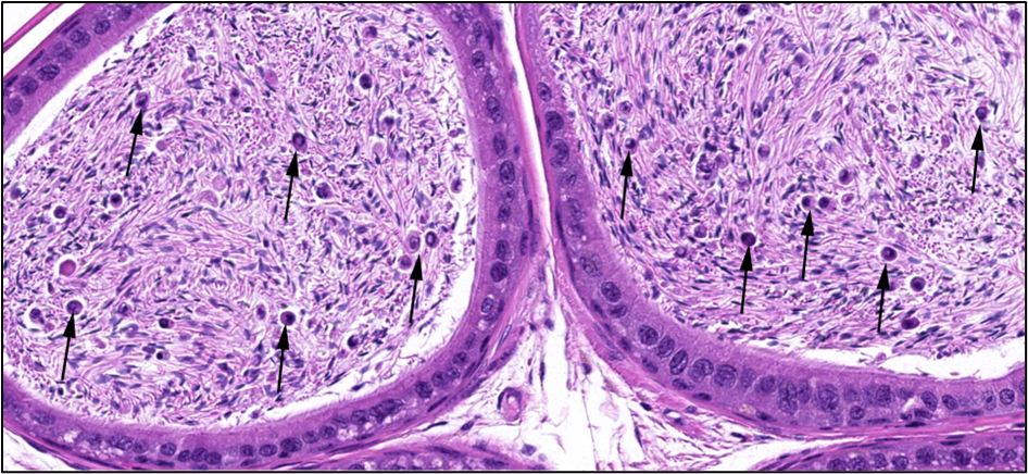
There were significantly increased incidences of degeneration and degeneration or atrophy (combined) in the germinal epithelium of the testis and exfoliated germ cells in the duct of the epididymis in the 400 ppm group.
α-Pinene and α-pinene oxide were present in blood and mammary gland.
Reproductive Performance in Rats and Mice
As described above, male Sprague Dawley rats (n = 25/exposure group), CD-1 mice (n = 25/exposure group), and B6C3F1/N mice (n = 10/exposure group) were exposed to 0, 100, 200, or 400 ppm α-pinene via whole-body inhalation (6 hours plus T90 per day for 5 days per week) for 3 months. Following exposure, male rats and CD-1 mice were mated with naïve females to evaluate reproductive performance. Epididymis and testis were weighed and evaluated histopathologically in all animals. Although collected, sperm parameters could not be evaluated because of artifacts (e.g., clumping of cells, too many sperm in sample).
Species differences in male reproductive tissue responses were noted with exposure-related effects observed in male rats and B6C3F1/N mice but not CD-1 mice. In male rats, absolute epididymis and testis weights were significantly decreased and nonneoplastic lesions in the testis were observed. While B6C3F1/N mice displayed significantly decreased absolute testis weights, no accompanying exposure-related histopathological lesions were found.
No exposure-related effects were seen in the number of females that were paired, mated, or became pregnant in rats or CD-1 mice. A negative trend was observed in the number of implantations per female in rats.
Three-month Investigative Study in Rats
Male and female Sprague Dawley rats (n = 10/sex/exposure group) were exposed to 0, 50, 100, or 200 ppm α-pinene via whole-body inhalation (6 hours plus T90 per day for 5 days per week) for 3 months to measure internal concentrations of α-pinene and α-pinene oxide and to collect tissue for development and analysis of early biomarkers of mammary gland carcinogenesis. In addition, select male reproductive tissues were weighed and examined histopathologically and vaginal cytology and sperm parameters were evaluated.
Blood α-pinene concentration increased with the exposure concentration in both male and female rats. Blood α-pinene oxide concentration was generally higher than the α-pinene concentration but did not increase with exposure concentration in male or female rats. In general, female rats had higher concentrations of α-pinene and α-pinene oxide than male rats. Mammary gland α-pinene and α-pinene oxide concentrations were much higher than those observed in blood.
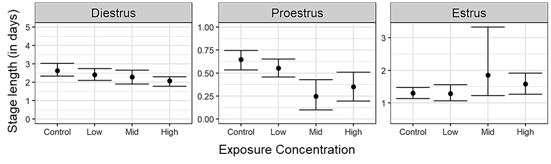
There was a marginal but significant decrease in proestrus cycle length in the 100 and 200 ppm groups compared to the control group.
Conclusions
Under the conditions of these 2-year inhalation studies, there was some evidence of carcinogenic activity of α-pinene in male Hsd:Sprague Dawley SD rats based on the higher incidence of urinary bladder papilloma. There was clear evidence of carcinogenic activity of α-pinene in female Hsd:Sprague Dawley SD rats based on the increased incidences of mammary gland adenocarcinoma and adenoma or adenocarcinoma (combined). Increased incidences of stromal polyp; adenocarcinoma; squamous cell carcinoma; and squamous cell papilloma, squamous cell carcinoma, adenoma, or adenocarcinoma (combined) in the uterus were also considered to be related to exposure.
There was clear evidence of carcinogenic activity of α-pinene in male B6C3F1/N mice based on the increased incidences of Harderian gland adenoma, adenocarcinoma, and adenoma or adenocarcinoma (combined); hepatocellular adenoma, carcinoma, and adenoma or carcinoma (combined); and alveolar/bronchiolar adenoma and adenoma or carcinoma (combined). Higher incidences of urinary bladder papilloma and forestomach papilloma were also considered to be related to exposure. There was clear evidence of carcinogenic activity of α-pinene in female B6C3F1/N mice based on the increased incidences of Harderian gland adenoma and adenoma or adenocarcinoma (combined); hepatocellular adenoma, carcinoma, and adenoma or carcinoma (combined); alveolar/bronchiolar adenoma, carcinoma, and adenoma or carcinoma (combined); and mammary gland adenocarcinoma and adenoma or adenocarcinoma (combined). Increased incidences of granulosa cell tumor, benign, malignant (combined) and tubulostromal adenoma in the ovary and a higher incidence of squamous cell carcinoma in the forestomach were also considered to be related to exposure.
Exposure to α-pinene resulted in increased incidences of nonneoplastic lesions in the epididymis, testis, adrenal gland, and liver of male rats; bone marrow, spleen, and uterus of female rats; epididymis, liver, lung, testis, and urinary bladder of male mice; and lung, ovary, urinary bladder, and stomach of female mice.
Under the conditions of these 3-month reproductive assessments in Hsd:Sprague Dawley SD rats and CD-1 mice, reproductive function was not affected by exposure.
Synonyms: acitene A; cyclic dexadiene; 2-pinene; 2,6,6-trimethylbicyclo[3.1.1]hept-2-ene
Summary of the Two-year Carcinogenesis Studies of α-Pinene
| Male Sprague Dawley Rats | Female Sprague Dawley Rats | Male B6C3F1/N Mice | Female B6C3F1/N Mice | |
|---|---|---|---|---|
| Concentrations in air | 0, 50, 100, or 200 ppm | 0, 50, 100, or 200 ppm | 0, 100, 200, or 400 ppm | 0, 100, 200, or 400 ppm |
| Survival rates | 22/50, 19/50, 21/50, 14/50 | 27/50, 15/50, 13/50, 5/50 | 39/50, 30/50, 34/50, 31/50 | 33/50, 29/50, 24/40, 24/50 |
| Body weights | ↓ (200 ppm group: 10% less than the control group at study termination) | ↓ (200 ppm group: <10% lower than the control group from study day 71 to 449) | ↓ (400 ppm group: 15.7% less than the control group at study termination) | No effect |
| Nonneoplastic effects | Testis: germinal epithelium, degeneration (includes bilateral) (12/50, 24/50, 20/50, 17/50); germinal epithelium, degeneration or atrophy (combined, includes bilateral) (13/50, 24/50, 20/50, 17/50) Epididymis: hypospermia (includes bilateral) (7/50, 3/50, 10/50, 14/50) Adrenal gland: medulla, hyperplasia, focal (includes bilateral) (9/50, 8/50, 12/50, 18/50) Liver: bile duct, hyperplasia (5/50, 25/50, 26/50, 19/50) | Uterus: hyperplasia, atypical (0/50, 0/49, 0/50, 3/49); endometrium, hyperplasia, stromal (0/50, 6/49, 3/50, 6/49) Bone marrow: hypercellularity (10/50, 22/50, 18/50, 24/48) Spleen: extramedullary hematopoiesis, increased (10/50, 28/49, 28/49, 34/50) | Liver: multinucleated hepatocyte (3/50, 11/50, 10/49, 23/50); basophilic focus (1/50, 2/50, 3/49, 7/50); necrosis (1/50, 3/50, 2/49, 7/50) Lung: alveolar/bronchiolar, epithelium, hyperplasia (2/50, 10/50, 20/50, 27/50) Urinary bladder: urothelium, hyperplasia (1/48, 7/43, 18/48, 15/45); infiltration cellular, lymphocyte (1/48, 2/43, 9/48, 9/45); inflammation, suppurative (0/48, 2/43, 0/48, 5/45) Testis: germinal epithelium, degeneration (includes bilateral) (14/50, 9/49, 15/50, 28/47); germinal epithelium, atrophy (includes bilateral) (2/50, 1/49, 3/50, 6/47); germinal epithelium, degeneration or atrophy (combined, includes bilateral) (14/50, 9/49, 16/50, 28/47) Epididymis: duct, exfoliated germ cell (includes bilateral) (2/50, 5/49, 2/50, 18/48) | Lung: alveolar/bronchiolar, epithelium, hyperplasia (0/50, 10/50, 6/39, 17/50) Urinary bladder: urothelium, hyperplasia (0/45, 5/43, 11/34, 20/46) Ovary: hyperplasia, tubulostromal (includes bilateral) (1/48, 18/49, 21/36, 28/46) Stomach, forestomach: epithelium, hyperplasia, focal (0/49, 0/49, 0/38, 5/49) |
| Neoplastic effects | Urinary bladder: papilloma (0/50, 0/50, 0/50, 3/50) | Mammary gland: adenocarcinoma (5/49, 7/50, 11/50, 25/50); adenoma or adenocarcinoma (combined) (6/49, 7/50, 15/50, 25/50) Uterus: adenocarcinoma (1/50, 7/49, 3/50, 4/49); squamous cell carcinoma (0/50, 0/49, 1/50, 3/49); squamous cell papilloma, squamous cell carcinoma, adenoma or adenocarcinoma (combined) (1/50, 7/49, 4/50, 6/49); polyp, stromal (5/50, 2/49, 4/50, 8/49) | Harderian gland: adenoma (includes bilateral) (6/50, 9/50, 11/50, 16/50); adenocarcinoma (includes bilateral) (0/50, 1/50, 3/50, 7/50); adenoma or adenocarcinoma (combined) (6/50, 9/50, 14/50, 23/50) Liver: hepatocellular adenoma (includes multiple) (23/50, 26/50, 31/49, 33/50); hepatocellular carcinoma (includes multiple) (18/50, 26/50, 17/49, 36/50); hepatocellular adenoma or hepatocellular carcinoma (combined, includes multiple) (33/50, 40/50, 40/49, 46/50) Lung: alveolar/bronchiolar adenoma (includes multiple) (5/50, 8/50, 10/50, 18/50); alveolar/bronchiolar adenoma or alveolar/bronchiolar carcinoma (combined, includes multiple) (14/50, 14/50, 21/50, 26/50) Urinary bladder: papilloma (0/48, 0/43, 0/48, 3/45) Stomach, forestomach: papilloma (0/49, 0/49, 2/50, 3/48) | Harderian gland: adenoma (includes bilateral) (3/50, 6/49, 11/40, 16/50); adenocarcinoma (includes bilateral) (1/50, 2/49, 3/40, 6/50); adenoma or adenocarcinoma (combined) (4/50, 8/49, 13/40, 22/50) Liver: hepatocellular adenoma (includes multiple) (6/50, 15/50, 20/38, 28/50); hepatocellular carcinoma (includes multiple) (7/50, 14/50, 11/38, 20/50); hepatocellular adenoma or hepatocellular carcinoma (combined, includes multiple) (12/50, 27/50, 24/38, 39/50) Lung: alveolar/bronchiolar adenoma (includes multiple) (2/50, 8/50, 9/39, 17/50); alveolar/bronchiolar carcinoma (includes multiple) (1/50, 2/50, 6/39, 23/50); alveolar/bronchiolar adenoma or alveolar/bronchiolar carcinoma (combined, includes multiple) (3/50, 10/50, 15/39, 37/50) Mammary gland: adenocarcinoma (0/48, 0/46, 0/40, 5/48); adenoma or adenocarcinoma (combined) (0/48, 0/46, 1/40, 5/48) Ovary: granulosa cell tumor, benign, malignant (combined) (0/48, 3/49, 2/36, 5/46); tubulostromal adenoma (0/48, 0/49, 1/36, 7/46) Stomach, forestomach: squamous cell carcinoma (0/49, 0/49, 0/38, 2/49) |
| Equivocal findings | Nonea | None | None | None |
| Level of evidence of carcinogenic activity | Some evidence | Clear evidence | Clear evidence | Clear evidence |
Introduction
Chemical and Physical Properties
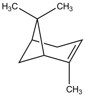
α-Pinene (Figure 1) is a bicyclic monoterpene within the diverse terpene class of compounds. It is a fragrant volatile organic compound emitted from natural sources as well as anthropogenic activities. α-Pinene exists naturally as a mixture of (+) and (−) enantiomers. It is a clear colorless liquid with a boiling point of 155°C and a vapor pressure of 4.75 mm Hg. α-Pinene is poorly soluble in water (1.82 × 10⁻⁵ mol/L) and has a logP (octanol:water partition coefficient) value of 4.83.8,9 Once in the atmosphere, α-pinene can react with ozone to create secondary organic aerosols.10
Production, Use, and Human Exposure
α-Pinene occurs naturally in a wide variety of plants, such as pine trees,11 rosemary and other herbs,12 and cannabis.13 Various essential oils (i.e., hydrophobic liquid fractions extracted from plants) contain α-pinene (e.g., turpentine contains 44% to 94% α-pinene) along with other volatile hydrocarbons.14 The ratio of (+) to (−) enantiomers of α-pinene can differ across plant species and plant parts, with ranges of 27%–62% (+) α-pinene and 38%–73% (−) α-pinene measured in Norway spruce (Picea abies), Scots pine (Pinus sylvestris), and juniper (Juniperus communis) in one study15 and ranges of 72%–89% (+) α-pinene and 11%–28% (−) α-pinene measured in loblolly pine (Pinus taeda) in another study.16 Much larger ranges of enantiomeric ratios were measured in cannabis (Cannabis sativa) at 0%–90% (+) α-pinene and 10%–100% (−) α-pinene.17 Turpentine has been used in complementary medicine as a topical antibacterial ointment,18 and pine bark extract is a common dietary supplement ingredient.19 α-Pinene, within essential oil mixtures or as a pure compound, is widely used as a flavor and fragrance ingredient. The daily per capita intake of α-pinene in the United States from food was estimated to be 317 µg/day.20 The fragrance profile of α-pinene has made it a popular ingredient in consumer products. It can be found in personal care products, air fresheners, and cleaners.21-23 The fragrance associated with α-pinene differs between enantiomers, with (−) α-pinene having a pine scent and (+) α-pinene having a slightly minty scent.24
A summary of literature reporting exposure to α-pinene from consumer products, occupational exposures, and indoor air environments is available in Appendix G. Occupational exposure to α-pinene in the lumber industry has been widely reported. In a Canadian softwood lumber mill, α-pinene levels were measured ranging from below the limit of detection (LOD; 0.3 µg/m3) to 5.1 mg/m3 (approximately 0.92 ppm).25 New Zealand plywood mill workers were exposed to α-pinene concentrations with geometric means ranging from 0.5 to 2.4 mg/m3 (approximately 0.09 to 0.43 ppm).26 Higher levels of α-pinene were measured in Finnish saw mills with arithmetic means in the range of 57 to 152 mg/m3 (approximately 10 to 27 ppm) and total monoterpenes (α-pinene, β-pinene, Δ3-carene, and limonene) reaching 326 mg/m3 (approximately 59 ppm).27 Similarly, total terpenes measured in a Swedish saw mill ranged from 100 to 550 mg/m3 (approximately 18 to 99 ppm), with an average of 254 mg/m3 (approximately 46 ppm).28 While α-pinene has been measured in other occupational settings with anticipated exposure (e.g., greenhouses or industries in which cleaning and consumer products containing α-pinene are widely used), levels appear to be an order of magnitude lower than in the lumber industry. For example, in a National Institute for Occupational Safety and Health (NIOSH) study measuring volatile organic compounds in health care workers, α-pinene exposures were in the low ppb range (median concentrations <0.25 ppb).29
α-Pinene is frequently detected in studies evaluating indoor air quality. For example, α-pinene was measured in 100% of indoor air samples from three cities in Michigan, with overall mean, median, and maximum values of 9.04, 3.16, and 139.2 µg/m3 (0.0016, 0.00057, and 0.025 ppm), respectively.30 In a study of emissions from a newly manufactured house, α-pinene was measured at 0.042 ppm.31 Concentrations of α-pinene inside taxis ranged from 0.2 to 1.8 µg/m3 (approximately 0.00004 to 0.00032 ppm).32 A review of the effects of excessive fragrances on indoor air quality cataloged mean and maximum concentrations of α-pinene ranging from 0.1 to 32 µg/m3 (approximately 0.00002 to 0.00574 ppm) and 0.7 to 854 µg/m3 (approximately 0.00013 to 0.15 ppm), respectively.33
In the United States, occupational exposure limits pertain to turpentine and are not specific to αpinene. The NIOSH-recommended exposure limit and the Occupational Safety and Health Administration (OSHA) permissible exposure limit for turpentine are both 100 ppm, whereas the American Conference of Governmental Industrial Hygienists (ACGIH) threshold limit value is 20 ppm.34-36 Belgium, Canada, and Switzerland each have an occupational exposure limit for α-pinene of 20 ppm, whereas Sweden’s limit is 25 ppm.37
Absorption, Distribution, Metabolism, and Excretion
Experimental Animals
As part of the NTP research program on α-pinene, the toxicokinetic (TK) behavior of α-pinene and its potential reactive metabolite, α-pinene oxide, was investigated in the blood and mammary gland following whole-body inhalation exposure of adult male and female Sprague Dawley (Hsd:Sprague Dawley SD) rats and B6C3F1/N mice to α-pinene for 6 hours per day for 7 days at 50 ppm (279 and 282 mg/m3 for rats and mice, respectively) or 100 ppm (557 and 561 mg/m3 for rats and mice, respectively).6 The focus on α-pinene oxide was based on its status as a metabolite of α-pinene38 and the potential reactivity based on its structure.
In both rats and mice, the systemic exposure to α-pinene increased with exposure concentration. Given the blood maximum concentration (Cmax) and area under the curve (AUC), the exposure in female rats was approximately 1.5-fold and 2-fold higher, respectively, than in male rats.6 However, unlike in rats, no sex difference in systemic exposure to α-pinene was found in mice. When the magnitude of systemic exposure to α-pinene was compared between rats and mice, male and female rats had higher exposures than male and female mice. α-Pinene was eliminated from blood with half-lives of 12.2–17.4 hours for rats and 6.18–19.4 hours for mice. The long elimination half-life suggests α-pinene has a high affinity for poorly perfused tissues.
α-Pinene oxide was measured in the blood of rats and mice of both sexes, and concentrations were lower than α-pinene. In general, α-pinene oxide increased with the exposure concentration of α-pinene in rats and mice. The ratio of αpinene oxide to α-pinene was higher in rats (Cmax ratio 0.200–0.237; AUC ratio 0.279–0.615) than in mice (Cmax ratio 0.060–0.086; AUC ratio 0.036–0.105) at 50 ppm, and females had higher values than males in both species. The ratio was lower at the higher exposure concentration of 100 ppm, pointing to saturation of pathways leading to the formation of α-pinene oxide, and the decrease was more evident in rats than in mice. There were no apparent species or sex differences in the ratio of α-pinene oxide to α-pinene at 100 ppm. The blood elimination half-life of α-pinene oxide in rats was 11.6–19.2 hours, with no apparent exposure concentration- or sex-related differences. The blood elimination half-life of α-pinene oxide in mice was shorter than in rats, with male mice having a longer half-life than female mice (7.70–7.91 hours versus 1.31–2.37 hours, respectively) and with no apparent exposure concentration-related difference.6
α-Pinene and α-pinene oxide were distributed to the mammary glands of rats and mice of both sexes. α-Pinene and α-pinene oxide concentrations in mammary glands were ≥23-fold higher than those in blood, demonstrating retention of these analytes in lipid-rich tissues. Many of the patterns described in blood were also observed in mammary glands, with female rats having higher concentrations than male rats and rats having higher concentrations than mice.
Humans
The TK behavior of α-pinene has been reported in male volunteers (n = 8) following a single inhalation exposure to 10, 225, or 450 mg/m3 (1.8, 40, or 81 ppm, respectively) of (+) αpinene for 2 hours during mild physical exercise.39 During exposure, α-pinene blood concentrations first increased rapidly, then leveled off, reaching a maximum at the termination of exposure, before declining postexposure. α-Pinene blood concentrations at the termination of exposure increased linearly with the exposure concentration. For the 10 and 225 mg/m3 exposures, blood α-pinene concentrations were below the LOD (10 nmol/L) 4 hours after exposure; hence, TK parameters were provided only for the 450 mg/m3 group. The elimination of α-pinene from blood was triphasic with alpha-, beta-, and gamma-phase half-lives of 4.8 minutes (0.08 hours), 38 minutes (0.63 hours), and 695 minutes (11.6 hours), respectively. The estimated clearance up to 21 hours after exposure at 450 mg/m3 was 1.09 L/h*kg. The authors reported no difference in the observed TK behavior following exposure to 450 mg/m3 (−) α-pinene enantiomer.39 In another investigation, following inhalation of α-pinene as turpentine, the observed TK behavior of α-pinene in male volunteers was demonstrated to be similar.40 Urinary metabolites identified following inhalation exposure in humans (sex not specified) were cis- and trans-verbenol and myrtenol.41,42
Toxicity
Experimental Animals
Previous NTP studies investigated the toxicity of α-pinene in male and female Fischer 344 (F344/N) rats and B6C3F1/N mice following whole-body inhalation exposure 6 hours per day, 5 days per week for 2 weeks and 3 months.2 In the 2-week studies, following exposure to 0, 100, 200, 400, 800, or 1,600 ppm α-pinene, a significant decrease in survival of male and female rats and mice in the 800 and 1,600 ppm groups was observed. Clinical signs of toxicity were observed in rats exposed to ≥400 ppm and mice exposed to ≥800 ppm, as well as significantly increased absolute and/or relative liver weights in both species. In the 3-month studies following exposure to 0, 25, 50, 100, 200, or 400 ppm, all animals survived to the end of the study except six out of ten 400 ppm female rats that died before study termination. The major target organs were the urinary system (kidney of rats and urinary bladder of mice) and the male reproductive system.2 In prenatal developmental studies with gavage administration of an essential oil containing α-pinene (20% to 25%), β-pinene (15% to 18%), and sabinene (38% to 42%), the number of live offspring per dam was significantly decreased at doses ≥120 and ≥56 mg/kg body weight/day (mg/kg/day) in CD-1 mice and Wistar rats, respectively.43 α-Pinene was positive in an acute dermal irritation assay.44 Results in skin sensitization assays were mixed. α-Pinene tested negative in a guinea pig maximization test44 and a murine local lymph node assay,45 but was positive in an open epicutaneous test in guinea pigs.46
Humans
In humans, reports of toxicity resulting from α-pinene alone or terpene mixtures containing αpinene indicate potential respiratory and skin irritation. Johard et al.47 assessed the effects of short-term inhalation exposure to a terpene mixture (α-pinene, β-pinene, and Δ3-carene) on bronchioalveolar lavage fluid from eight healthy volunteers and found that macrophage and mast cell counts increased following exposure to 450 mg/m3. Irritation of the eyes, nose, and throat has been observed in volunteers exposed to 450 mg/m3 α-pinene.39 Dermal exposure to α-pinene has been associated with an allergic response.48
Reproductive and Developmental Toxicity
Limited data are available on the reproductive and developmental toxicity of α-pinene. In the NTP 3-month toxicity inhalation studies of α-pinene in male and female F344/N rats and B6C3F1/N mice, significantly lower numbers of sperm per cauda compared to the chamber control group were observed in the 200 and 400 ppm male rats (19% lower) and 100, 200, and 400 ppm male mice (24%, 33%, and 40% lower, respectively). Mouse testis weight in the 400 ppm group was 7% lower than that of the control group.2 In a screening-level hazard characterization of bicyclic terpene hydrocarbons, the U.S. Environmental Protection Agency referenced a series of developmental toxicity studies in CD-1 mice, Wistar Han rats, and golden hamsters dosed via gavage on gestation day (GD) 6 through GD 15 with an essential oil containing 20%–25% α-pinene, 15%–18% β-pinene, and 38%–42% sabinene.43 Significant decreases in live offspring per dam were reported in mice at 120 and 560 mg/kg/day and rats at 56 and 260 mg/kg/day but not in hamsters.43
Carcinogenicity
Limited carcinogenicity data are available for α-pinene. Two epidemiological studies have examined associations between turpentine or terpene exposure in occupational settings and cancer outcomes. In a case-control study of Finnish woodworkers, a weak association (odds ratio [OR] = 1.33; 95% confidence interval [CI]: 0.78, 2.27 for any exposure to terpenes lasting over 1 month) was found between respiratory cancer and exposure to terpenes (primarily α-pinene and Δ3-carene) and other heating products of pine and spruce.49 Another case-control study found an association between paternal exposure to turpentine and neuroblastoma in offspring (OR = 1.9; CI: 1.0, 3.6 to 10.4; CI: 2.4, 44.8, depending on methods for exposure categorization).50 The literature contains limited studies on the chronic toxicity of α-pinene. A single study in rats, aimed at characterizing potential chronic nephrotoxicity associated with turpentine exposure, used a rudimentary inhalation chamber to expose animals to turpentine at unknown concentrations and found no exposure-related histopathological changes in the kidney following up to 293 days of exposure.51
Genetic Toxicity
Results from previous NTP genotoxicity tests, including the original Ames test with α-pinene at doses up to 10,000 μg/plate in three strains of bacteria, as well as in vivo peripheral blood micronucleus tests conducted in male and female B6C3F1/N mice that were integrated into the 3-month NTP toxicity studies, were reported previously in TOX 81.2 Both tests yielded negative results, indicating that in these two assays, α-pinene was not mutagenic and did not induce chromosomal alterations.
In contrast, a recent study demonstrated that the metabolite α-pinene oxide was highly mutagenic in the Ames test.5 The authors proposed that α-pinene is metabolized to α-pinene oxide in the standard Ames test when in the presence of rat liver S9, but the concentrations of α-pinene oxide generated by S9 are approximately threefold lower than the lowest dose (25 μg/plate) of α-pinene oxide that showed mutagenic activity. Therefore, metabolism may be a factor to consider in all in vitro assays conducted with the parent compound α-pinene.
Consistent with the earlier NTP data, results of several other mutation assays with α-pinene that used multiple strains of bacteria, with and without rat liver S9 mix, were also negative.52-54 Two other monoterpenes, β-myrcene and α-terpinene, were shown to be nonmutagenic in several strains of Salmonella typhimurium, with and without rat liver S9.54,55 In contrast, pine bark extract induced mutations in S. typhimurium strains TA100 (only with S9) and TA98 (with and without S9),56 two strains that detect different mechanisms of mutation. Pine bark extract is the only monoterpene-related substance tested in NTP studies that has shown mutational potential in bacteria and, for strain TA98, both directly (without S9) and after metabolic activation (with S9). However, the increases in mutational events induced by pine bark extract were seen only at high doses that generally exceeded the Organisation for Economic Co-operation and Development (OECD)-recommended assay limit dose.56,57
No in vivo studies have been reported for α-pinene aside from the NTP in vivo micronucleus study referenced above, but a few reports from in vitro mammalian cell assays have examined DNA damage and chromosomal alterations following exposure to α-pinene. No induction of DNA damage, as measured by the comet assay, was detected in human lung A549 cells exposed to α-pinene in air in a closed system at concentrations ranging from 1 to 1,800 mg/m3.58 In contrast, exposure of Chinese hamster V79-C13 cells to α-pinene in cell culture medium was reported to induce genetic damage in the form of DNA damage (comet assay), micronuclei, and chromosome breaks; the effective concentrations of α-pinene ranged from 25 to 35 µM.59 These authors also reported an increase in endoreduplication and aberrant mitotic figures in α-pinene-treated cells, indicating interference with microtubule functions. A similar study measured DNA damage, micronucleus induction, and chromosomal damage in cultured human lymphocytes treated with α-pinene concentrations ranging from 0 to 200 µg/mL; in this study, no increases in any of the three endpoints of genotoxicity were observed, despite a decrease in cell viability at 200 µg/mL.60
The negative results with α-pinene in the in vitro human cell studies are interesting in light of the results from a recent human biomonitoring study of artists who were exposed daily to turpentine used as an oil paint thinner.61 In this study, blood samples were obtained from turpentine-exposed subjects and from nonexposed age- and sex-matched control subjects, and lymphocytes were cultured and evaluated in the cytokinesis-block micronucleus assay. A significant increase in micronucleated binucleated lymphocytes was observed in the turpentine-exposed group, along with other indicators of nuclear damage, such as nuclear buds, which are evaluated in this assay. Cytotoxicity was also significantly higher in the cell cultures derived from the exposed group compared with the nonexposed control samples. The researchers further stratified their exposed subjects by duration of turpentine use in preparing their paints and determined that micronucleus frequencies increased significantly with prolonged exposure.
It is possible that cytogenetic damage in exposed humans may be induced by certain constituents of turpentine or reactive metabolites of turpentine such as α-pinene oxide. α-Pinene oxide was also shown to be a potent mutagen in the Ames test, in contrast to the lack of mutagenicity observed with the parent compound α-pinene.5 Reactive metabolites may not be produced in sufficient amounts in human cell cultures to induce measurable genotoxicity.
Study Rationale
The International Union of the United Auto Workers originally nominated turpentine to NTP for comprehensive assessment because of widespread exposure potential and a lack of chronic toxicity data. α-Pinene was selected for study because it is both a major constituent of turpentine and has a wider exposure profile from its use as a fragrance and flavoring ingredient. An inhalation route was chosen to reflect occupational exposure in the lumber and cleaning industries, as well as exposure through the use of consumer products such as air fresheners and perfumes. Previously reported NTP 2-week and 3-month inhalation toxicity studies with α-pinene in male and female F344/N rats and B6C3F1/N mice revealed an exposure-related significant increase in hyperplasia in the urinary bladder of mice, a potentially preneoplastic lesion, and significantly decreased cauda epididymal sperm in both rats and mice.2 Based on these findings, 2-year inhalation studies in Sprague Dawley rats and B6C3F1/N mice and a reproductive assessment following a 90-day exposure in male Sprague Dawley rats and CD-1 mice that were mated with naïve females were conducted. During the chronic study, early deaths observed in female rats were attributed to mammary masses or nodules and prompted a follow-up investigative 3-month study in male and female rats to evaluate early biomarkers of carcinogenicity in the mammary gland and measure internal concentrations of α-pinene and α-pinene oxide. Additionally, sperm parameters were evaluated because of the lack of sperm data from the reproductive assessment.
Materials and Methods
Procurement and Characterization of α-Pinene
α-Pinene was obtained from The John Walsh Company, Inc. (Ringwood, NJ) in one lot (A-9211). Identity, purity, and stability analyses were conducted by the analytical chemistry laboratory at RTI International (Research Triangle Park, NC) and the study laboratory at Battelle (Columbus, OH) (Appendix A). Reports on analyses performed in support of α-pinene studies are on file at the National Institute of Environmental Health Sciences (NIEHS).
Lot A-9211, a clear oily liquid at room temperature, was identified as α-pinene by the analytical chemistry laboratory using gas chromatography (GC) with mass spectrometry (MS) detection and by the study laboratory using infrared (IR) and 1H and 13C nuclear magnetic resonance (NMR) spectroscopies. All spectra were consistent with reference spectra and the anticipated structures of the test article (Appendix A). Enantiomeric composition was determined to be approximately 68% (+) α-pinene [(1R)-(+)-α-pinene] and approximately 32% (−) α-pinene [(1S)-(−)-α-pinene] with chiral GC/MS and optical polarimetry by the analytical chemistry laboratory and with GC with flame ionization detection (FID) by the study laboratory. Elemental analysis was performed by Galbraith Laboratories, Inc. (Knoxville, TN) to aid in identification. The elemental compositions were within 4% of theoretical values for α-pinene.
Purity of lot A-9211 was evaluated by GC/MS at both laboratories and by GC/FID at the study laboratory. Four reportable impurities with peak areas between 0.09% and 0.63% of the total integrated peak area were detected. Three of the reportable impurities were identified as camphene, tricyclene, and β-pinene. The fourth was not conclusively identified. Lot A-9211 was also analyzed by GC/MS for butylated hydroxytoluene (BHT), a commonly used antioxidant for preventing radical-mediated oxidation of compounds, and it was confirmed to be BHT-free. Moisture content was determined by Karl Fischer titration at Galbraith Laboratories, Inc. (Knoxville, TN) and yielded an average water content of <0.38%. The overall purity was determined to be >98%.
During the studies and between the 2-year and 3-month investigative rat studies, bulk α-pinene was stored in the original shipping containers at room temperature. Periodic reanalysis of the bulk chemical was performed by the study laboratory before and after the study, at regular intervals during each chronic study, and during all studies using GC/FID, and no degradation was detected.
Vapor Generation and Exposure System
A diagram of the vapor generation and delivery system used in the studies is shown in Figure A-5. The test chemical, α-pinene, was pumped from an 8-gallon stainless steel reservoir into a heated glass vaporizer column filled with glass beads and completely wrapped with heat tape. A waste collection flask was connected to the bottom of the vaporizer column to collect residual chemical not completely vaporized.
Preheated nitrogen entered the vaporizer column from below, vaporized the test chemical, and carried the vapor from the generator cabinet to the distribution manifold through a heated chemical transport line. The nitrogen-chemical mixture was diluted with heated air before entering the distribution manifold. Concentration in the manifold was determined by the chemical pump rate, nitrogen flow rate, and dilution air flow rate. The pressure in the distribution manifold was kept fixed to ensure constant flow rates through the manifold and into all exposure chambers as the flow of vapor to each chamber was adjusted.
Individual heated Teflon delivery lines carried the vapor from the distribution manifold to three-way exposure valves at the chamber inlets. The chamber exposure valves diverted vapor delivery to the manifold exhaust until the generation system stabilized and exposure could proceed. The delivery rate to each chamber was controlled by a precision metering valve at the manifold. To initiate exposure, the chamber exposure valves were rotated to direct α-pinene vapor into the chamber inlet, where it was diluted with conditioned, filtered, and temperature-controlled air to achieve the desired exposure concentration.
The study laboratory designed the inhalation exposure chamber (Lab Products, Inc.; Seaford, DE) so that uniform vapor concentrations could be maintained throughout the chambers. The total active mixing volume of each chamber was 1.7 m3. A condensation particle detector (Model 3022A; TSI, Inc.; St. Paul, MN) was used in the exposure chambers before and during animal exposure to ensure that α-pinene vapors (and not aerosols) were produced. Particle counts <200 particles/cm3 are typical of an exposure atmosphere when no generation is occurring. Particle counts above this level suggest a contribution to the aerosol concentration due to the generation system. Particle counts above 200 particles/cm3 were detected at one target concentration before exposure and at seven target concentrations after exposure in a test prior to the studies, indicating particle contamination was due to generation. This observation was in contrast to the previously published 3-month studies in Fischer 344 (F344/N) rats and B6C3F1/N mice in which particle counts were consistently below 200 particles/cm3.2 Particle counts were collected from all chambers at regular intervals during all studies, and particle counts were frequently >200 particles/cm3 except for the 400 ppm chamber for Sprague Dawley (Hsd:Sprague Dawley SD) rats in the 3-month reproductive study. The source and identity of the particles were undetermined. Using a scanning mobility particle sizer (SMPS; TSI, Model 3036), the particles were determined to have a mass median aerodynamic diameter of approximately 0.2 microns. Using this diameter and assuming a density of 1 g/cm3, the total mass of the particles was calculated in each exposure chamber atmosphere as a percentage by weight of the amount of α-pinene. Across all studies and in all chambers, the contribution of particle formation on the overall exposure was very small, with the maximum particle concentrations by weight being less than 0.1% of the target α-pinene concentration. Based on this consideration, there was not a concern for the particle formation to affect the overall interpretation because impurities less than or equal to 0.1% of the total concentration are considered acceptable and do not need to be reported.
Vapor Concentration Monitoring
Exposure chamber and room concentrations of α-pinene were monitored using an online GC/FID (Table A-2). Samples from exposure and control chambers were drawn approximately two times per hour during each exposure period. Samples were drawn through Teflon tubing connected to each exposure chamber’s sampling line using a 16-port Hastelloy-C stream-select valve that directs a continuous stream of sampled atmosphere to a 6-port Hastelloy-C gas-sampling valve with a 1 mL Silcosteel sample loop. Both valves and the sampling loop were mounted in a dedicated valve oven. A vacuum regulator maintained a constant vacuum in the sample loop to compensate for variations in sample line pressure. An in-line flow meter between the vacuum regulator and GC allowed for digital measurement of sample flow. Summaries of the chamber vapor concentrations are given in Table A-3 through Table A-7. The mean measured chamber concentrations were within the acceptance criteria of 10% for all exposure groups of every study. The number of acceptable samples was ≥99% for all exposure groups of every study except for the 100 ppm group of the 3-month reproductive study in Sprague Dawley rats, which was 98%.
Chamber Atmosphere Characterization
Buildup and decay rates for chamber vapor concentrations were determined with and without animals present in the chambers. At a chamber airflow rate of 15 air changes per hour, the theoretical value for the time to achieve 90% of the target concentration after the beginning of vapor generation (T90) and the time for the chamber concentration to decay to 10% of the target concentration after vapor generation was terminated (T10) was approximately 9 minutes. T90 and T10 values ranged from 9 to 10 minutes without animals present and from 9 to 12 minutes with animals present. Because the presence of animals may have had a slight effect on the time for the vapor concentration to build up and decay, a value of 12 minutes was selected for T90 during in-life exposure.
The persistence of α-pinene in the chambers after vapor delivery ended was determined by monitoring α-pinene concentrations in the 200 ppm rat chambers and 400 ppm mouse chambers with and without animals present. The time for the chamber concentration to decay to <1% of the starting concentration after vapor generation was terminated (T1) was 21 minutes for both chambers without animals present and 28 minutes (200 ppm rat chamber) and 31 minutes (400 ppm mouse chamber) with animals present.
The uniformity of α-pinene vapor concentration in the chambers was evaluated with and without animals present. Vapor concentrations were measured using the online GC/FID at 12 chamber positions: one in front and one in the back for each of the six possible animal cage unit positions per chamber. The uniformity of the vapor in the chambers without animals was within the acceptable range of ≤5% relative standard deviation (RSD). During the studies, concentrations were measured at the regular monitoring port and from chamber positions where animals were present. Chamber concentration uniformity was maintained throughout the studies.
To measure stability and purity of the test article in the generation and delivery system prior to the study, samples of the test atmosphere from the distribution line, generator reservoir, and low and high exposure concentration chambers for each species were collected at the beginning and end of the exposure day, with and without animals present. The atmospheric samples were collected with sorbent gas-sampling tubes, and an additional liquid sample was collected from the generator reservoir. All samples were analyzed for purity and enantiomeric composition. No evidence of degradation or change in enantiomeric composition was noted in any part of the exposure system. Overall, purity of α-pinene in the exposure chambers reflected the purity of the bulk test chemical. α-Pinene was stable under the generation and exposure conditions used during the studies.
Animal Source
Male and female Sprague Dawley rats were obtained from Envigo (formerly Harlan Laboratories, Inc.; Indianapolis, IN) for the 2-year and 3-month reproductive studies. Male and female Sprague Dawley rats were obtained from Envigo (Haslett, MI) for the 3-month investigative study. Male and female B6C3F1/N mice were obtained from the National Toxicology Program (NTP) colony maintained by Taconic Biosciences, Inc. (Germantown, NY), and CD-1 mice were obtained from Charles River (Kingston, NY).
Animal Welfare
Animal care and use were in accordance with the Public Health Service Policy on Humane Care and Use of Animals. All animal studies were conducted in an animal facility accredited by AAALAC International. Studies were approved by the Battelle (West Jefferson, OH) Animal Care and Use Committee and conducted in accordance with all relevant National Institutes of Health (NIH) and NTP animal care and use policies and applicable federal, state, and local regulations and guidelines.
Exposure Concentration Selection Rationale
Exposure concentration selection for the 2-year studies was based primarily on findings from the 3-month studies in F344/N rats and B6C3F1/N mice.2 In the previous 3-month studies, six out of ten female F344/N rats in the 400 ppm group died prior to study termination, indicating overt toxicity. All other signs of toxicity observed in male and female F344/N rats at the 200 ppm exposure concentration in the 3-month studies (i.e., increased liver and kidney weights in male and female rats and decreased sperm in male rats) were considered not to be exposure limiting. Therefore, a high exposure concentration of 200 ppm was selected for the 2-year study using Sprague Dawley rats under the assumption that they would exhibit a similar level of sensitivity to α-pinene as F344/N rats. Male F344/N rats in the previous 3-month study exhibited similar patterns of effect between the 200 and 400 ppm exposure concentrations; therefore, 200 ppm was selected as the high exposure concentration for both male and female Sprague Dawley rats in the 2-year study to facilitate comparison across sexes. Exposure concentrations for the 2-year Sprague Dawley rat study were 0, 50, 100, and 200 ppm α-pinene. There were no premature deaths in the previous 3-month studies in B6C3F1/N mice, and all observed toxicity findings at the 400 ppm exposure concentration were considered not to be exposure limiting. Therefore, 400 ppm was selected as the high exposure concentration for the 2-year studies in male and female B6C3F1/N mice. Exposure concentrations for the 2-year B6C3F1/N mice study were 0, 100, 200, and 400 ppm α-pinene. These exposure concentrations (0, 100, 200, and 400 ppm) were also used for the 3-month reproductive studies in male Sprague Dawley rats and CD-1 mice and for the 3-month study in B6C3F1/N mice to maximize the likelihood of detecting functional effects on reproduction following exposure of male animals to α-pinene and to allow for direct comparison with the findings in the previous 3-month studies in F344/N rats and B6C3F1/N mice. In the 3-month investigative study in male and female Sprague Dawley rats, exposure concentrations matching those used in the 2-year studies (0, 50, 100, and 200 ppm) were included to allow for measurement of interim α-pinene concentrations and to investigate early biomarkers of carcinogenesis.
Study Design for Rats
Two-year Study in Rats
Male and female rats were approximately 4 weeks old upon receipt and were quarantined for 11 days before study start. Rats were randomly assigned to one of four exposure groups (n = 50 rats/sex/exposure group). Randomization was stratified by body weight that produced similar group mean weights using NTP Provantis software (Instem, Stone, UK). Rats were exposed to α-pinene via whole-body inhalation for 6 hours plus T90 per day for 5 days per week (excluding holidays) at one of four target concentrations (0, 50, 100, or 200 ppm) for 2 years.
Twenty male and 20 female rats were randomly selected for parasite evaluation and gross observation of disease. The health of the rats was monitored during the study according to the protocols of the NTP Sentinel Animal Program (Appendix C). All test results were negative.
Rats were housed individually. Feed and water were available ad libitum, except during exposure when feed was removed. Cages and racks were changed and rotated at least once weekly. Further details of animal maintenance are given in Table 1. Information on feed composition and contaminants is given in Appendix B.
Three-month Reproductive Study in Rats
Male rats were approximately 4 weeks old upon receipt and were quarantined for 11 days before study start. Male rats were randomly assigned to one of four exposure groups (n = 25 rats/exposure group). Randomization was stratified by body weight that produced similar group mean weights using NTP Provantis software (Instem, Stone, UK). Male rats were exposed to α-pinene via whole-body inhalation for 6 hours plus T90 per day for 5 days per week (excluding holidays) at one of four target concentrations (0, 100, 200, or 400 ppm) for 3 months.
Following 3 months of exposure, male rats were removed from exposure, transferred to the room housing unexposed naïve female rats, and housed individually for 3 days before cohabitation. Unexposed females were approximately 12 weeks old upon receipt and were quarantined for 11 days before study start. Female rats were randomly assigned to male rats for mating purposes only and were never administered α-pinene. The first day of cohabitation marked reproductive study day 1, when unexposed females were mated with exposed males. Reproductive study rats remained paired until evidence of mating was confirmed by daily examination (vaginal copulatory plug or presence of sperm in vaginal lavage sample) or until reproductive study day 15, whichever came first. Upon confirmation of copulation, male and female rats were housed individually until study termination, and this was considered gestation day (GD) 0. During the mating period, individual body weights of male and female animals were obtained weekly, beginning on the first day of cohabitation through reproductive study day 15 and again before study termination. Following confirmation of mating, female rats were weighed twice weekly, beginning on GD 0 through GD 14.
Two diets were used in the 3-month reproductive study: (1) NTP-2000 during the 13-week exposure phase for males and (2) NIH-07 during cohabitation until termination. The NIH-07 diet is a higher protein diet that supports reproduction and lactation in rodents, whereas the NTP-2000 diet is a lower protein diet that decreases the incidence of chronic nephropathy in adult rats. Information on feed composition and contaminants for both diets is provided in Appendix B.
Study Design for Mice
Two-year Study in B6C3F1/N Mice
Male and female B6C3F1/N mice were approximately 4 weeks old upon receipt and were quarantined for 11 days before study start. Mice were randomly assigned to one of four exposure groups (n = 50 mice/sex/exposure group). Randomization was stratified by body weight that produced similar group mean weights using NTP Provantis software (Instem, Stone, UK). Mice were exposed to α-pinene via whole-body inhalation for 6 hours plus T90 per day for 5 days per week (excluding holidays) at one of four target concentrations (0, 100, 200, or 400 ppm) for 2 years.
Twenty male and 20 female B6C3F1/N mice were randomly selected for parasite evaluation and gross observation of disease. The health of the mice was monitored during the study according to the protocols of the NTP Sentinel Animal Program (Appendix C). All test results were negative.
Mice were housed individually. Feed and water were available ad libitum, except during exposure when feed was removed. Cages and racks were changed and rotated at least once weekly. Further details of animal maintenance are given in Table 1. Information on feed composition and contaminants is given in Appendix B.
Three-month Study in B6C3F1/N Mice
Male B6C3F1/N mice were approximately 4 weeks old upon receipt and were quarantined for 11 days before study start. Male B6C3F1/N mice were randomly assigned to one of four exposure groups (n = 10 mice/exposure group). Randomization was stratified by body weight that produced similar group mean weights using NTP Provantis software (Instem, Stone, UK). Male B6C3F1/N mice were exposed to α-pinene via whole-body inhalation for 6 hours plus T90 per day for 5 days per week (excluding holidays) at one of four target concentrations (0, 100, 200, or 400 ppm) for 3 months. Following 3 months of exposure, male B6C3F1/N mice were euthanized.
Mice were housed individually. Feed and water were available ad libitum, except during exposure when feed was removed. Cages and racks were changed and rotated at least once weekly. Further details of animal maintenance are given in Table 1. Information on feed composition and contaminants is given in Appendix B.
Three-month Reproductive Study in CD-1 Mice
Male CD-1 mice were approximately 4 weeks old upon receipt and were quarantined for 11 days before study start. Male CD-1 mice were randomly assigned to one of four exposure groups (n = 25 mice/exposure group). Randomization was stratified by body weight that produced similar group mean weights using NTP Provantis software (Instem, Stone, UK). Male CD-1 mice were exposed to α-pinene via whole-body inhalation for 6 hours plus T90 per day for 5 days per week (excluding holidays) at one of four target concentrations (0, 100, 200, or 400 ppm) for 3 months.
Five CD-1 mice were randomly selected for parasite evaluation and gross observation of disease. The health of the mice was monitored during the study according to the protocols of the NTP Sentinel Animal Program (Appendix C). All test results were negative.
Following 3 months of exposure, male CD-1 mice were removed from exposure, transferred to the room housing unexposed naïve female CD-1 mice, and housed individually for 2 days before cohabitation. Unexposed CD-1 females were approximately 12 weeks old upon receipt and were quarantined for 11 days before study start. Female CD-1 mice were randomly assigned to male CD-1 mice for mating purposes only and were never administered α-pinene. The first day of cohabitation marked reproductive study day 1, when unexposed CD-1 females were mated with exposed CD-1 males. Reproductive study CD-1 mice remained paired until evidence of mating was confirmed by daily examination (vaginal copulatory plug or presence of sperm in vaginal lavage) or until reproductive study day 15, whichever came first. Upon confirmation of copulation, CD-1 males and females were housed individually until study termination, and this was considered GD 0. During the mating period, individual body weights of male and female CD-1 mice were obtained weekly, beginning on the first day of cohabitation through reproductive study day 15 and again before study termination. Following confirmation of mating, female CD-1 mice were weighed twice weekly, beginning on GD 0 through GD 14.
Two diets were used in the 3-month reproductive study: (1) NTP-2000 during the 13-week exposure phase for CD-1 males and (2) NIH-07 during cohabitation until termination. The NIH-07 diet is a higher protein diet that supports reproduction and lactation in rodents, whereas the NTP-2000 diet is a lower protein diet that decreases the incidence of chronic nephropathy in adult mice. Information on feed composition and contaminants for both diets is provided in Appendix B.
Clinical Examinations and Pathology for Two-year and Three-month Studies
In the 2-year studies in rats and mice, animals were observed twice daily for signs of morbidity and moribundity and were weighed initially, weekly for the next 13 weeks, every 4 weeks thereafter, and at study termination. Clinical observations were recorded every 4 weeks, coinciding with body weight collection days, and at study termination.
Necropsies were performed on the 3-month study male B6C3F1/N mice following 3 months of exposure and the 3-month reproductive study male rats and CD-1 mice following 3 months of exposure and subsequent cohabitation and potential breeding with naïve females. Organ weights were recorded for the left and right testes and left and right epididymides. Samples were collected for sperm motility and count evaluations from 3-month study male B6C3F1/N mice and the 3-month reproductive study male rats and CD-1 mice. The parameters evaluated are listed in Table 1. The tail of the epididymis (cauda epididymis) was removed and weighed. An incision was made in the distal region of the left cauda epididymis, and the cauda was placed in a beaker containing M199 solution (maintained at approximately 37°C) with 0.5% bovine serum albumin (BSA). A small sample of the diluted sperm was loaded into an 80 µm-chambered slide for determination of motility. After completion of sperm motility estimates, the remainder of the diluted sperm and cauda epididymis in M199/BSA solution and the left testis were stored frozen (approximately −70°C) until enumeration of sperm concentration was performed. The left cauda epididymis in M199/BSA solution was thawed, homogenized, and evaluated for epididymal sperm numbers. The left testis was thawed and homogenized in 0.9% saline with 0.05% Triton-X 100. Homogenized samples were mixed with a DNA-specific fluorescent dye (IDENT) to allow for sperm identification under fluorescent illumination. Given the artifacts identified (e.g., clumping of cells, too many sperm in sample), meaningful interpretation of sperm motility and count data was not possible, nor was enumeration of epididymal and testicular sperm concentration and sperm production rate. A gross and histopathological examination of the urinary bladder and kidneys was performed on 10 randomly selected 3-month reproductive study male rats per exposure group. A gross and histopathological examination of the urinary bladder was performed for all 3-month study B6C3F1/N male mice and for 10 designated 3-month reproductive study CD-1 male mice per exposure group. For all male rats and CD-1 mice in the 3-month reproductive and B6C3F1/N mice in the 3-month studies, the right testis, right epididymis, and the remainder of the left epididymis (for rats) were examined. The right testis, right epididymis, and remainder of the left epididymis were first fixed in modified Davidson’s solution and then preserved in 10% neutral buffered formalin (NBF). Tissues were processed and trimmed, embedded in paraffin, sectioned at a thickness of 4–6 μm, and stained with H&E for microscopic evaluation. For molecular pathology, a sample of grossly normal urinary bladder and half of any gross lesions of the urinary bladder were collected and frozen.
Necropsies were also performed on the 3-month reproductive study female rats and CD-1 mice following the mating period (for females with no evidence of mating, with the exception of 11 rats and four CD-1 mice that were kept on the study because of body weight changes indicative of pregnancy) or at study termination on GD 14 (for females exhibiting evidence of mating) for gross examination, and the number of viable and nonviable embryos, early resorptions, and the total number of implantations were recorded. The number of corpora lutea on each ovary was recorded for gravid females that did not deliver. Uteri that appeared nongravid upon macroscopic evaluation were opened (for rats only) and placed in a 10% (rats) or 11.5% (CD-1 mice) ammonium sulfide solution for detection of early implantation loss.
Blood was collected from the heart of up to 10 (rats) or 11 (mice) randomly selected animals per sex per exposure group to determine the feasibility of internal concentration assessment at the end of the 2-year studies for rats and B6C3F1/N mice (Appendix D). Animals were anesthetized with a 70% carbon dioxide/30% oxygen mixture and bled in a random order. Blood was collected into tubes containing tripotassium ethylenediaminetetraacetic acid (K3 EDTA). Two 100 μL aliquots of whole blood were transferred into separate tubes and stored frozen (approximately −70°C). Remaining blood was processed to plasma, and three aliquots (at least 100 μL each) of plasma were collected and stored frozen (approximately −70°C). Tissues from mammary glands 1, 2, and 3 were collected from up to 16 (rats) or 26 (mice) female animals and B6C3F1/N mice per exposure group. Each side was stored separately, with one side used for internal concentration assessment and the other side used for molecular pathology. Both sides were snap frozen in liquid nitrogen and stored frozen (approximately −70°C). For internal concentration assessment, α-pinene and α-pinene oxide concentrations were determined in whole blood samples using a qualified method (Appendix D) and in the mammary gland using a validated method.62,63
Complete necropsies and microscopic examinations were performed on all rats and B6C3F1/N mice at the end of the 2-year studies. All organs and tissues were examined for grossly visible lesions, and all major tissues were fixed and preserved in 10% NBF, except for the eyes, which were first fixed in Davidson’s solution, and testes, vaginal tunics, and epididymides, which were first fixed in modified Davidson’s solution. Lungs were fixed using a Marriott bottle at approximately 25 cm water pressure or until fixative flow stopped due to equalization of pressure. The uterus/cervix/vagina and ovaries were mounted on cardstock before fixation. Rat mammary and mouse liver and lung tumors >5 mm in diameter were dissected in half, and one-half was collected in 10% NBF and the other half was snap frozen in liquid nitrogen and stored at −80°C until processed for a molecular pathology study (Appendix E). Tissues were processed and trimmed, embedded in paraffin, sectioned at a thickness of 4–6 μm, and stained with hematoxylin and eosin (H&E) for microscopic examination. Complete histopathological examinations were performed by the study laboratory pathologist on all organs with gross lesions and on all tissues collected from all rats and mice in the 2-year studies. Tissues examined microscopically are listed in Table 1.
Microscopic evaluations were completed by a board-certified veterinary pathologist, and the pathology data were entered into the NTP Provantis software (Instem, Stone, UK). The report, slides, paraffin blocks, residual wet tissues, and pathology data were sent to the NTP Archives for inventory and storage. An audit of pathology specimens was conducted wherein the wet tissues, blocks, and slides were examined for quality and adherence to the NTP Specifications (published in 2011)64 by technical staff, and the wet tissues were examined by a team of pathologists to ensure all tissues were sampled according to NTP Specifications. The slide and tissue counts were also verified. Slide-mounted, H&E-stained slides were evaluated for accuracy and consistency of diagnoses by a team of quality assessment (QA) pathologists at a pathology laboratory independent of the study laboratory. The histotechnique was also evaluated. For the 2-year studies, QA pathologists evaluated slides from all tumors and all potential target organs and other slides as identified during the pathology data review, which included the Harderian gland (B6C3F1/N mice only), liver (B6C3F1/N mice only), nose, larynx, trachea, lung, bone marrow (rats only), stomach (forestomach; B6C3F1/N mice only), testis (B6C3F1/N mice only), epididymis (B6C3F1/N mice only), mammary gland (females only), ovary (B6C3F1/N mice only), and urinary bladder (B6C3F1/N mice only) of core animals, as well as the kidney (rats only), testis, epididymis, and urinary bladder (B6C3F1/N and CD-1 mice only) of the 3-month study male B6C3F1/N mice and 3-month reproductive study male rats and CD-1 mice.
The QA report and the reviewed slides were submitted to the Pathology Working Group (PWG) coordinator, who reviewed the selected tissues and addressed any inconsistencies in the diagnoses made by the laboratory and QA pathologists. Representative histopathology slides containing examples of lesions related to chemical administration, examples of disagreements in diagnoses between the laboratory and QA pathologists, or lesions of general interest were presented by the coordinator to the PWG for review. The PWG consisted of the QA pathologist and other pathologists experienced in rodent toxicological pathology. When the PWG consensus diagnosis differed from that of the laboratory pathologist, the diagnosis was changed. The study pathologist and QA pathologist read the slides in an informed manner (with knowledge of the animal numbers and which exposure groups the animals were from), but the PWG members reviewed the slides in a blinded fashion (with no knowledge of exposure groups). The rationale for this is presented in Sills et al.65 Final diagnoses for reviewed lesions represent a consensus between the laboratory pathologist, reviewing pathologist(s), and the PWG. Details of these review procedures have been described, in part, by Maronpot and Boorman66 and Boorman et al.67 For subsequent analyses of the pathology data, the decision of whether to evaluate the diagnosed lesions for each tissue type separately or combined was generally based on the guidelines of Brix et al.68
Three-month Investigative Study in Rats
Study Design
Male and female Sprague Dawley rats were approximately 4 weeks old on receipt and were quarantined for 13–15 days before study start. Rats were randomly assigned to one of four exposure groups (core group: n = 10 rats/sex/exposure group; biosample group: n = 5 rats/sex/exposure group). Randomization was stratified by body weight that produced similar group mean weights using NTP Provantis software (Instem, Stone, UK). Rats were exposed to α-pinene via whole-body inhalation for 6 hours plus T90 per day for 5 days per week (excluding holidays) at one of four target concentrations (0, 50, 100, or 200 ppm) for 3 months.
Five male and five female rats were randomly selected for parasite evaluation and gross observation of disease. The health of the rats was monitored during the study according to the protocols of the NTP Sentinel Animal Program (Appendix C). All test results were negative.
Rats were housed individually. Feed and water were available ad libitum, except during exposure when feed was removed. Cages and racks were changed and rotated at least once weekly. Further details of animal maintenance are given in Table 1. Information on feed composition and contaminants is given in Appendix B.
Clinical Examinations and Pathology
In the 3-month investigative study, rats were observed twice daily for signs of morbidity or moribundity and were weighed on the first day of exposure, weekly thereafter for the next 3 months, and at study termination. Clinical observations were recorded at least weekly and at study termination.
On the last day of exposure, animals designated for biosampling were removed immediately following exposure for tissue and blood collection. Animals were anesthetized with a 70% carbon dioxide/30% oxygen mixture and bled in a random order. Blood was collected from the retroorbital plexus into tubes containing K3 EDTA, aliquoted as appropriate for analysis of α-pinene and α-pinene oxide (Appendix D), and stored between −85°C and −60°C. Immediately following terminal blood collection, samples of the fourth and/or fifth mammary gland were excised, trimmed of excess adipose tissue, divided as appropriate for analysis, flash frozen in liquid nitrogen, and stored between −85°C and −60°C. For internal concentration assessment, whole blood and mammary gland samples were analyzed for α-pinene and α-pinene oxide concentrations using a previously validated method.62,63
Samples were collected for vaginal cytology and sperm motility evaluations and a limited necropsy was performed on all rats. Vaginal cytology smears were prepared for all female core group rats for 17 consecutive days up to and including the day of scheduled necropsy and for all female biosample group rats only on the day of necropsy. The estrous stage was determined for females on the basis of microscopic evaluation of the uterine and vaginal epithelial tissue. Organ weights were recorded for the left testis and left epididymis. The cauda epididymis was removed and weighed. Sperm motility and count were determined in the core males at the time of necropsy, using the Tox IVOS analysis system. At necropsy, all organs and tissues were examined for grossly visible lesions. The tissues collected (epididymides, mammary glands, testes, uterus, cervix, and vagina) were fixed and preserved in 10% NBF except for the testis and epididymis, which were first fixed in modified Davidson’s solution. Tissues were processed and trimmed, embedded in paraffin, sectioned at a thickness of 4–6 μm, and stained with H&E for microscopic examination. The parameters evaluated are listed in Table 1.
For the female core rats, the fourth and fifth mammary glands were removed as a single tissue. The mammary gland was slide-mounted, fixed, stained in carmine alum, and made transparent for 3-dimensional evaluation of the epithelial structures. Due to issues with sample quality, the slides were not evaluated.
Statistical Methods
For all analyses, p values ≤0.05 were considered statistically significant. Statistical significance is one component of the “weight-of-evidence” approach to evaluate carcinogenicity (described in the Explanation of Levels of Evidence of Carcinogenic Activity section).
Survival Analyses
The probability of survival in the chronic studies was estimated by the product-limit procedure of Kaplan and Meier69 and is presented graphically. Animals surviving to the end of the observation period are treated as censored observations (i.e., included in analysis while accounting for unknown true lifespan), as are animals dying from unnatural causes (e.g., via laboratory accident) within the observation period. Animals dying from natural causes (i.e., found dead or moribund) are included in analyses and are treated as uncensored observations. Exposure concentration-related trends are identified with Tarone’s life-table test,70 and pairwise exposure concentration-related effects are assessed using Cox’s method.71 All reported p values for the survival analyses are two-sided.
Calculation of Incidence
The incidences of neoplasms or nonneoplastic lesions are presented as the numbers of animals bearing such lesions at a specific anatomic site. For calculation of incidence rates, the denominator for most neoplasms and all nonneoplastic lesions is the number of animals for which the site was examined microscopically. When neoplasms had multiple potential sites of occurrence (e.g., leukemia or lymphoma), the denominator consists of the number of animals on which a necropsy was performed. Additional study data also give the survival-adjusted neoplasm rate for each group and each site-specific neoplasm in the chronic studies. This survival-adjusted rate (based on the Poly-3 method described below) accounts for differential mortality by assigning a reduced risk of neoplasm, proportional to the third power of the fraction of time on study, only to site-specific, lesion-free animals that do not reach terminal euthanasia.
Analysis of Neoplasm and Nonneoplastic Lesion Incidence
The Poly-k test72-74 was used to assess neoplasm and nonneoplastic lesion prevalence. This test is a survival-adjusted quantal-response procedure that modifies the Cochran-Armitage linear trend test to account for survival differences. More specifically, this method modifies the denominator in the quantal estimate of lesion incidence to approximate more closely the total number of animal years at risk. For analysis of a given site, each animal is assigned a risk weight. This value is 1 if the animal had a lesion at that site or if it survived until terminal euthanasia; if the animal died before terminal euthanasia and did not have a lesion at that site, its risk weight is the fraction of the entire study time that it survived, raised to the kth power.
This method yields a lesion prevalence rate that depends only on the choice of a shape parameter for a Weibull hazard function describing cumulative lesion incidence over time.72 Unless otherwise specified, a value of k = 3 was used in the analysis of site-specific lesions. This value was recommended by Bailer and Portier72 after an evaluation of neoplasm onset time distributions for a variety of site-specific neoplasms in control Fischer 344 (F344) rats and B6C3F1 mice.75 Bailer and Portier72 showed that the Poly-3 test gave valid results if the true value of k was anywhere in the range from 1 to 5. A further advantage of the Poly-3 method is that it does not require lesion lethality assumptions. Variation introduced by the use of risk weights, which reflect differential mortality, was accommodated by adjusting the variance of the Poly-3 statistic as recommended by Bieler and Williams.76 Poly-3 tests used the continuity correction described by Nam.77
Tests of significance included pairwise comparisons of each exposed group with control groups and a test for an overall exposure concentration-related trend. Continuity-corrected Poly-3 tests were used in the analysis of lesion incidence in the chronic studies. No Poly-3 adjustment is needed for subchronic studies, so Cochran-Armitage trend tests and Fisher’s exact test were used. All reported p values are one-sided.
Analysis of Continuous Variables
Before statistical analysis, outliers identified using the Dixon and Massey test78 for small samples (n < 20) and Tukey’s outer fences method79 for large samples (n ≥ 20) were examined by Division of Translational Toxicology (DTT) personnel, and biologically implausible values (likely due to experimental error) were eliminated from the analysis. Organ and body weight measurements, which historically have approximately normal distributions, were analyzed with the parametric multiple comparison procedures of Dunnett80 and Williams.81,82 Per-litter endpoints (live embryos, dead embryos, early/late resorptions, total resorptions), precoital interval, corpora lutea per female, pre/postimplantation loss, and tissue concentration data were analyzed using the nonparametric multiple comparison methods of Shirley83 (as modified by Williams84) and Dunn,85 given that these endpoints typically have skewed distributions. For all quantitative endpoints unaffected by litter structure, the Jonckheere test86 was used to assess the significance of the exposure concentration-related trends and to determine at the 0.01 level of significance whether a trend-sensitive test (the Williams or Shirley test) was more appropriate for pairwise comparisons than a test that does not assume a monotonic exposure concentration-related trend (the Dunnett or Dunn test). P values for these analyses are two-sided.
Analysis of Gestational and Fertility Indices
Cochran-Armitage trend tests were used to test the significance of trends in gestational and fertility indices across exposure groups. Fisher’s exact test was used to conduct pairwise comparisons of each exposed group with the control group. P values for these analyses are two-sided.
Analysis of Vaginal Cytology Data
Vaginal cytology data consist of daily observations of estrous cycle stages over a 17-day period. Differences from the control group for cycle length and number of cycles were analyzed using a Dunn’s test or a Shirley’s test as determined by the results of a Jonckheere trend test.
To identify disruptions in estrous cyclicity, a continuous-time Markov chain model (multistate model) was fit using a maximum likelihood approach,87 producing estimates of stage lengths for each exposure group, or these estimates were obtained using bootstrap sampling of the individual animal cycle sequences. Stage lengths that were significantly different from the control group were identified using permutation testing with a Hommel adjustment.
Historical Control Data
The concurrent control group is the most valid comparison to the exposure groups and is the only control group analyzed statistically in NTP bioassays. However, historical control data are often helpful in interpreting potential exposure-related effects, particularly for uncommon or rare neoplasm types. For meaningful comparisons, the conditions for studies in the historical control data must be generally similar. Significant factors affecting the background incidence of neoplasms at a variety of sites are diet, sex, strain/stock, and route of exposure. The NTP historical control database contains all 2-year studies for each species, sex, and strain/stock with histopathology findings in control animals completed within the most recent 5-year period,88-90 including the concurrent control for comparison across multiple technical reports. In general, the historical control data for a given study include studies using the same route of administration, and the overall incidence of neoplasms in control groups for all routes of administration is included for comparison, including the current study. Note that in this report, the historical control data for the inhalation route of exposure consist of three studies, one of which is the current study.
Quality Assurance Methods
The 2-year and 3-month rat and 2-year mouse studies were conducted in compliance with U.S. Food and Drug Administration Good Laboratory Practice Regulations.91 In addition, the study reports were audited retrospectively by an independent QA contractor against study records submitted to the NTP Archives. Separate audits covered completeness and accuracy of the pathology data, pathology specimens, final pathology tables, and a draft of this NTP Technical Report. Audit procedures and findings are presented in the reports and are on file at NIEHS. The audit findings were reviewed and assessed by DTT staff, and all comments were resolved or otherwise addressed during the preparation of this Technical Report.
Results
Data Availability
All study data were evaluated. Data relevant for evaluating toxicological findings are presented here. All study data are available in the National Toxicology Program (NTP) Chemical Effects in Biological Systems (CEBS) database: https://doi.org/10.22427/NTP-DATA-TR-606.92
Rats
Three-month Reproductive Study
All male rats survived to the end of the study. There were no exposure-related clinical observations, nor were there significant effects on body weight attributed to α-pinene exposure in male rats. A subset of 10 male rats was selected for histopathological evaluation of the kidney and urinary bladder. There were no exposure-related histopathological findings in the examined tissues (Appendix G).
Two-year Study
Estimates of the 2-year survival probabilities for male and female rats are shown in Table 2 and in the Kaplan-Meier survival curves (Figure 2). Chronic exposure to α-pinene did not significantly affect the survival of male rats. However, survival of female rats was significantly decreased compared to the control group in all exposed groups. The decrease in survival was evident at approximately week 40 of exposure across all exposed groups. The most frequently noted cause of early death for female rats was mammary gland mass(es) or nodule(s) (Appendix G).
Negative trends in body weights were observed in male and female rats, with decreases (≤10%) reaching statistical significance noted inconsistently throughout the study period in the 100 and 200 ppm groups compared to the control group (Table 3, Table 4; Figure 3). The body weights of the 200 ppm female rats were generally within 10% of the control group until study termination, at which point the body weights were within 12% of the control group and were not significant.
In male rats, the clinical observations noted during the 2-year study were mostly sporadic and not exposure related, with the possible exception of a finding of thinness in 4, 7, 8, and 8 male rats in the 0, 50, 100, and 200 ppm groups, respectively. In female rats, clinical observations included brown and red vaginal discharge in 0, 2, 3, and 4 rats and 2, 12, 10, and 13 rats, respectively, and animals noted as pale in 3, 5, 11, and 15 rats in the 0, 50, 100, and 200 ppm groups, respectively (Appendix G).
Histopathology
This section describes the statistically significant or biologically noteworthy changes in the incidence of neoplasms and/or nonneoplastic lesions of the mammary gland, uterus, urinary bladder, testis, epididymis, adrenal gland, bone marrow, liver, and spleen.
Mammary gland: In female rats, there was a positive trend and an exposure-related significant increase in the incidence of adenocarcinoma in the 100 and 200 ppm groups (Table 5). The incidence was outside the historical control range for all routes of exposure. The incidence of adenoma or adenocarcinoma (combined) was also significantly increased in the 100 and 200 ppm groups with a positive trend.
Figure 4A and Figure 4B illustrate the histologic architecture of normal mammary gland in control female rats. In comparison, mammary gland adenomas occurred as variably-sized, nodular, expansive masses that were well demarcated from the surrounding normal mammary parenchyma and distorted the underlying architecture (Figure 4C). They were composed of variably dilated, single and multiloculated, cystic, glandular-like spaces that were empty or contained variable amounts of flocculent to vacuolated, variably eosinophilic secretory material. The glandular spaces were usually lined by well-differentiated, cuboidal, and often vacuolated epithelial cells but often had focal to expanded areas of disorganized, proliferative epithelium extending from the basement membrane (Figure 4D). In some adenomas, this proliferative epithelium formed solid, expansive sheets with the cells arranged as papillary or tight alveolar to tubular-like structures. In these areas, the cells were uniform with minimal atypia. Mammary gland adenocarcinomas were irregularly nodular to expansive, frequently invasive masses that completely effaced the underlying architecture (Figure 4E). They were composed of densely crowded and/or stratified epithelial cuboidal to columnar cells that formed solid sheets, large and small acinar structures, ribbons, tubules, atypical anastomosing duct-like structures (partially lined by atypical hyperplastic epithelial cells), papillary structures, and tubulopapillary structures within a delicate background of well-vascularized fibrous stroma, with variably sized areas of necrosis, inflammation, and/or dissecting hemorrhage (Figure 4F). Constituent neoplastic epithelial cells exhibited round to oval nuclei with coarsely clumped heterochromatin, one to two prominent nucleoli, pale basophilic vacuolated cytoplasm, with variable nuclear-to-cytoplasmic ratios.
Uterus: In female rats, the incidences of adenocarcinoma and squamous cell papilloma, squamous cell carcinoma, adenoma, or adenocarcinoma (combined) were significantly increased in the 50 and 200 ppm groups, whereas the incidence of squamous cell carcinoma was significantly increased in the 200 ppm group (Table 6). Both squamous cell papilloma, squamous cell carcinoma, adenoma, or adenocarcinoma (combined) and squamous cell carcinoma exhibited a positive trend.
Adenocarcinomas of the uterus were characterized by lobular masses that effaced the glandular endometrial epithelium and protruded into the uterine lumen (Figure 5A) and/or effaced the uterus. They were composed of haphazardly arranged tubules and anastomosing cords lined by neoplastic cuboidal to columnar epithelial cells (Figure 5B). Occasionally, masses comprised solid areas composed of sheets or packets of polygonal cells (carcinomas). Variation in the size and shape (pleomorphism) of neoplastic cells was moderate to marked. Mitoses were frequent. There were often areas of hemorrhage and necrosis.
Squamous cell carcinomas of the uterus were characterized by replacement of the glandular endometrial epithelium by variably thick layers of keratinized stratified squamous epithelium (Figure 6A, Figure 6B). There was often abundant keratin within the uterine lumen associated with the neoplastic epithelium (Figure 6A, Figure 6B). Neutrophilic inflammation was associated with the mass of keratin. Variation in the size and shape (pleomorphism) of neoplastic cells was mild. Mitoses were few.
There was a positive trend in the incidence of stromal uterine polyps, and the increase was significant in the 200 ppm group. Stromal polyps of the uterus were characterized by noninvasive polypoid masses that extended into the uterine lumen (Figure 7A). The predominant bulk of the polyps was composed of stromal spindle-shaped or stellate cells with few embedded endometrial glands (Figure 7B). The polyps were covered by cuboidal to columnar epithelium, which was continuous with the endometrial lining epithelium.
There was a positive trend in the incidence of atypical hyperplasia with a significant increase in the 200 ppm group. The incidence of stromal endometrium hyperplasia was significantly increased in the 50 and 200 ppm groups with a positive trend.
Additional nonneoplastic lesions observed in the uterus included hemorrhage and thrombus wherein a positive trend was observed for each endpoint. A significant increase was observed in the incidence of hemorrhage in the 100 and 200 ppm groups and in the incidence of thrombus in the 50 and 200 ppm groups.
Urinary bladder: In male rats, there was a positive trend, and the incidence of papilloma was higher in the 200 ppm group relative to the control group (Table 7). Although not statistically significant, the incidence was greater than the historical control range for all routes of exposure. Papillomas in the urinary bladder were composed of exophytic, pedunculated masses lined by uniform urothelial cells and supported by fibrovascular stroma that projected into the lumen of the urinary bladder.
Testis: The incidences of degeneration and degeneration or atrophy (combined) in the germinal epithelium of the testis were higher in all exposed groups with a significant increase in the 50 ppm group (Table 8). Degeneration of the germinal epithelium of the testis was characterized by tubular vacuolation, partial or segmental depletion of germ cells, degenerating (multinucleated or apoptotic) germ cells, and disordered arrangement of the germ cell layers.
Epididymis: There was a positive trend in the incidence of hypospermia that was significantly increased in male rats exposed to 200 ppm α-pinene (Table 8). Hypospermia of the epididymis was characterized by depletion of spermatids in the ductular lumen.
Adrenal gland: There was a positive trend, and the incidence of focal hyperplasia in the adrenal gland medulla was significantly increased in male rats exposed to 200 ppm α-pinene (Table 9). Focal hyperplasia of the adrenal gland medulla was characterized by a localized increase in the number of medullary cells with absent to minimal compression of the surrounding tissue (Figure 8A). The hyperplastic cells tended to be smaller and sometimes more basophilic than normal adrenal gland medullary epithelial cells (Figure 8B).
Bone marrow: In female rats, the incidence of bone marrow hypercellularity was significantly increased in all exposed groups with a positive trend (Table 9). Compared to the bone marrow of control animals (Figure 9A, Figure 9B) hypercellularity of the bone marrow was characterized by an increase of hematopoietic cells relative to marrow fat in the exposed groups compared to concurrent control animals (Figure 9C, Figure 9D).
Liver: In male rats, the incidence of bile duct hyperplasia was significantly increased in all exposed groups with a positive trend (Table 9). Bile duct hyperplasia in the liver was minimal in severity and was characterized by an increased number of small bile ducts frequently arising in the portal region (Figure 10). The biliary epithelium was well differentiated, forming normal ducts.
Spleen: In female rats, the incidence of increased extramedullary hematopoiesis was significantly increased in all exposed groups with a positive trend (Table 9). Increased extramedullary hematopoiesis was characterized by increased hematopoietic cell numbers in the splenic red pulp (Figure 11A, Figure 11B). An increase in the myeloid precursor cell population was observed more commonly than an increase in the erythroid precursor cell population. Normal distribution and a mixture of hematopoietic cell types were observed.
Other tissues: In addition to the lesions described above, significant increases in nonneoplastic lesions were observed in the mesenteric lymph nodes and glandular stomach in male and female rats; the lung, pancreas, and skin in male rats; and the adrenal gland cortex, bronchial lymph node, kidney, ovary, and pars distalis of the pituitary gland in female rats (Appendix G). The biological and toxicological significance of these lesions is not known.
Mice
Three-month Study in B6C3F1/N Mice
All male mice survived to the end of the study. There were no exposure-related clinical observations, and there were no significant effects on body weight attributed to α-pinene exposure (Appendix G).
Histopathology
This section describes the statistically significant or biologically noteworthy changes in the incidence of nonneoplastic lesions in the urinary bladder.
Urinary bladder: There was a positive trend, and the incidence of urothelium hyperplasia was significantly increased in male mice in the 200 and 400 ppm groups relative to the control group (Table 10). Urothelium hyperplasia of the urinary bladder was of minimal severity and was characterized by increased numbers of urothelial cells (particularly in the basal cell layer), increased cytoplasmic basophilia, and/or increased mucosal thickness (>5 cells thick) compared to the urothelium of control animals.
The incidence of lymphocytic cellular infiltration was significantly increased in male mice in the 200 and 400 ppm groups with a positive trend (Table 10). Lymphocytic cellular infiltration in the urinary bladder was of minimal severity and was characterized by increased numbers of submucosal perivascular lymphocytes or less frequent clusters or nodular aggregates of lymphocytes within the submucosa.
There was a positive trend, and the incidence of cytoplasmic vacuolation in the urinary bladder was significantly increased in all exposed groups of male mice (Table 10). Cytoplasmic vacuolation in the urinary bladder was of minimal severity and was primarily observed in the superficial umbrella cells. These vacuoles were either empty or contained finely granular to flocculant eosinophilic material and occasionally pyknotic debris.
The incidence of single cell death in the urinary bladder was significantly increased in all exposed groups of male mice with a positive trend (Table 10). Single cell death in the urinary bladder was of minimal severity and was characterized by vacuoles within urothelial cells that contained pale pink flocculent material or were filled with either whole pyknotic cells or cellular debris (Figure 12).
Three-month Reproductive Study in CD-1 Mice
All male mice survived to the end of the study. There were no exposure-related clinical observations, nor were there any effects on body weight attributed to α-pinene exposure in male mice (Appendix G). Ten male mice were selected for histopathological examination of the urinary bladder, and no exposure-related histopathological changes were observed (Appendix G).
Two-year Study
Estimates of the 2-year survival probabilities for male and female mice are shown in Table 11 and in the Kaplan-Meier survival curves (Figure 13). Survival was not significantly decreased with α-pinene exposure. No clinical observations were associated with α-pinene exposure in male or female mice (Appendix G).
On study day 92, 14 female mice in the 200 ppm group were found dead. The deaths were attributed to an accidental lack of feed over a 24-hour period. To partially mitigate the loss of animals in the 200 ppm group, four sentinel animals housed in the 200 ppm exposure chamber were reassigned to the chronic study on study day 94, resulting in a sample size of 40 for this group.
In male mice, there was a negative trend and significant decreases in body weight in the 400 ppm group beginning on approximately study day 80 and persisting until study termination (Table 12; Figure 14). Body weights were decreased by 16% compared to the control group on study day 703. In female mice, there were significant decreases in body weight that were sporadic and not attributed to α-pinene exposure (Table 13; Figure 14).
Histopathology
This section describes the statistically significant or biologically noteworthy changes in the incidence of neoplasms and/or nonneoplastic lesions of the Harderian gland, liver, lung, mammary gland, urinary bladder, ovary, stomach, testis, and epididymis.
Harderian gland: There were positive trends in the incidences of Harderian gland adenoma, adenocarcinoma, and adenoma or adenocarcinoma (combined) in male and female mice (Table 14). The incidence of Harderian gland adenoma was significantly increased in male mice in the 400 ppm group and in female mice exposed to 200 or 400 ppm α-pinene compared to their respective control groups. The incidence of Harderian gland adenocarcinoma was significantly increased in male mice in the 400 ppm group. The incidence of Harderian gland adenoma or adenocarcinoma (combined) was significantly increased in the 200 and 400 ppm groups for both male and female mice. In male mice, the incidence of hyperplasia was higher in all exposed groups and significantly increased in the 100 ppm group, whereas in female mice, the incidence of hyperplasia was higher in the 100 and 400 ppm groups.
Harderian gland adenomas were well demarcated, compressive masses composed of neoplastic acinar cells that formed multiple, papillary structures supported by a thin fibrovascular stroma, occasionally bordered by a fibrous capsule (Figure 15A). Neoplastic acinar cells lining papillary structures were generally one cell layer thick and had basal round nuclei and finely vacuolated basophilic cytoplasm (Figure 15B and Figure 15C).
Harderian gland adenocarcinomas were poorly demarcated, infiltrative masses that variably expanded and effaced the Harderian gland (Figure 16A; Figure 16B). Neoplastic acinar cells formed multiple, disarrayed papillary structures supported by fibrovascular stroma and/or grew in solid sheets. Neoplastic cells lining papillary structures were similar to those in the adenomas, but those in the solid sheets were disorganized and lacked the finely vacuolated cytoplasm. The neoplastic cells were pleomorphic, mitoses were increased, and areas of tissue invasion were often present (Figure 16C).
Harderian gland hyperplasia was characterized by focal to multifocal regions in which there were increased numbers of crowded acinar epithelial cells with slight tinctorial differences when compared to the surrounding parenchyma (Figure 17). Occasionally, papillary infolding of hyperplastic acinar cells was evident. Regions of Harderian gland hyperplasia were well demarcated and noncompressive.
Liver: There was a positive trend and exposure-related significant increase in the incidence of hepatocellular adenoma in male and female mice (Table 15). The incidence of hepatocellular adenoma was significantly increased in male mice exposed to 200 or 400 ppm α-pinene and in all exposed groups of female mice compared to the control group. The incidence of multiple hepatocellular adenomas was higher in male and female mice exposed to 200 or 400 ppm. There was a positive trend, and the incidence of hepatocellular carcinoma was significantly increased in the 400 ppm group for both male and female mice. The incidence of multiple hepatocellular carcinomas was higher in all male and female mice exposed to 400 ppm. The incidence of hepatocellular adenoma or hepatocellular carcinoma (combined) was significantly increased in all exposed groups of male and female mice with a positive trend.
Hepatocellular adenomas were well-demarcated, compressive masses composed of hepatocytes arranged in disorganized cords (1–2 hepatocytes thick) with increased mitotic activity, occasional necrosis, variable cytoplasmic vacuolation, and minimal pleomorphism. Neoplastic hepatocytes often resembled normal hepatocytes apart from a disorganized growth pattern. Hepatocellular adenomas occasionally bulged from the capsular surface, had distinct transitions from neoplastic tissue to adjacent normal tissue, and lacked lobular structures with portal triads (Figure 18A, Figure 18B, Figure 18C).
Hepatocellular carcinomas were generally larger, poorly demarcated expansile masses that occasionally effaced entire liver sections (Figure 19A, Figure 19B). Neoplastic hepatocytes were polygonal with poorly defined cell margins and had round nuclei with coarsely clumped chromatin and variable amounts of occasionally vacuolated eosinophilic cytoplasm. Neoplastic hepatocytes demonstrated significant pleomorphism and grew in acinar and/or trabecular patterns (trabeculae ≥3 cells thick), often forming islands of neoplastic hepatocytes separated by irregular dilated spaces.
The incidence of multinucleated hepatocytes was significantly increased in all exposed groups of male mice with a positive trend (Table 15). Multinucleated hepatocytes were of minimal severity and enlarged, polyhedral, or rhomboid in shape; contained 6–12 round to oval nuclei; and were randomly distributed within nontumor hepatic parenchyma (Figure 20).
Additional nonneoplastic lesions observed in the liver of male mice included basophilic focus and necrosis, which occurred with positive trends (Table 15). For both basophilic focus and necrosis, there was a significant increase in incidence in the 400 ppm group. Basophilic foci were generally round or oval and ranged in size from approximately 1 to 3 mm diameter. Hepatocytes had basophilic cytoplasm and variably sized vesicular nuclei that had prominent nucleoli. Necrosis consisted of focal to focally extensive areas of coagulative necrosis, occurring within and/or adjacent to hepatocellular carcinomas often associated with minimal to mild acute to chronic active inflammation.
Lung: There was a positive trend, and the incidence of alveolar/bronchiolar adenoma was significantly increased in male mice exposed to 400 ppm α-pinene and in all exposed groups of female mice; the incidence of multiple alveolar/bronchiolar adenoma was higher in male and female mice exposed to 400 ppm α-pinene (Table 16). The incidence of alveolar/bronchiolar carcinoma was significantly increased in female mice exposed to 200 and 400 ppm α-pinene and exhibited a positive trend; the incidence of multiple alveolar/bronchiolar carcinoma was higher in female mice exposed to 400 ppm α-pinene. The incidence of alveolar/bronchiolar adenoma or alveolar/bronchiolar carcinoma (combined) was significantly increased in the 200 and 400 ppm groups in male mice and in all exposed groups in female mice with a positive trend.
Alveolar/bronchiolar adenomas were well-demarcated neoplasms composed of cuboidal to columnar epithelial cells with solid or papillary growth patterns, scant fibrovascular stroma, and little to no mitotic activity (Figure 21). Alveolar/bronchiolar adenomas compressed the adjacent pulmonary parenchyma and demonstrated loss of the normal alveolar/bronchiolar architecture.
Alveolar/bronchiolar carcinomas tended to be larger than adenomas, involving much of the lung lobe, sometimes creating gross distortion of the tissue (Figure 22A). Alveolar/bronchiolar carcinomas were further distinguished from adenomas by stratification or piling up of cells, the presence of multiple growth patterns within the same neoplasm, cellular pleomorphism, increased mitotic activity, and/or the presence of extrapulmonary metastases (Figure 22B, Figure 22C). Occasionally, alveolar/bronchiolar carcinomas exhibited squamous differentiation.
The incidence of alveolar/bronchiolar epithelium hyperplasia was significantly increased in all exposed groups of male and female mice with a positive trend (Table 16). In female mice, the average severity grade increased with increasing exposure concentration. Hyperplasia of the alveolar/bronchiolar epithelium was a focal or multifocal lesion that did not disrupt the alveolar architecture and demonstrated little or no compression of the adjacent pulmonary parenchyma. Alveolar/bronchiolar epithelial hyperplasia exhibited the following growth patterns: extension of cuboidal to low columnar bronchiolar-type epithelium into peribronchiolar alveoli (bronchiolization), proliferation of terminal bronchiolar epithelium resulting in formation of short papillary fronds that projected into bronchiolar lumina, and/or hyperplasia of cuboidal type II pneumocytes (Figure 23A, Figure 23B) with occasional binucleation.
Mammary gland: In female mice, there was a positive trend, and the incidence of mammary gland adenocarcinoma was significantly increased in the 400 ppm group and exceeded the historical control rate (Table 17). The incidence of adenoma or adenocarcinoma (combined) was also significantly increased in the 400 ppm group with a positive trend.
Mammary gland adenocarcinomas were poorly demarcated, expansile masses composed of neoplastic glandular epithelial cells arranged as variably sized and shaped tubulo-acini and/or solid structures within a fibrous to fibrovascular stroma (Figure 24A). Disorganized neoplastic glandular epithelial cells were highly pleomorphic with poorly delineated cell margins, had euchromatic oval nuclei and moderate amounts of eosinophilic cytoplasm, and demonstrated increased mitotic activity. There were often areas of neoplastic capsular invasion and/or necrosis (Figure 24B).
Urinary bladder: In male mice, there was a positive trend and higher incidence of urinary bladder papilloma in the 400 ppm group (Table 18). Although not statistically significant, the incidence exceeded the historical control rate for all routes of exposure. Urinary papillomas were exophytic, pedunculated masses that extended from the urinary bladder mucosa into the urinary bladder lumen. They were composed of uniform urothelial cells arranged in papillary fronds supported by a core of fibrovascular stroma (Figure 25).
The incidence of urinary bladder urothelium hyperplasia was significantly increased in all exposed groups of male and female mice with a positive trend (Table 18). Urothelium hyperplasia of the urinary bladder was characterized by increased numbers of urothelial cells (particularly in the basal cell layer), increased cytoplasmic basophilia, and/or increased mucosal thickness (>5 cells thick) compared to the urothelium of control animals (Figure 26).
The incidence of lymphocytic cellular infiltration in the urinary bladder was significantly increased in male mice exposed to 200 or 400 ppm α-pinene and exhibited a positive trend (Table 18). Lymphocytic cellular infiltration in the urinary bladder was characterized by increased numbers of submucosal perivascular lymphocytes or, less frequently, clusters or nodular aggregates of lymphocytes within the submucosa (Figure 26). There was a positive trend in suppurative inflammation in the urinary bladder that was significantly increased in male mice exposed to 400 ppm α-pinene.
Ovary: There were higher incidences of benign granulosa cell tumor in the ovary in all exposed groups of female mice (Table 19). There was a positive trend, and the incidence of malignant granulosa cell tumor in the ovary was higher in the 400 ppm group; however, the incidence was not statistically significant. The incidence of granulosa cell tumor, benign, malignant (combined) was significantly increased in female mice exposed to 400 ppm α-pinene and exhibited a positive trend.
Benign granulosa cell tumors were expansile masses composed of polygonal cells with round to oval nuclei and scant eosinophilic cytoplasm resembling granulosa cells arranged in cords and trabeculae supported by scant fibrovascular stroma. Occasionally, fluid or blood-filled cystic spaces were present within the neoplasms. Malignant granulosa cell tumors were expansile masses similarly composed of polygonal cells with round to oval nuclei and scant eosinophilic cytoplasm resembling granulosa cells. Malignant granulosa cell tumors were distinguished from benign granulosa cell tumors by the presence of cellular atypia and pleomorphism, multiple growth patterns, and a high mitotic rate. Focal areas of necrosis and hemorrhage and/or local invasion may be present.
There was a positive trend, and the incidence of tubulostromal adenoma was significantly increased in the 400 ppm group (Table 19). Ovarian tubulostromal adenomas were characterized by nodular structures consisting of delicate tubules lined by low cuboidal epithelium that was continuous with that of the ovary surface epithelium (Figure 27A). Tubules were densely packed (Figure 27B) or separated by cords and packets of sex cord stromal cells and/or macrophages. The incidence of tubulostromal hyperplasia was significantly increased in all exposed groups of female mice with a positive trend (Table 19). Ovarian tubulostromal adenomas were distinguished from marked tubulostromal hyperplasia by lesion size (larger than a normal corpus luteum) and/or compression or replacement of the remaining ovarian tissue.
Ovarian tubulostromal hyperplasia was minimal to mild in severity and was characterized by the extension of the ovarian surface epithelium into the ovary that formed ribbons and fine tubules of low cuboidal epithelium (Figure 28A). The tubules or ribbons were either densely packed or separated by cords and packets of sex cord stromal cells and/or macrophages of varying quantities (Figure 28B).
Stomach, forestomach: In male mice, there was a positive trend and higher incidence of forestomach papilloma in the 200 and 400 ppm groups (Table 20). Papillomas of the forestomach were exophytic proliferations of the mucosal squamous epithelium that projected from the mucosal surface as frond-like proliferations supported by a fibrous stalk (Figure 29A).
In female mice, there was a positive trend and higher incidence of squamous cell carcinoma in the 400 ppm group. The incidence of focal hyperplasia in the forestomach epithelium was significantly increased in the 400 ppm group and exhibited a positive trend (Table 20). Squamous cell carcinomas were masses composed of dysplastic, anaplastic squamous epithelium that invaded and effaced the mucosa and underlying submucosal tissue (lamina propria), in one case extending into the muscle layers to the serosal surface of the forestomach (Figure 29B). Focal hyperplasia of the forestomach epithelium was characterized by variably well-demarcated areas of increased mucosal thickness, sometimes resulting in the formation of short papillary projections into the lumen and/or rete peg-like structures extending into the submucosa (lamina propria) (Figure 29C). Hyperkeratosis was often seen when hyperplasia of the mucosa was present. Inflammation in the submucosa may also be present (Figure 29D).
Testis: The incidences of degeneration and degeneration or atrophy (combined) in the germinal epithelium of the testis exhibited a positive trend and were significantly increased in male mice exposed to 400 ppm α-pinene (Table 21). There was a positive trend in the incidence of atrophy in the germinal epithelium of the testis. Degeneration of the germinal epithelium of the testis was characterized by disorganization/depletion of the germinal epithelium with or without multinucleated cells and vacuolation (Figure 30A, Figure 30B).
Epididymis: In male mice, the incidence of exfoliated germ cells in the epididymal ducts was significantly increased in the 400 ppm group with a positive trend (Table 21). Exfoliated germ cells in the duct of the epididymis were minimal in severity and were characterized by the presence of sloughed testicular germ cells and/or cell debris within the ductular lumen (Figure 31).
Other lesions: In addition to the lesions described above, significant increases in nonneoplastic lesions were observed in the preputial gland in male mice (Appendix G). The biological and toxicological significance of these lesions is not known.
Reproductive Assessment in Rats and Mice
Three-month Reproductive Study in Rats
As described in the methods, sperm parameter data are not presented for rats because of the presence of artifacts (e.g., clumping of cells, too many sperm in sample).
Negative trends were observed for both left and right absolute epididymis and testis weights (Table 22). There were significant decreases in the 400 ppm group for absolute left and right epididymis (6%) and testis weights (8%–9%) with corresponding significant decreases in the relative left and right testis weights. There was a positive trend in the incidences of atrophy and degeneration or atrophy (combined) in the germinal epithelium of the testis with no incidences in any group except the 400 ppm group, which had two (Appendix G). No histopathological lesions in the epididymis were attributed to α-pinene exposure.
Three-month Study in B6C3F1/N Mice and Three-month Reproductive Study in CD-1 Mice
As described in the methods, sperm parameter data are not presented for B6C3F1/N or CD-1 mice because of the presence of artifacts (e.g., clumping of cells, too many sperm in sample).
In CD-1 mice, there were significant increases in the relative left epididymis and relative left and right testis weights in the 400 ppm group, but they were considered secondary to the marginally lower body weight (Table 23). No histopathological lesions in the testis or epididymis of CD-1 mice were attributed to α-pinene exposure (Appendix G). In B6C3F1/N mice, absolute left and right testis weights were significantly decreased in the 200 and 400 ppm groups (by 7%–8% and 16%, respectively) compared to the control group, and there was a negative trend in the absolute and relative left and right testis weights (Table 23). There was also a negative trend in the absolute right epididymis weights. However, there were no corresponding histopathological lesions in the testis or epididymis of B6C3F1/N mice attributed to α-pinene exposure (Appendix G).
Reproductive Performance in Rats and CD-1 Mice
Twenty-five male Sprague Dawley rats and CD-1 mice were paired with their respective naïve females to determine the reproductive performance of male rats and mice exposed to α-pinene. No exposure-related effects were observed in mating (mated/paired) or pregnancy (pregnant/mated) rates in Sprague Dawley rats or CD-1 mice (Appendix G).
After examination of pregnant female rats on gestation day (GD) 14, there was a negative trend in the number of implantations, and while a smaller litter size (live embryos/litter) was observed in exposed groups, there was no statistical pairwise difference in litter size compared to the control group or in the trend test (Appendix G).
In pregnant CD-1 female mice on GD 14, there were no effects on implantation numbers, pre- or postimplantation loss, or live embryos per litter (Appendix G). In all groups in which the males were exposed, a significant decrease in the number of corpora lutea per female was observed. This decrease reflected the natural variability in the CD-1 model, as females were not exposed to α-pinene and the number of corpora lutea per female is independent of male influence.
Three-month Investigative Study in Rats
The 3-month investigative study in male and female rats (n = 10) was undertaken to collect samples for evaluation of early biomarkers associated with mammary gland carcinogenesis observed in the 2-year study and evaluation of vaginal cytology and sperm parameters. In addition, blood and mammary gland were collected from male and female rats (n = 5) for evaluation of internal concentrations of α-pinene and α-pinene oxide.
One male rat in the 100 ppm group was euthanized as moribund on study day 84. All other animals survived to study termination (Appendix G). No clinical observations were attributed to α-pinene exposure in male or female rats.
In male rats from study day 21 through study day 77, there was a negative trend in body weights, with minor (5%–7%) but significant decreases in all exposed groups during that period, with few exceptions (Table 24; Figure 32). No exposure-related changes in the body weights of female rats were observed (Table 25; Figure 32).
In male rats, the absolute left testis and epididymis weights in the 50 ppm group were significantly decreased (approximately 10%) relative to the control group but were not associated with histopathological changes and were therefore not attributed to α-pinene exposure (Appendix G). In male and female rats, there were no exposure-related histopathological findings in the tissues examined (Appendix G).
Assessments of sperm parameters in males and estrous cyclicity in females were conducted. No exposure-related changes were noted in sperm parameters (Appendix G). The numbers of cauda sperm in the 50 and 200 ppm groups were lower by 24% and 21%, respectively, compared to the control group, but no significant trend was observed, and the differences were not significant (Table 26).
The number of females cycling and overall estrous cycle length were not affected by exposure. However, a marginal but significant decrease was observed in proestrus cycle length in the 100 and 200 ppm groups compared to the control group (Table 27; Figure 33).
Mammary gland samples were collected for a mammary whole-mount assessment, but artifacts introduced during slide preparation prevented evaluation.
Internal Concentration Assessment in Rats and Mice
Blood and/or mammary gland samples were collected for the measurement of α-pinene and the purported reactive metabolite, α-pinene oxide. Samples from the 2-year studies were collected at study termination to determine the feasibility of this assessment—specific sample collection and handling protocols for volatile/reactive analytes were not developed at the time of this sample collection. Blood was analyzed using qualified analytical methods (Appendix D), whereas mammary gland was analyzed using validated analytical methods (Appendix D).62,63 To perform a definitive assessment, animals were added to the subsequent 3-month investigative rat study, wherein samples were collected immediately following exposure on the last exposure day using an established sample collection and handling protocol for collection of volatile/reactive analytes (Appendix D) and analyzed using validated analytical methods.62,63
α-Pinene and α-pinene oxide were detected in blood and mammary gland following exposure to α-pinene in both the 2-year and 3-month investigative studies. Blood data are reported as ng/mL, whereas mammary gland data are presented as both ng/g mammary gland and ng/g lipid. For comparisons between mammary gland and blood, ng/g mammary gland and ng/mL blood data, respectively, were used, assuming a density of 1 g/mL for mammary gland. For comparison of mammary gland data between analytes, species, and sexes, ng/g lipid data were used.
Two-year Study in Rats
Cause-of-death information from early removals in the 2-year rat study revealed the mammary gland was a target tissue. To best leverage the available study animals, blood and mammary gland were collected from the remaining 2-year study rats to obtain preliminary information on blood and tissue concentrations of α-pinene and to investigate potential formation of reactive metabolites (e.g., α-pinene oxide). However, at the time of collection, specific protocols for sample collection and handling of volatile and/or reactive constituents were not available. Because of the number of exposure-related early deaths, few samples were available for analysis in female rats. The primary goal of the 2-year study was to generate data related to chronic toxicity and carcinogenicity; therefore, the number of animals with blood and tissue available for the internal concentration determination was variable across groups and may have been impacted by the need to prioritize other endpoints (e.g., histopathology). Because sample collection procedures were not adapted for volatile or reactive metabolites, the internal concentration data from the 2-year study (Table 28) may not reflect accurate measurements of the α-pinene and α-pinene oxide internal concentrations. Blood α-pinene and α-pinene oxide concentrations increased less than proportionally to the exposure concentration in male and female rats. α-Pinene and α-pinene oxide concentrations were similar between male and female rats, indicating no apparent sex differences (Table 28).
In mammary gland samples collected from female rats, negligible levels of α-pinene were detected in control rat samples. Lipid adjusted α-pinene levels in control rats were approximately 820-fold lower than those observed for the lowest exposure group of 50 ppm and were therefore unlikely to impact study interpretation. α-Pinene and α-pinene oxide concentrations in mammary gland samples were much higher than those observed in blood. Unlike with blood, the concentration of α-pinene was much higher than the oxide. There was no distinct exposure concentration-related increase in either α-pinene or the oxide.
Two-year Study in Mice
Blood α-pinene and α-pinene oxide concentrations increased less than proportionally to the exposure concentration in male and female mice. The concentration of α-pinene was slightly higher than α-pinene oxide in both sexes. α-Pinene and α-pinene oxide concentrations in male and female mice were similar except at the highest exposure concentration of 400 ppm, where concentrations in females were higher than in males (Table 29).
Similar to the rats, in mammary gland samples collected from female mice, negligible levels of α-pinene were detected in control mouse samples. Lipid-adjusted α-pinene levels were approximately 290-fold lower than those observed for the lowest exposure group of 100 ppm and hence unlikely to affect study interpretation. α-Pinene and α-pinene oxide concentrations in mammary gland samples were much higher than those observed in blood. As observed with blood, the concentration of α-pinene was higher than α-pinene oxide. α-Pinene and α-pinene oxide concentrations increased with increasing exposure concentration; while α-pinene concentrations increased more than proportionally to the exposure concentration, the increase in α-pinene oxide was less than proportional.
Three-month Investigative Study in Rats
Blood α-pinene concentration increased with the exposure concentration in both male and female rats, although the increase in general was less than proportional. Blood α-pinene oxide concentration was generally higher than the α-pinene concentration, although unlike α-pinene, α-pinene oxide did not increase with exposure concentration in male or female rats. In general, female rats had higher concentrations of α-pinene and α-pinene oxide than male rats (Table 30).
Mammary gland α-pinene and α-pinene oxide concentrations were much higher than those observed in blood. α-Pinene concentration increased proportionally to the exposure concentration in male and female rats, except in the 200 ppm female rats. Mammary α-pinene oxide concentrations were lower than α-pinene concentrations and increased less than proportionally to the exposure concentration. As observed with blood, female rats had higher concentrations of α-pinene and α-pinene oxide than male rats in mammary gland.
α-Pinene and α-pinene oxide concentrations in the 3-month investigative rat study were higher than those in the 2-year rat study, and the difference was not consistent across matrices and analytes. Up to 4-fold and 5- to 15-fold higher concentrations were observed in male and female blood, respectively, and 2- to 43-fold higher concentrations were observed in female mammary gland. Because both α-pinene and α-pinene oxide have long half-lives in rats,6 the observed higher concentrations in the 3-month study cannot be entirely due to the differences in sample collection timing between the two studies. Inadequate sample collection and storage procedures used during the 2-year study leading to loss/instability of analytes, in addition to differences in sample analysis, may have also played a role in the observed lower concentrations and variability between matrices and analytes.
Discussion
α-Pinene is a naturally occurring monoterpene produced by some plants (e.g., pine trees, rosemary, cannabis) that is commonly used as a fragrance and flavor ingredient and has been measured at exposure levels up to 27 ppm (α-pinene) or 99 ppm (total terpenes) in certain occupational settings, such as softwood lumber processing. α-Pinene is the main constituent of turpentine, which is distilled from the resin of pine trees and used as a paint thinner61 and in complementary medicine as a treatment for intestinal and skin ailments.18,93 Turpentine was originally nominated for National Toxicology Program (NTP) evaluation by the International Union of the United Auto Workers, but after review, α-pinene was selected because of its broader exposure potential and the decreasing use of turpentine as a general solvent in favor of cheaper petroleum-based products. The α-pinene used in the current studies was comprised of 68% (+) α-pinene and 32% (−) α-pinene. This is within the range of enantiomeric percentages observed in different plant species.15-17 While some studies show that the (+) α-pinene enantiomer is more biologically active than the (−) α-pinene enantiomer,94,95 it is unclear whether this difference in activity applies for the mechanism(s) of action responsible for the observed toxicological and carcinogenic findings in the studies reported herein.
In light of the widespread exposure potential, a lack of safety data on chronic exposure to α-pinene or turpentine, and signals of potential toxicity in the urinary system (kidney of rats and urinary bladder of mice) and male reproductive system during 3-month studies,2 2-year inhalation carcinogenicity studies were conducted in male and female rats and mice. An evaluation of reproductive performance following inhalation exposure of male rats and mice was included to further investigate whether decreased sperm quality observed in the 3-month studies2 could affect reproductive function. An inhalation route of exposure was selected because of its relevance to human occupational exposures. The range of exposure concentrations evaluated in rats (0, 50, 100, or 200 ppm in the 2-year study and 0, 100, 200, or 400 ppm in the 3-month reproductive study) and mice (0, 100, 200, and 400 ppm) was higher than those recorded in European saw mills, where α-pinene was measured at 10–27 ppm27 and total terpenes were measured at 18–99 ppm.28 The high end of measured occupational exposures overlaps with current international exposure limits, wherein the international exposure limits range from 20 ppm for α-pinene up to 100 ppm for turpentine (corresponding to 44–94 ppm α-pinene, according to the α-pinene content in turpentine).37,96 Toxicokinetic (TK) data indicate that for a unit dose of α-pinene exposure, systemic exposure to α-pinene in humans is expected to be higher than that in rats (16–32 fold) and mice (41–51 fold) based on their respective area under the curve (AUC).6 Taken together, the exposure concentrations used in the studies reported here are within the range of human occupational exposure levels recorded in the lumber industry and current occupational exposure limits.
As noted above, the urinary system was identified as a target of α-pinene toxicity in previous 3-month inhalation studies.2 The urinary bladder transitional epithelium hyperplasia observed in the previous 3-month studies in male and female B6C3F1/N mice2 was replicated in the current 3-month study in B6C3F1/N male mice, wherein similar exposure-related significant increases in the incidence of hyperplasia (now termed urothelium hyperplasia) were observed. Significant increases in the incidences of cytoplasmic vacuolation, lymphocytic cellular infiltration, and single cell death were also observed in the urinary bladders of male B6C3F1/N mice in the current study. Unlike the B6C3F1/N mice, nonneoplastic lesions in the urinary bladder of CD-1 mice and Sprague Dawley (Hsd:Sprague Dawley SD) rats were sporadic and not attributable to α-pinene exposure. Additionally, the kidney lesions observed in the previous 3-month studies in male Fischer 344 (F344/N) rats (i.e., granular casts, hyaline droplet accumulation) were not observed in the current 3-month studies in male Sprague Dawley rats. Male F344 and Sprague Dawley rats have been shown to exhibit similar responses to decalin through an α2u-globulin-mediated mechanism,97 ruling out species differences in α2u-globulin as an explanation for the lack of response in the kidney seen in male Sprague Dawley rats in the current study.
In the 2-year studies, both male rats and mice displayed a positive trend in the incidence of urinary bladder papilloma, with an incidence of three in each of the high exposure concentration groups (200 and 400 ppm in rats and mice, respectively). Although not statistically significant, the incidence of urinary bladder papilloma exceeded the historical control rates in both rats and mice. While male rats did not exhibit accompanying nonneoplastic lesions in the urinary bladder, male B6C3F1/N mice displayed an exposure-related significant increase in the incidence of urothelium hyperplasia (considered to be a precursor to papilloma) in all exposed groups and a significant increase in the incidence of lymphocytic cellular infiltration in the 200 and 400 ppm groups and suppurative inflammation in the 400 ppm group. The urinary bladder papillomas were considered to be related to exposure in both male rats and mice but did not rise to clear evidence, as the increase was seen in only the high exposure concentration and did not reach statistical significance. Incidences of papilloma were not observed in the urinary bladder of female rats or mice, but there was an exposure-related significant increase in urothelium hyperplasia in female mice in all exposed groups. While the current studies confirm that urinary bladder is a target following α-pinene exposure, sex, strain, and species differences were noted in sensitivity and the types of nonneoplastic lesions observed at this target site. In humans, men are more likely to develop urinary bladder cancer than women, but the mechanism for the sex difference has yet to be fully elucidated.98
In general, female rats in the chronic study appeared to be more sensitive to α-pinene exposure than male rats, and this result is consistent with the observed higher internal concentrations in female rat blood and mammary gland compared to male rats in the 3-month investigative study and higher systemic exposure (blood maximum concentration [Cmax] and AUC) observed in female rats compared to male rats following inhalation exposure in previous TK studies with Sprague Dawley rats.6 Female rats exposed to α-pinene displayed a significant increase in early mortality, which was attributed to mammary gland masses or nodules. Correspondingly, there was an exposure-related significant increase in the incidences of mammary gland adenocarcinoma and adenoma or adenocarcinoma (combined) in the 100 and 200 ppm groups that exceeded historical control rates. While the incidence of adenoma was not significantly increased at any exposure concentration, the overall pattern of neoplasms was consistent with the expected progression of carcinogenesis: increasing malignancy with increasing exposure concentration. Early deaths attributed to mammary nodules or masses, an exposure-related increased incidence of adenocarcinomas that exceeded historical control rates, evidence of malignancy progression with exposure concentration, and consistency of findings across species contributed to the determination of clear evidence of carcinogenic activity. Whole-genome sequencing of fresh frozen rat mammary tumors revealed an exposure concentration-dependent increase in mutation burden and an exposure-specific mutation signature (single base substitution 21 [SBS21] Catalogue of Somatic Mutations in Cancer [COSMIC] signature) that is enriched for T>C transitions (Appendix E). SBS21 was seen in some human breast cancers and is related to DNA mismatch repair deficiency.99 Further evaluation of rat mammary gland from both the 2-year study and the 3-month investigative study is underway to identify early biomarkers of mutagenesis and carcinogenesis using error-corrected duplex sequencing technology.
An exposure-related significant increase in the incidence of uterine neoplasms was also observed in female rats. Significant increases were seen in the incidences of adenocarcinoma and squamous cell carcinoma and in the combined incidence of squamous cell papilloma, squamous cell carcinoma, or adenoma or adenocarcinoma. In addition, stromal polyps in the uterus were increased in the highest exposure group. Accompanying nonneoplastic lesions in the uterus included atypical hyperplasia and stromal endometrium hyperplasia. Increased incidences of hemorrhage and thrombus observed in the sections were considered to be secondary to the presence of uterine neoplasms. The uterine neoplasms were considered to be related to exposure in female rats but did not meet the criteria for clear evidence because there was not a consistent exposure-related increase and the incidences were relatively low. There were no preneoplastic lesions observed in either the mammary gland or uterus in female rats or mice from the previous 3-month studies, suggesting these findings would not have been predicted from shorter-term studies.2
Mammary and uterine neoplasms are known to be sensitive to hormones in both rats and humans.100,101 For example, mammary neoplasms can be induced in rats by prolonged exogenous estrogen exposure. Higher prolactin plasma concentrations at diestrus, but not estrogen or progesterone levels, were found to correspond to a higher mammary tumor-induction rate following exposure of two Sprague Dawley rat stocks to the carcinogen 7,12-dimethylbenzanthracene.102 Contrastingly, decreased prolactin levels in rats have been associated with increased uterine neoplasms.103 Hormone levels were not measured in the current study; however, future analysis of the effects of α-pinene on hormone concentrations could provide insight into their potential involvement in tumor development.
Additional nonneoplastic lesions observed in female rats included significant increases in the incidences of hypercellularity in the bone marrow and increased extramedullary hematopoiesis in the spleen. Nonneoplastic lesions observed in male rats included significant increases in the incidences of focal hyperplasia in the adrenal gland medulla, bile duct hyperplasia in the liver, hypospermia in the epididymis, and degeneration and degeneration or atrophy (combined) in the germinal epithelium of the testis.
Male and female B6C3F1/N mice in the chronic study displayed a similar pattern of carcinogenic responses in the Harderian gland, liver, and lung following chronic exposure to α-pinene. Unlike rats, male and female mice did not display a sex difference in systemic α-pinene concentrations following exposure,6 which is reflected in the similar carcinogenic response observed in male and female mice. In male mice, there were significant increases in the incidences of Harderian gland adenoma and adenocarcinoma in the 400 ppm group and adenoma or adenocarcinoma (combined) in the 200 and 400 ppm groups. In female mice, there were significant increases in the incidences of Harderian gland adenoma and adenoma or adenocarcinoma (combined) in the 200 and 400 ppm groups. A significant increase in Harderian gland hyperplasia was limited to male mice in the 100 ppm group. While the Harderian gland is not present in humans, it is a common target for multi-site chemical carcinogens in rodent studies.104
The incidences of hepatocellular adenoma, carcinoma, and adenoma or carcinoma (combined) were significantly increased in male and female mice, with the combination significantly increased at all exposure concentrations and exceeding historical control rates at the lowest exposure concentration of 100 ppm. In male mice, there were also significantly increased incidences of nonneoplastic lesions in the liver, including basophilic focus, multinucleated hepatocyte, and liver necrosis. Basophilic foci are considered a precursor to hepatocellular neoplasms.
The incidences of alveolar/bronchiolar adenoma and alveolar/bronchiolar adenoma or carcinoma (combined) in the lung were also significantly increased in male and female mice. Female mice demonstrated a slightly greater response, with adenoma and adenoma or carcinoma (combined) reaching statistical significance in all exposed groups and exceeding historical control rates, including at the lowest exposure concentration. A significant increase in alveolar/bronchiolar carcinoma was also observed in female mice in the 200 and 400 ppm groups. Male mice displayed significant increases in the incidence of adenoma at 400 ppm and adenoma or carcinoma (combined) at ≥200 ppm. A significant increase in alveolar/bronchiolar epithelium hyperplasia was observed at all exposure concentrations in both male and female mice.
Exposure-related increases in the incidences of neoplasms in the Harderian gland, liver, and lung that generally exceeded historical control rates, increased incidences of related nonneoplastic lesions, and consistency of findings across sex contributed to the determination of clear evidence of carcinogenic activity in male and female mice at each of these target tissues.
Female mice had significantly increased incidences of mammary gland adenocarcinoma and adenoma or adenocarcinoma (combined) at the high exposure concentration, which exceeded historical control rates. The consistency of mammary neoplasms across species, as well as progressing malignancy with adenoma in the middle exposure concentration and adenocarcinoma at the high exposure concentration, contributed to a finding of clear evidence of carcinogenic activity in female mice. The significant increases in the incidences of granulosa cell tumor, benign or malignant (combined) and tubulostromal adenoma in the ovary in the 400 ppm group were related to exposure, but the response was not as strong as in the Harderian gland, liver, and lung. A significantly increased incidence in tubulostromal hyperplasia in the ovary at all exposure concentrations was observed. In male mice, there was a positive trend and higher incidence of forestomach papilloma in the 200 and 400 ppm groups. Although not statistically significant, the incidence of forestomach papilloma exceeded the historical control rate. This trend was not noted in females, although there was a positive trend and higher incidence of squamous cell carcinoma and a significant increase in the incidence of hyperplasia of the forestomach epithelium in the 400 ppm group. There was a consistency across sexes in forestomach lesions that fall along a continuum from hyperplasia to papilloma to carcinoma, along with incidences that exceeded those of the historical controls. Taken together, the positive trend and higher incidences in forestomach papilloma and carcinoma were considered to be related to exposure in male and female mice, respectively, in the 2-year studies. The rodent forestomach does not have a human homolog. However, review of the literature on human and animal carcinogens suggests that chemicals that act through genotoxic mechanisms and elicit tumors at multiple sites, including the forestomach, are likely to be human carcinogens.105 Additional nonneoplastic lesions observed in male mice included significant increases in the incidences of degeneration and degeneration or atrophy (combined) in the germinal epithelium of the testis and exfoliated germ cells in the epididymal ducts.
In the previous 3-month study, male F344/N rats exposed to 200 or 400 ppm α-pinene exhibited a significant decrease in the number of cauda sperm (19%).2 In the current 3-month investigative study in Sprague Dawley rats, the numbers of cauda sperm in the 50 and 200 ppm groups were lower (by 24% and 21%, respectively) compared to the control group, but no significant trend was observed, and the differences were not significant due to greater variability in this study versus the previous study. Although the absolute left testis and epididymis weights were significantly decreased in the 50 ppm group, there were no accompanying nonneoplastic lesions. A subtle change in estrous cyclicity limited to decreased time in proestrus was observed in female rats exposed to 100 or 200 ppm α-pinene for 3 months, which by itself was not sufficient to indicate reproductive toxicity but could warrant further evaluation. In the previous NTP 3-month study in F344/N rats, an increase in overall estrous cycle length was observed in the 400 ppm group, which was thought to be secondary to overt toxicity in female rats at that exposure concentration.2
The constellation of mild signals of reproductive toxicity associated with α-pinene exposure included significant decreases in sperm counts in the previous studies,2 as well as significant decreases in absolute epididymis and testis weights in male Sprague Dawley rats, significant decreases in absolute testis weights in male B6C3F1/N mice, a positive trend in the incidences of atrophy and degeneration or atrophy (combined) in the germinal epithelium in the testis of male rats, and a negative trend in the number of implantations in female rats in the current 3-month studies. However, no effects on reproductive outcomes were noted in the number of females that paired, mated, or became pregnant. In general, the relatively minor reproductive effects occurred at higher concentrations than the neoplastic effects. Additionally, there appeared to be some differences in species and strain sensitivity, with rats and B6C3F1/N mice being more sensitive than CD-1 mice to the effects of α-pinene exposure on male reproductive tissues. While there are some epidemiological studies linking occupational exposure in painters (with potential exposure to α-pinene via turpentine use61,106) to decreased semen quality,107,108 there are no direct measures of α-pinene or turpentine exposure in those studies. The mechanism of action for reproductive effects is not known, and further work would be needed to evaluate whether genotoxicity to germ cells could explain the reproductive effects. Evaluation of effects following developmental exposure to α-pinene is warranted.
The target sites identified in the current studies, including the mammary gland in rats and mice and the Harderian gland, liver, and lung in mice only, are consistent with the common target sites for carcinogenic epoxides and epoxide precursors including acrylonitrile, benzene, 1,3-butadiene, chloroprene, glycidol, isoprene, and vinyl fluoride.109 Evaluation of α-pinene using standard mutagenicity assays has consistently displayed negative results in the Ames assay2,52-54 and the in vivo peripheral blood micronucleus test.2 Given the structure of α-pinene, the epoxide α-pinene oxide was identified as a potential active metabolite and was found to be present in rats and mice exposed to α-pinene.6 Furthermore, we conducted a comparative in vitro investigation of the metabolism of α-pinene to α-pinene oxide in rat and human microsomes and hepatocytes.5 α-Pinene oxide was formed by both human and rat microsomes and hepatocytes.5 The formation of α-pinene oxide was found to be 2- to 4-fold higher in rats than in humans, whereas clearance of α-pinene oxide was similar in rat and human hepatocytes. Finally, unlike α-pinene, α-pinene oxide was positive for mutagenicity when evaluated in the Ames assay.5 Investigation into the lack of mutagenicity of α-pinene despite the presence of metabolic enzymes in rat liver S9 pointed to formation of inadequate levels of the genotoxic metabolite under the conditions of the assay.5 The human relevance of the genotoxic mechanism of α-pinene is further supported by a recent study that showed increased micronucleus formation in people occupationally exposed to turpentine.61
A significant increase in neoplastic lesions was observed in multiple tissues, sexes, and species at an exposure concentration of 100 ppm α-pinene, and a significant increase in nonneoplastic lesions was observed in the rat study at the low exposure concentration of 50 ppm. According to comparisons of human and rodent TK data, human systemic exposure would be 16- to 32-fold higher than rats and 41- to 51-fold higher than mice given the same unit dose (i.e., same exposure concentration).6 However, in vitro studies indicate that the likely active metabolite α-pinene oxide is formed at a higher level in rats than in humans (based on AUC comparison) and is cleared at a similar rate by rat and human hepatocytes.5 Whole-genome sequencing of mouse hepatocellular carcinomas demonstrated an exposure-related increase in the mutational burden when compared to the corresponding tumors arising spontaneously due to aging (Appendix E). The mutation burden in mouse hepatocellular carcinomas was higher than in mouse alveolar/bronchiolar carcinomas despite the fact that inhalation was the route of exposure. These data support a mutagenic mode of carcinogenesis, wherein α-pinene is metabolized to a reactive intermediate, α-pinene oxide, in the liver that induces a higher mutation burden compared to the lung. This interpretation is consistent with the findings of Waidyanatha et al.,5 wherein α-pinene oxide was shown to be the mutagenic metabolite. This type of work can further the understanding of the mechanism of genotoxicity and carcinogenicity involved and help to relate findings in rodent studies to human exposure conditions.
The toxicity and carcinogenicity studies described herein provide important hazard characterization information that will aid in regulatory decision making. However, several areas that could benefit from further research should be noted. First, the studies did not include exposure concentrations low enough to determine a no-observed-adverse-effect level. Second, because of concerns with occupational exposures, the studies were designed to focus on adult-only exposures. Therefore, it is not known if developmental exposures could lead to different effects or effects at lower exposure concentrations. In addition, a complete reproductive assessment was not performed; the assessment instead focused on whether exposure to male rats and mice would impact reproductive success. While an opportunistic evaluation of α-pinene and α-pinene oxide levels was conducted in blood and mammary glands from the 2-year study, the collection and analysis methods were not validated, and mammary gland samples were limited to one female rat in the highest exposure group. Therefore, internal concentration data from the 2-year studies should be viewed with caution. However, this was partially mitigated by collecting internal concentration data from the 3-month investigative study. Preliminary data from an analysis of mutation signatures in select tissues were also provided (Appendix E), but additional work is needed to evaluate the sensitivity of the analysis and account for possible artifacts. Finally, several of the intended endpoints (e.g., mammary whole mount, sperm evaluation from the reproductive studies) could not be evaluated or reported because of sample preparation issues.
Monoterpenes such as α-pinene are composed of two isoprene units. Structural similarity among monoterpenes has motivated read across efforts to fill in data gaps, as in the evaluation of aliphatic and aromatic monoterpene hydrocarbons for generally recognized as safe (GRAS) status by an expert panel of the Flavor and Extract Manufacturer’s Association.20 The expert panel reaffirmed the GRAS status of the substances, including α-pinene, β-pinene, camphene, d-limonene, and β-myrcene, among others, based on wide margins of safety between doses eliciting toxicity and human exposure levels and a lack of genotoxic and mutagenic potential.20 NTP studies have evaluated other monoterpenes for chronic toxicity and carcinogenicity, including citral,110 geranyl acetate,111 d-limonene,112 β-myrcene,55 and α,β-thujone.113 This group of structurally related compounds does not appear to share similar effect patterns. Key findings from chronic d-limonene exposure via gavage in F344/N rats and B6C3F1 mice were limited to α2μ-globulin-mediated nephropathy accompanied by tubular cell adenoma and carcinoma in the kidneys of male rats, with no neoplastic effects observed in female rats or male and female mice.112 Contrastingly, α,β-thujone exposure via gavage elicited seizures in F344/N rats and B6C3F1 mice at the high dose (50 mg/kg/day for rats and 25 mg/kg/day for mice) and was carcinogenic to male rats, as evidenced by neoplasms in the preputial gland. Robust findings of carcinogenicity in both sexes and species evaluated in the current α-pinene studies, along with the in vitro mutagenicity studies with α-pinene oxide, warrant re-evaluation of existing assumptions about the safety for this class of compounds.
References
1. National Information Standards Organization (NISO). CRediT (Contributor Roles Taxonomy). Baltimore, MD: National Information Standards Organization; 2024. [Accessed: July 26, 2024]. https://credit.niso.org/
2. National Toxicology Program (NTP). NTP technical report on the toxicity studies of α-pinene (CAS No. 80-56-8) administered by inhalation to F344/N rats and B6C3F1/N mice. Research Triangle Park, NC: U.S. Department of Health and Human Services, Public Health Service, National Toxicology Program; 2016. NTP Toxicity Report No. 81. DOI: 10.22427/NTP-TOX-81 PubMed: 33530655
3. King-Herbert A, Thayer K. NTP workshop: Animal models for the NTP rodent cancer bioassay: Stocks and strains—should we switch? Toxicol Pathol. 2006; 34(6):802-805. DOI: 10.1080/01926230600935938 PubMed: 17162538
4. King-Herbert AP, Sills RC, Bucher JR. Commentary: Update on animal models for NTP studies. Toxicol Pathol. 2010; 38(1):180-181. DOI: 10.1177/0192623309356450 PubMed: 20019353
5. Waidyanatha S, Black SR, Witt KL, Fennell TR, Swartz C, Recio L, Watson SL, Patel P, Fernando RA, Rider CV. The common indoor air pollutant α-pinene is metabolised to a genotoxic metabolite α-pinene oxide. Xenobiotica. 2022; 52(3):301-311. DOI: 10.1080/00498254.2022.2070047 PubMed: 35473450
6. Waidyanatha S, Hackett M, Black SR, Stout MD, Fennell TR, Silinski MR, Watson SL, Licause J, Robinson VG, Sparrow B, et al. Toxicokinetic evaluation of the common indoor air pollutant, α-pinene, and its potential reactive metabolite, α-pinene oxide, following inhalation exposure in rodents. Toxicol Appl Pharmacol. 2021; 418:115496. DOI: 10.1016/j.taap.2021.115496 PubMed: 33744279
7. Waidyanatha S, Fennell TR, Black SR, Silinski MR, Knudsen GA, Fernando RA, Rider CV. Oral toxicokinetics of the indoor air pollutant, α-pinene, and its genotoxic metabolite, α-pinene oxide, in rodents and comparison to inhalation route of exposure. Toxicol Appl Pharmacol. 2026; 513:117893. DOI: 10.1016/j.taap.2026.117893 PubMed: 42217586
8. National Center for Biotechnology Information (NCBI). PubChem compound summary for CID 6654, (+-)-alpha-pinene. Bethesda, MD: U.S. Department of Health and Human Services, National Institutes of Health, National Library of Medicine, National Center for Biotechnology Information; 2024. [Accessed: May 30, 2024]. https://pubchem.ncbi.nlm.nih.gov/compound/alpha-PINENE
9. U.S. Environmental Protection Agency (USEPA). CompTox Chemicals Dashboard: alpha-Pinene: 80-56-8 | DTXSID4026501: Properties. Washington, DC: U.S. Environmental Protection Agency; 2024. [Accessed: May 6, 2024]. https://comptox.epa.gov/dashboard/chemical/properties/DTXSID4026501
10. Bagchi A, Yu Y, Huang JH, Tsai CC, Hu WP, Wang CC. Evidence and evolution of Criegee intermediates, hydroperoxides and secondary organic aerosols formed via ozonolysis of α-pinene. Phys Chem Chem Phys. 2020; 22(12):6528-6537. DOI: 10.1039/C9CP06306D PubMed: 32091071
11. Geron C, Rasmussen R, Arnts RR, Guenther A. A review and synthesis of monoterpene speciation from forests in the United States. Atmos Environ. 2000; 34(11):1761-1781. DOI: 10.1016/S1352-2310(99)00364-7
12. Gachkar L, Yadegari D, Rezaei MB, Taghizadeh M, Astaneh SA, Rasooli I. Chemical and biological characteristics of Cuminum cyminum and Rosmarinus officinalis essential oils. Food Chem. 2007; 102(3):898-904. DOI: 10.1016/j.foodchem.2006.06.035
13. Booth JK, Page JE, Bohlmann J. Terpene synthases from Cannabis sativa. PLoS One. 2017; 12(3):e0173911. DOI: 10.1371/journal.pone.0173911 PubMed: 28355238
14. Tisserand R, Young R. Essential oil composition. In: Essential Oil Safety: A Guide for Health Care Professionals. 2nd ed. Edinburgh, Scotland: Churchill Livingstone/Elsevier; 2014. p. 5-22. DOI: 10.1016/B978-0-443-06241-4.00002-3
15. Wibe A, Borg-Karlson AK, Persson M, Norin T, Mustaparta H. Enantiomeric composition of monoterpene hydrocarbons in some conifers and receptor neuron discrimination of α-pinene and limonene enantiomers in the pine weevil, Hylobius abietis. J Chem Ecol. 1998; 24(2):273-287. DOI: 10.1023/A:1022580308414
16. Phillips MA, Savage TJ, Croteau R. Monoterpene synthases of loblolly pine (Pinus taeda) produce pinene isomers and enantiomers. Arch Biochem Biophys. 1999; 372(1):197-204. DOI: 10.1006/abbi.1999.1467 PubMed: 10562434
17. Raeber J, Bajor B, Poetzsch M, Steuer C. Comprehensive analysis of chemical and enantiomeric stability of terpenes in Cannabis sativa L. flowers. Phytochem Anal. 2025; 36(1):205-217. DOI: 10.1002/pca.3432 PubMed: 39103248
18. Fuchs-Algrim J, Lorenz H, Zimmermann C, Günnewich N, Schwarzensteiner I, Kaiser PM, Tronnier H. Turpentine ointment in bacterial skin infections: A randomized, placebo-controlled, double-blind clinical trial. Complement Med Res. 2023; 30(1):56-62. DOI: 10.1159/000528220 PubMed: 36417842
19. Maimoona A, Naeem I, Saddiqe Z, Jameel K. A review on biological, nutraceutical and clinical aspects of French maritime pine bark extract. J Ethnopharmacol. 2011; 133(2):261-277. DOI: 10.1016/j.jep.2010.10.041 PubMed: 21044675
20. Adams TB, Gavin CL, McGowen MM, Waddell WJ, Cohen SM, Feron VJ, Marnett LJ, Munro IC, Portoghese PS, Rietjens IMCM, et al. The FEMA GRAS assessment of aliphatic and aromatic terpene hydrocarbons used as flavor ingredients. Food Chem Toxicol. 2011; 49(10):2471-2494. DOI: 10.1016/j.fct.2011.06.011 PubMed: 21726592
21. Nazaroff WW, Weschler CJ. Cleaning products and air fresheners: Exposure to primary and secondary air pollutants. Atmos Environ. 2004; 38(18):2841-2865. DOI: 10.1016/j.atmosenv.2004.02.040
22. Kwon KD, Jo WK, Lim HJ, Jeong WS. Characterization of emissions composition for selected household products available in Korea. J Hazard Mater. 2007; 148(1-2):192-198. DOI: 10.1016/j.jhazmat.2007.02.025 PubMed: 17376591
23. Rastogi SC, Heydorn S, Johansen JD, Basketter DA. Fragrance chemicals in domestic and occupational products. Contact Dermatitis. 2001; 45(4):221-225. DOI: 10.1034/j.1600-0536.2001.450406.x PubMed: 11683833
24. de Carvalho CCCR, da Fonseca MMR. Biotransformation of terpenes. Biotechnol Adv. 2006; 24(2):134-142. DOI: 10.1016/j.biotechadv.2005.08.004 PubMed: 16169182
25. Demers PA, Teschke K, Davies HW, Kennedy SM, Leung V. Exposure to dust, resin acids, and monoterpenes in softwood lumber mills. AIHAJ. 2000; 61(4):521-528. DOI: 10.1080/15298660008984564 PubMed: 10976682
26. Fransman W, McLean D, Douwes J, Demers PA, Leung V, Pearce N. Respiratory symptoms and occupational exposures in New Zealand plywood mill workers. Ann Occup Hyg. 2003; 47(4):287-295. PubMed: 12765869
27. Rosenberg C, Liukkonen T, Kallas-Tarpila T, Ruonakangas A, Ranta R, Nurminen M, Welling I, Jäppinen P. Monoterpene and wood dust exposures: Work-related symptoms among Finnish sawmill workers. Am J Ind Med. 2002; 41(1):38-53. DOI: 10.1002/ajim.10033 PubMed: 11757054
28. Hedenstierna G, Alexandersson R, Wimander K, Rosén G. Exposure to terpenes: Effects on pulmonary function. Int Arch Occup Environ Health. 1983; 51(3):191-198. DOI: 10.1007/BF00377751 PubMed: 6852927
29. Su FC, Friesen MC, Stefaniak AB, Henneberger PK, LeBouf RF, Stanton ML, Liang X, Humann M, Virji MA. Exposures to volatile organic compounds among healthcare workers: Modeling the effects of cleaning tasks and product use. Ann Work Expo Health. 2018; 62(7):852-870. DOI: 10.1093/annweh/wxy055 PubMed: 29931140
30. Jia C, Batterman S, Godwin C. VOCs in industrial, urban and suburban neighborhoods, part 1: Indoor and outdoor concentrations, variation, and risk drivers. Atmos Environ. 2008; 42(9):2083-2100. DOI: 10.1016/j.atmosenv.2007.11.055
31. Hodgson AT, Beal D, McIlvaine JER. Sources of formaldehyde, other aldehydes and terpenes in a new manufactured house. Indoor Air. 2002; 12(4):235-242. DOI: 10.1034/j.1600-0668.2002.01129.x PubMed: 12532755
32. Moreno T, Pacitto A, Fernández A, Amato F, Marco E, Grimalt JO, Buonanno G, Querol X. Vehicle interior air quality conditions when travelling by taxi. Environ Res. 2019; 172:529-542. DOI: 10.1016/j.envres.2019.02.042 PubMed: 30852456
33. Wolkoff P, Nielsen GD. Effects by inhalation of abundant fragrances in indoor air – An overview. Environ Int. 2017; 101:96-107. DOI: 10.1016/j.envint.2017.01.013 PubMed: 28126407
34. American Conference of Governmental Industrial Hygienists (ACGIH). 2014 TLVs and BEIs: Based on the documentation of the threshold limit values for chemical substances and physical agents & biological exposure indices. Cincinnati, OH: American Conference of Governmental Industrial Hygienists; 2014.
35. Occupational Safety and Health Administration (OSHA). Occupational safety and health guideline for turpentine. Washington, DC: U.S. Department of Labor, Occupational Safety and Health Administration; 2013. [Accessed: January 4, 2024]. https://web.archive.org/web/20130216085929/http://www.osha.gov/SLTC/healthguidelines/turpentine/recognition.html
36. National Institute for Occupational Safety and Health (NIOSH). Recommended exposure level to turpentine-Air. U.S. Department of Health and Human Services, Public Health Service, Centers for Disease Control and Prevention, National Institute for Occupational Safety and Health; 1992.
37. Institut für Arbeitsschultz der Deutschen Gesetzlichen Unfallversicherung (IFA). GESTIS - International limit values: Substance list. Sankt Augustin, Germany: Institut für Arbeitsschultz der Deutschen Gesetzlichen Unfallversicherung; 2024. [Accessed: March 19, 2024]. https://ilv.ifa.dguv.de/substances
38. White RA, Agosin M. Metabolism of α-pinene by rat liver reconstituted cytochrome p-450 systems. In: Gustafsson JA, Carlstedt-Duke J, Mode A, Rafter J, editors. Biochemistry, Biophysics, and Regulation of Cytochrome P-450: Proceedings of the Third European Meeting on Cytochrome P-450, held in Saltsj̈obaden, Sweden, June 16-19, 1980. Amsterdam, The Netherlands: Elsevier/North-Holland Biomedical Press; 1980.
39. Falk AA, Hagberg MT, Lof AE, Wigaeus-Hjelm EM, Wang ZP. Uptake, distribution and elimination of alpha-pinene in man after exposure by inhalation. Scand J Work Environ Health. 1990; 16(5):372-378. DOI: 10.5271/sjweh.1771 PubMed: 2255878
40. Filipsson AF. Short term inhalation exposure to turpentine: Toxicokinetics and acute effects in men. Occup Environ Med. 1996; 53(2):100-105. DOI: 10.1136/oem.53.2.100 PubMed: 8777445
41. Eriksson K, Levin JO. Identification of cis- and trans-verbenol in human urine after occupational exposure to terpenes. Int Arch Occup Environ Health. 1990; 62(5):379-383. DOI: 10.1007/BF00381368 PubMed: 2228258
42. Eriksson K, Levin JO. Gas chromatographic-mass spectrometric identification of metabolites from alpha-pinene in human urine after occupational exposure to sawing fumes. J Chromatogr B Biomed Appl. 1996; 677(1):85-98. DOI: 10.1016/0378-4347(95)00435-1 PubMed: 8925106
43. U.S. Environmental Protection Agency (USEPA). Screening-level hazard characterization: Bicyclic terpene hydrocarbons category. Washington, DC: U.S. Environmental Protection Agency; 2010.
44. Wei Q, Harada K, Ohmori S, Minamoto K, Wei C, Ueda A. Toxicity study of the volatile constituents of myoga utilizing acute dermal irritation assays and the Guinea-Pig Maximization Test. J Occup Health. 2006; 48(6):480-486. DOI: 10.1539/joh.48.480 PubMed: 17179641
45. Wei QJ, Wei CN, Harada K, Minamoto K, Okamoto Y, Otsuka M, Ueda A. Evaluation of allergenicity of constituents of myoga using the murine local lymph node assay. Int J Immunopathol Pharmacol. 2010; 23(2):463-470. DOI: 10.1177/039463201002300208 PubMed: 20646341
46. Klecak G. The Freund's complete adjuvant test and the open epicutaneous test: A complementary test procedure for realistic assessment of allergenic potential. In: Andersen KE, Maibach HI, editors. Contact Allergy Predictive Tests in Guinea Pigs. Basel, Switzerland: Karger; 1985. p. 152-171. DOI: 10.1159/000411610
47. Johard U, Larsson K, Löf A, Eklund A. Controlled short-time terpene exposure induces an increase of the macrophages and the mast cells in bronchoalveolar lavage fluid. Am J Ind Med. 1993; 23(5):793-799. DOI: 10.1002/ajim.4700230512 PubMed: 8506856
48. Cachão P, Menezes Brandão F, Carmo M, Frazão S, Silva M. Allergy to oil of turpentine in Portugal. Contact Dermatitis. 1986; 14(4):205-208. DOI: 10.1111/j.1600-0536.1986.tb01225.x PubMed: 3720296
49. Kauppinen TP, Partanen TJ, Hernberg SG, Nickels JI, Luukkonen RA, Hakulinen TR, Pukkala EI. Chemical exposures and respiratory cancer among Finnish woodworkers. Br J Ind Med. 1993; 50(2):143-148. DOI: 10.1136/oem.50.2.143 PubMed: 8435346
50. De Roos AJ, Olshan AF, Teschke K, Poole C, Savitz DA, Blatt J, Bondy ML, Pollock BH. Parental occupational exposures to chemicals and incidence of neuroblastoma in offspring. Am J Epidemiol. 2001; 154(2):106-114. DOI: 10.1093/aje/154.2.106 PubMed: 11447042
51. Chapman EM. Observations on the effect of paint on the kidneys with particular reference to the role of turpentine. J Ind Hyg Toxicol. 1941; 23(7):277-289.
52. Florin I, Rutberg L, Curvall M, Enzell CR. Screening of tobacco smoke constituents for mutagenicity using the Ames’ test. Toxicology. 1980; 15(3):219-232. DOI: 10.1016/0300-483X(80)90055-4 PubMed: 7008261
53. Connor TH, Theiss JC, Hanna HA, Monteith DK, Matney TS. Genotoxicity of organic-chemicals frequently found in the air of mobile homes. Toxicol Lett. 1985; 25(1):33-40. DOI: 10.1016/0378-4274(85)90097-9 PubMed: 3887653
54. Gomes-Carneiro MR, Viana MES, Felzenszwalb I, Paumgartten FJR. Evaluation of beta-myrcene, alpha-terpinene and (+)- and (-)-alpha-pinene in the Salmonella/microsome assay. Food Chem Toxicol. 2005; 43(2):247-252. DOI: 10.1016/j.fct.2004.09.011 PubMed: 15621337
55. National Toxicology Program (NTP). NTP technical report on the toxicology and carcinogenesis studies of β-myrcene (CAS No. 123-35-3) in F344/N rats and B6C3F1 mice (gavage studies). Research Triangle Park, NC: U.S. Department of Health and Human Services, Public Health Service, National Institutes of Health, National Toxicology Program; 2010. NTP Technical Report No. 557. NIH Publication No. 11-5898. [Accessed: February 12, 2024]. https://ntp.niehs.nih.gov/go/tr557abs PubMed: 21415873
56. National Toxicology Program (NTP). Pine bark extract (PINEBARKEXT). Chemical Effects in Biological Systems (CEBS). Research Triangle Park, NC: U.S. Department of Health and Human Services, Public Health Service, National Toxicology Program; 2024. DOI: 10.22427/NTP-DATA-DTXSID2031908
57. Organisation for Economic Co-operation and Development (OECD). Test No. 471: Bacterial reverse mutation test. (OECD guidelines for the testing of chemicals, section 4). Paris, France: OECD Publishing; 2020. DOI: 10.1787/9789264071247-en
58. Gminski R, Tang T, Mersch-Sundermann V. Cytotoxicity and genotoxicity in human lung epithelial A549 cells caused by airborne volatile organic compounds emitted from pine wood and oriented strand boards. Toxicol Lett. 2010; 196(1):33-41. DOI: 10.1016/j.toxlet.2010.03.015 PubMed: 20362040
59. Catanzaro I, Caradonna F, Barbata G, Saverini M, Mauro M, Sciandrello G. Genomic instability induced by α-pinene in Chinese hamster cell line. Mutagenesis. 2012; 27(4):463-469. DOI: 10.1093/mutage/ges005 PubMed: 22379123
60. Türkez H, Aydin E. In vitro assessment of cytogenetic and oxidative effects of α-pinene. Toxicol Ind Health. 2016; 32(1):168-176. DOI: 10.1177/0748233713498456 PubMed: 24081629
61. Kević Dešić S, Viljetić B, Wagner J. Assessment of the genotoxic and cytotoxic effects of turpentine in painters. Life (Basel). 2023; 13(2):530. PubMed: 36836885
62. Fernando RA, Fennell TR, Watson SL, Silinski MAR, Blake JC, Robinson VG, Waidyanatha S. Development and validation of an analytical method for quantitation of alpha-pinene oxide in rodent blood and mammary glands by GC–MS. J Anal Toxicol. 2022; 46(3):270-276. DOI: 10.1093/jat/bkab007 PubMed: 33438723
63. Silinski MAR, Licause J, Uenoyama T, Blake JC, Fernando RA, Robinson VG, Waidyanatha S. Development and validation of an analytical method for quantitation of alpha-pinene in rodent blood and mammary gland by headspace GC–MS. J Anal Toxicol. 2022; 46(2):128-134. DOI: 10.1093/jat/bkaa195 PubMed: 33336684
64. National Toxicology Program (NTP). Specifications for the conduct of studies to evaluate the toxic and carcinogenic potential of chemical, biological and physical agents in laboratory animals for the National Toxicology Program (NTP). Research Triangle Park, NC: U.S. Department of Health and Human Services, Public Health Service, National Institutes of Health, National Toxicology Program; 2011.
65. Sills RC, Cesta MF, Willson CJ, Brix AE, Berridge BR. National Toxicology Program position statement on informed (“nonblinded”) analysis in toxicologic pathology evaluation. Toxicol Pathol. 2019; 47(7):887-890. DOI: 10.1177/0192623319873974 PubMed: 31522628
66. Maronpot RR, Boorman GA. Interpretation of rodent hepatocellular proliferative alterations and hepatocellular tumors in chemical safety assessment. Toxicol Pathol. 1982; 10(2):71-78. DOI: 10.1177/019262338201000210 PubMed: 28094716
67. Boorman GA, Haseman JK, Waters MD, Hardisty JF, Sills RC. Quality review procedures necessary for rodent pathology databases and toxicogenomic studies: The National Toxicology Program experience. Toxicol Pathol. 2002; 30(1):88-92. DOI: 10.1080/01926230252824752 PubMed: 11890481
68. Brix AE, Hardisty JF, McConnell EE. Combining neoplasms for evaluation of rodent carcinogenesis studies. In: Hsu CH, Stedeford T, editors. Cancer Risk Assessment: Chemical Carcinogenesis, Hazard Evaluation, and Risk Quantification. Hoboken, NJ: Wiley; 2010. p. 699-715. DOI: 10.1002/9780470622728.ch28
69. Kaplan EL, Meier P. Nonparametric estimation from incomplete observations. J Am Stat Assoc. 1958; 53(282):457-481. DOI: 10.1080/01621459.1958.10501452
70. Tarone RE. Tests for trend in life table analysis. Biometrika. 1975; 62(3):679-690. DOI: 10.1093/biomet/62.3.679
71. Cox DR. Regression models and life‐tables. J R Stat Soc Series B Stat Methodol. 1972; 34(2):187-202. DOI: 10.1111/j.2517-6161.1972.tb00899.x
72. Bailer AJ, Portier CJ. Effects of treatment-induced mortality and tumor-induced mortality on tests for carcinogenicity in small samples. Biometrics. 1988; 44(2):417-431. DOI: 10.2307/2531856 PubMed: 3390507
73. Piegorsch WW, Bailer AJ. Statistics for environmental biology and toxicology: Section 6.3.2. London, UK: Chapman and Hall; 1997.
74. Portier CJ, Bailer AJ. Testing for increased carcinogenicity using a survival-adjusted quantal response test. Fundam Appl Toxicol. 1989; 12(4):731-737. DOI: 10.1016/0272-0590(89)90004-3 PubMed: 2744275
75. Portier CJ, Hedges JC, Hoel DG. Age-specific models of mortality and tumor onset for historical control animals in the National Toxicology Program’s carcinogenicity experiments. Cancer Res. 1986; 46(9):4372-4378. PubMed: 3731095
76. Bieler GS, Williams RL. Ratio estimates, the delta method, and quantal response tests for increased carcinogenicity. Biometrics. 1993; 49(3):793-801. DOI: 10.2307/2532200 PubMed: 8241374
77. Nam JM. A simple approximation for calculating sample sizes for detecting linear trend in proportions. Biometrics. 1987; 43(3):701-705. DOI: 10.2307/2532006 PubMed: 3663825
78. Dixon WJ, Massey FJ. Introduction to statistical analysis. 2nd ed. New York, NY: McGraw-Hill; 1957.
79. Tukey JW. Easy summaries--numerical and graphical. In: Exploratory Data Analysis. Reading, MA: Addison-Wesley; 1977. p. 27-56.
80. Dunnett CW. A multiple comparison procedure for comparing several treatments with a control. J Am Stat Assoc. 1955; 50(272):1096-1121. DOI: 10.1080/01621459.1955.10501294
81. Williams DA. A test for differences between treatment means when several dose levels are compared with a zero dose control. Biometrics. 1971; 27(1):103-117. DOI: 10.2307/2528930 PubMed: 5547548
82. Williams DA. The comparison of several dose levels with a zero dose control. Biometrics. 1972; 28(2):519-531. DOI: 10.2307/2556164 PubMed: 5037867
83. Shirley E. A non-parametric equivalent of Williams’ test for contrasting increasing dose levels of a treatment. Biometrics. 1977; 33(2):386-389. DOI: 10.2307/2529789 PubMed: 884197
84. Williams DA. A note on Shirley’s nonparametric test for comparing several dose levels with a zero-dose control. Biometrics. 1986; 42(1):183-186. DOI: 10.2307/2531254 PubMed: 3719054
85. Dunn OJ. Multiple comparisons using rank sums. Technometrics. 1964; 6(3):241-252. DOI: 10.1080/00401706.1964.10490181
86. Jonckheere AR. A distribution-free k-sample test against ordered alternatives. Biometrika. 1954; 41(1-2):133-145. DOI: 10.1093/biomet/41.1-2.133
87. Kalbfleisch JD, Lawless JF. The analysis of panel data under a Markov assumption. J Am Stat Assoc. 1985; 80(392):863-871. DOI: 10.1080/01621459.1985.10478195
88. Haseman JK. Value of historical controls in the interpretation of rodent tumor data. Drug Inf J. 1992; 26(2):191-200. DOI: 10.1177/009286159202600210
89. Haseman JK. Data analysis: Statistical analysis and use of historical control data. Regul Toxicol Pharmacol. 1995; 21(1):52-59. DOI: 10.1006/rtph.1995.1009 PubMed: 7784636
90. Haseman JK, Rao GN. Effects of corn oil, time-related changes, and inter-laboratory variability on tumor occurrence in control Fischer 344 (F344/N) rats. Toxicol Pathol. 1992; 20(1):52-60. DOI: 10.1177/019262339202000107 PubMed: 1411131
91. U.S. Food and Drug Administration (FDA). 21 CFR Part 58. https://www.ecfr.gov/current/title-21/chapter-I/subchapter-A/part-58
92. National Toxicology Program (NTP). TR-606: Technical report pathology tables and curves. Research Triangle Park, NC: U.S. Department of Health and Human Services, Public Health Service, National Toxicology Program; 2026. DOI: 10.22427/NTP-DATA-TR-606
93. Mercier B, Prost J, Prost M. The essential oil of turpentine and its major volatile fraction (alpha- and beta-pinenes): A review. Int J Occup Med Environ Health. 2009; 22(4):331-342. PubMed: 20197260
94. Rivas da Silva AC, Lopes PM, Barros de Azevedo MM, Costa DCM, Alviano CS, Alviano DS. Biological activities of α-pinene and β-pinene enantiomers. Molecules. 2012; 17(6):6305-6316. DOI: 10.3390/molecules17066305 PubMed: 22634841
95. Rufino AT, Ribeiro M, Judas F, Salgueiro L, Lopes MC, Cavaleiro C, Mendes AF. Anti-inflammatory and chondroprotective activity of (+)-α-pinene: Structural and enantiomeric selectivity. J Nat Prod. 2014; 77(2):264-269. DOI: 10.1021/np400828x PubMed: 24455984
96. Tisserand R, Young R. Essential oil profiles. In: Essential Oil Safety: A Guide for Health Care Professionals. 2nd ed. Edinburgh, Scotland: Churchill Livingstone/Elsevier; 2014. p. 187-482. DOI: 10.1016/B978-0-443-06241-4.00013-8
97. Ridder GM, Von Bargen EC, Alden CL, Parker RD. Increased hyaline droplet formation in male rats exposed to decalin is dependent on the presence of alpha 2u-globulin. Fundam Appl Toxicol. 1990; 15(4):732-743. DOI: 10.1016/0272-0590(90)90189-Q PubMed: 1707839
98. Lam CM, Li Z, Theodorescu D, Li X. Mechanism of sex differences in bladder cancer: Evident and elusive sex-biasing factors. Bladder Cancer. 2022; 8(3):241-254. DOI: 10.3233/BLC-211658 PubMed: 36277328
99. Wellcome Sanger Institute. COSMIC: Mutational signatures (v3.4 - October 2023): SBS21, GRCh37, COSMIC v102. Cambridge, UK: Wellcome Sanger Institute; 2025. [Accessed: September 19, 2025]. https://cancer.sanger.ac.uk/signatures/sbs/sbs21/
100. Cline JM. Neoplasms of the reproductive tract: The role of hormone exposure. ILAR J. 2004; 45(2):179-188. DOI: 10.1093/ilar.45.2.179 PubMed: 15111737
101. Nandi S, Guzman RC, Yang J. Hormones and mammary carcinogenesis in mice, rats, and humans: A unifying hypothesis. Proc Natl Acad Sci USA. 1995; 92(9):3650-3657. DOI: 10.1073/pnas.92.9.3650 PubMed: 7731959
102. Hawkins RA, Drewitt D, Freedman B, Killin E, Jenner DA, Cameron EH. Plasma hormone levels and the incidence of carcinogen-induced mammary tumours in two strains of rat. Br J Cancer. 1976; 34(5):546-549. DOI: 10.1038/bjc.1976.209 PubMed: 826265
103. Harleman JH, Hargreaves A, Andersson H, Kirk S. A review of the incidence and coincidence of uterine and mammary tumors in Wistar and Sprague-Dawley rats based on the RITA database and the role of prolactin. Toxicol Pathol. 2012; 40(6):926-930. DOI: 10.1177/0192623312444621 PubMed: 22585942
104. Albert DM, Frayer WC, Black HE, Massicotte SJ, Sang DN, Soque J. The harderian gland: Its tumors and its relevance to humans. Trans Am Ophthalmol Soc. 1986; 84:321-341. PubMed: 3296405
105. Proctor DM, Gatto NM, Hong SJ, Allamneni KP. Mode-of-action framework for evaluating the relevance of rodent forestomach tumors in cancer risk assessment. Toxicol Sci. 2007; 98(2):313-326. DOI: 10.1093/toxsci/kfm075 PubMed: 17426108
106. Karlberg AT, Lepoittevin JP. One hundred years of allergic contact dermatitis due to oxidized terpenes: What we can learn from old research on turpentine allergy. Contact Dermatitis. 2021; 85(6):627-636. DOI: 10.1111/cod.13962 PubMed: 34453446
107. Kenkel S, Rolf C, Nieschlag E. Occupational risks for male fertility: An analysis of patients attending a tertiary referral centre. Int J Androl. 2001; 24(6):318-326. DOI: 10.1111/j.1365-2605.2001.00304.x PubMed: 11737412
108. Ould Hamouda S, Perrin J, Achard V, Courbière B, Grillo JM, Sari-Minodier I. [Association between sperm abnormalities and occupational environment among male consulting for couple infertility]. J Gynecol Obstet Biol Reprod (Paris). 2016; 45(1):1-10. DOI: 10.1016/j.jgyn.2015.08.011 PubMed: 26387599
109. Melnick RL. Carcinogenicity and mechanistic insights on the behavior of epoxides and epoxide-forming chemicals. Ann N Y Acad Sci. 2002; 982(1):177-189. DOI: 10.1111/j.1749-6632.2002.tb04932.x PubMed: 12562636
110. National Toxicology Program (NTP). NTP technical report on the toxicology and carcinogenesis studies of citral (microencapsulated) (CAS No. 5392-40-5) in F344/N rats and B6C3F1 mice (feed studies). Research Triangle Park, NC: U.S. Department of Health and Human Services, Public Health Service, National Institutes of Health, National Toxicology Program; 2003. NTP Technical Report No. 505. NIH Publication No. 03-4439. [Accessed: February 12, 2024]. https://ntp.niehs.nih.gov/go/tr505abs PubMed: 12637974
111. National Toxicology Program (NTP). NTP technical report on the carcinogenesis studies of food grade geranyl acetate (71% geranyl acetate, 29% citronellyl acetate) (CAS No. 105-87-3) in F344/N rats and B6C3F1 mice (gavage study). Research Triangle Park, NC: U.S. Department of Health and Human Services, Public Health Service, National Institutes of Health, National Toxicology Program; 1987. NTP Technical Report No. 252. NIH Publication No. 88-2508. [Accessed: February 12, 2024]. https://ntp.niehs.nih.gov/go/tr252abs PubMed: 12748693
112. National Toxicology Program (NTP). NTP technical report on the toxicology and carcinogenesis studies of d-limonene (CAS No. 5989-27-5) in F344/N rats and B6C3F1 mice (gavage studies). Research Triangle Park, NC: U.S. Department of Health and Human Services, Public Health Service, National Institutes of Health, National Toxicology Program; 1990. NTP Technical Report No. 347. NIH Publication No. 90-2802. [Accessed: February 12, 2024]. https://ntp.niehs.nih.gov/go/tr347abs PubMed: 12704437
113. National Toxicology Program (NTP). NTP technical report on the toxicology and carcinogenesis studies of α,β-thujone (CAS No. 76231-76-0) in F344/N rats and B6C3F1 mice (gavage studies). Research Triangle Park, NC: U.S. Department of Health and Human Services, Public Health Service, National Institutes of Health, National Toxicology Program; 2011. NTP Technical Report No. 570. NIH Publication No. 12-5912. [Accessed: February 12, 2024]. https://ntp.niehs.nih.gov/go/tr570abs PubMed: 22127327
114. Johnson KR, Ellis G, Toothill C. The sulfophosphovanillin reaction for serum lipids: A reappraisal. Clin Chem. 1977; 23(9):1669-1678. DOI: 10.1093/clinchem/23.9.1669 PubMed: 556319
115. Li H, Handsaker B, Wysoker A, Fennell T, Ruan J, Homer N, Marth G, Abecasis G, Durbin R. 1000 Genome Project Data Processing Subgroup. The Sequence Alignment/Map format and SAMtools. Bioinformatics. 2009; 25(16):2078-2079. DOI: 10.1093/bioinformatics/btp352 PubMed: 19505943
116. McKenna A, Hanna M, Banks E, Sivachenko A, Cibulskis K, Kernytsky A, Garimella K, Altshuler D, Gabriel S, Daly M, et al. The Genome Analysis Toolkit: A MapReduce framework for analyzing next-generation DNA sequencing data. Genome Res. 2010; 20(9):1297-1303. DOI: 10.1101/gr.107524.110 PubMed: 20644199
117. Van der Auwera GA, O’Connor BD. Genomics in the cloud: Using Docker, GATK, and WDL in Terra. Sebastopol, CA: O'Reilly Media; 2020.
118. Danecek P, Bonfield JK, Liddle J, Marshall J, Ohan V, Pollard MO, Whitwham A, Keane T, McCarthy SA, Davies RM, et al. Twelve years of SAMtools and BCFtools. Gigascience. 2021; 10(2):giab008. DOI: 10.1093/gigascience/giab008 PubMed: 33590861
119. Gileta AF, Fitzpatrick CJ, Chitre AS, St. Pierre CL, Joyce EV, Maguire RJ, McLeod AM, Gonzales NM, Williams AE, Morrow JD, et al. Genetic characterization of outbred Sprague Dawley rats and utility for genome-wide association studies. PLoS Genet. 2022; 18(5):e1010234. DOI: 10.1371/journal.pgen.1010234 PubMed: 35639796
120. Sherry ST, Ward M, Sirotkin K. dbSNP-database for single nucleotide polymorphisms and other classes of minor genetic variation. Genome Res. 1999; 9(8):677-679. DOI: 10.1101/gr.9.8.677 PubMed: 10447503
121. Bergstrom EN, Huang MN, Mahto U, Barnes M, Stratton MR, Rozen SG, Alexandrov LB. SigProfilerMatrixGenerator: A tool for visualizing and exploring patterns of small mutational events. BMC Genomics. 2019; 20(1):685. DOI: 10.1186/s12864-019-6041-2 PubMed: 31470794
122. Islam SMA, Díaz-Gay M, Wu Y, Barnes M, Vangara R, Bergstrom EN, He Y, Vella M, Wang J, Teague JW, et al. Uncovering novel mutational signatures by de novo extraction with SigProfilerExtractor. Cell Genom. 2022; 2(11):100179. DOI: 10.1016/j.xgen.2022.100179 PubMed: 36388765
Conclusions
Under the conditions of these 2-year inhalation studies, there was some evidence of carcinogenic activity of α-pinene in male Hsd:Sprague Dawley SD rats based on the higher incidence of urinary bladder papilloma. There was clear evidence of carcinogenic activity of α-pinene in female Hsd:Sprague Dawley SD rats based on the increased incidences of mammary gland adenocarcinoma and adenoma or adenocarcinoma (combined). Increased incidences of stromal polyp; adenocarcinoma; squamous cell carcinoma; and squamous cell papilloma, squamous cell carcinoma, adenoma, or adenocarcinoma (combined) in the uterus were also considered to be related to exposure.
There was clear evidence of carcinogenic activity of α-pinene in male B6C3F1/N mice based on the increased incidences of Harderian gland adenoma, adenocarcinoma, and adenoma or adenocarcinoma (combined); hepatocellular adenoma, carcinoma, and adenoma or carcinoma (combined); and alveolar/bronchiolar adenoma and adenoma or carcinoma (combined). Higher incidences of urinary bladder papilloma and forestomach papilloma were also considered to be related to exposure. There was clear evidence of carcinogenic activity of α-pinene in female B6C3F1/N mice based on the increased incidences of Harderian gland adenoma and adenoma or adenocarcinoma (combined); hepatocellular adenoma, carcinoma, and adenoma or carcinoma (combined); alveolar/bronchiolar adenoma, carcinoma, and adenoma or carcinoma (combined); and mammary gland adenocarcinoma and adenoma or adenocarcinoma (combined). Increased incidences of granulosa cell tumor, benign, malignant (combined) and tubulostromal adenoma in the ovary and a higher incidence of squamous cell carcinoma in the forestomach were also considered to be related to exposure.
Exposure to α-pinene resulted in increased incidences of nonneoplastic lesions in the epididymis, testis, adrenal gland, and liver of male rats; bone marrow, spleen, and uterus of female rats; epididymis, liver, lung, testis, and urinary bladder of male mice; and lung, ovary, urinary bladder, and stomach of female mice.
Under the conditions of these 3-month reproductive assessments in Hsd:Sprague Dawley SD rats and CD-1 mice, reproductive function was not affected by exposure.
Appendices
Appendix A. Generation of Chamber Concentrations
A.1. Procurement and Characterization of α-Pinene
α-Pinene was obtained from The John Walsh Company, Inc. (Ringwood, NJ) in one lot (A-9211). Identity, purity, and stability analyses were conducted by the analytical chemistry laboratory at RTI International (Research Triangle Park, NC) and the study laboratory at Battelle (Columbus, OH). Reports on analyses performed in support of α-pinene studies are on file at the National Institute of Environmental Health Sciences (NIEHS).
Lot A-9211, a clear oily liquid at room temperature, was first received by the analytical chemistry laboratory in 17 drums. At the time of receipt, identity and purity were determined for samples from five drums chosen at random using gas chromatography (GC) with mass spectrometry (MS) detection, GC/MS using a chiral column, and optical polarimetry to confirm the identity of α-pinene and measure enantiomeric composition.
Seven drums of lot A-9211 were later received by the study laboratory from the analytical chemistry laboratory and used in support of the 2-year and 3-month investigative and reproductive Sprague Dawley (Hsd:Sprague Dawley SD) rat studies, the 2-year and 3-month studies in B6C3F1/N mice, and the 3-month reproductive study in CD-1 mice in this report. Identification was determined by the study laboratory using infrared (IR) and 1H and 13C nuclear magnetic resonance (NMR) spectroscopies (Figure A-1, Figure A-2, and Figure A-3, respectively). The IR spectrum was consistent with a reference spectrum (Sadtler Library, HLX #132, Bio-Rad, Hercules CA). 1H and 13C NMR spectra of the test article were also consistent with the structure of α-pinene and with reference spectra (Spectral Database for Organic Compounds, SDBSWeb, National Institutes of Advanced Industrial Science and Technology, Tokyo, Japan). Minor unidentified impurities were present in the 1H and 13C NMR spectra of the test article. Elemental analysis was performed by Galbraith Laboratories, Inc. (Knoxville, TN) to aid in identification. The relative amounts of carbon (87.04%), hydrogen (12.24%), and nitrogen (<0.05%) were within 4% of theoretical values (88.16%, 11.84%, and 0.00%, respectively). Enantiomeric composition measured using GC with flame ionization detection (FID) (Table A-2, System A) was determined to be 68% (+) α-pinene [(1R)-(+)-α-pinene] and 32% (−) α-pinene [(1S)-(−)-α-pinene] (Figure A-4), which was consistent with previous results obtained at the analytical laboratory.
Purity of lot A-9211, evaluated by the study laboratory using GC/FID (Table A-2, System B) and GC/MS (Table A-2, System C), was determined to be approximately 98.7%. Four reportable impurities with peak areas between 0.09% and 0.63% of the total integrated peak area were detected, and additional minor impurities were present at <0.1% peak area. Three of the reportable impurities were identified as camphene (0.63%), tricyclene (0.32%), and β-pinene (0.09%). The fourth (0.24%) was not conclusively identified. Lot A-9211 was also analyzed for butylated hydroxytoluene (BHT), a known inhibitor of α-pinene, using GC/MS (Table A-2, System C). The test article was confirmed BHT free. Moisture content was determined by Karl Fischer titration at Galbraith Laboratories, Inc. (Knoxville, TN), which yielded an average water content of <0.38%. The overall purity of lot A-9211 was determined to be >98%.
Bulk α-pinene was stored in the original shipping containers at room temperature without homogenization between drums. Reanalysis of the bulk chemical was performed by the study laboratory, using GC/FID, within 30 days prior to the start of each study, at regular intervals during each chronic study, and 30 to 60 days after termination of the last animal in each study. Purity (Table A-2, System F) and enantiomeric composition (Table A-2, System A) were measured relative to the frozen reference standard of the same lot that was collected during the initial chemical handling and stored at approximately −20°C. The purity and enantiomeric composition were consistent with the frozen reference standard, and no degradation was detected.
A.2. Vapor Generation and Exposure System
A diagram of the vapor generation and delivery system used in the studies is shown in Figure A-5. The test chemical, α-pinene, was pumped from an 8-gallon stainless steel reservoir into a heated glass column (approximately 300°F) filled with glass beads and completely wrapped with heat tape. A waste collection flask was connected to the bottom of the vaporizer column to collect residual chemical not completely vaporized.
Preheated nitrogen (approximately 300°F) entered the vaporizer column from below, vaporized the test chemical, and carried the vapor from the generator cabinet in the exposure suite to the distribution manifold in the exposure room through a heated chemical transport line (approximately 120°F). The nitrogen-chemical mixture was diluted with heated air (approximately 120°F) before entering the distribution manifold. Concentration in the manifold was determined by the chemical pump rate, nitrogen flow rate, and dilution air flow rate. The pressure in the distribution manifold was kept fixed (approximately 7 psi) to ensure constant flow rates through the manifold and into all exposure chambers as the flow of vapor to each chamber was adjusted.
Individual heated Teflon delivery lines (approximately 90°F) carried the vapor from the distribution manifold to three-way exposure valves at the chamber inlets. The chamber exposure valves diverted vapor delivery to the manifold exhaust until the generation system stabilized and exposure could proceed. The delivery rate to each chamber was controlled by a precision metering valve at the manifold. To initiate exposure, the chamber exposure valves were rotated to direct α-pinene vapor into the chamber inlet, where it was diluted with conditioned air to achieve the desired exposure concentration. Conditioned air was a temperature-controlled and filtered mix of air derived from each exposure chamber’s wet and dry air duct supplies. The temperature was adjusted by passage over a temperature-controlled radiator after sequential treatment with Purafil, charcoal, and HEPA filters. Target dew point temperatures of the wet and dry ducts were 63°F and 12°F, respectively. Air for the ducts was either passed over desiccant beds to lower the dew point (dry duct) or injected with clean steam to raise the dew point (wet duct).
The study laboratory designed the inhalation exposure chamber (Lab Products, Inc.; Seaford, DE) so that uniform vapor concentrations could be maintained throughout the chamber with catch pans in place. The total volume of the chamber was 2.3 m3 with an active mixing volume of 1.7 m3. A condensation particle detector (Model 3022A; TSI, Inc.; St. Paul, MN) was used in the exposure chambers before the start and during generation.
Prior to the studies, the chambers were tested for particle counts before and during exposure atmosphere generation. Particle counts <200 particles/cm3 are typical of an exposure atmosphere when no generation is occurring. Particle counts above this level, especially if the counts increase with exposure concentration and are above this level during the off-exposure period, suggest a contribution from the aerosol due to the generation system. Particle counts were detected above 200 particles/cm3 during the prestudy evaluations at one target concentration before exposure (1,410 particles/cm3) and at seven target concentrations during generation of the exposure atmosphere (1,210 to 16,000 particles/cm3), indicating the increase in particle counts was due to generation, although the particle counts were not proportional to the exposure concentration. To attempt to mitigate aerosol particle formation, different temperatures were tested in the vaporizer, column, and delivery line, but none of these temperature conditions affected the particle counts. Particle counts in the distribution manifold and delivery lines were measured and were below 200 particles/cm3, indicating particles were not sourced from the lines. Particle counts above 200 particles/cm3 were not present in the previously published 3-month studies conducted in Fischer 344 (F344/N) rats and B6C3F1/N mice, which used a different test article lot.2 To determine if the test article lot was the cause of particle formation, a small volume of the previous test article lot was evaluated in the current exposure system. No differences were observed in the high particle counts for the different lots. The chemical composition of particles was evaluated after collection on filter paper and none of the constituents correlated with the test article exposure. The source of particle formation remained undetermined.
As a final consideration to evaluate the effect of the particle formation on the experiment, the weight percentage of the aerosol contribution at the target concentrations was calculated. Using a scanning mobility particle sizer (SMPS; TSI, Model 3036), the particles were determined to have a mass median aerodynamic diameter of approximately 0.2 microns. Using this diameter and assuming a density of 1 g/cm3, the total mass of the particles was calculated in each exposure chamber atmosphere as a percentage by weight of the amount of α-pinene. Measurements were collected over the duration of a 6-hour test generation. During this test, the highest particle concentration (11,000 particles/cm3) was observed in the 50 ppm chamber, resulting in a weight percent of particles of 0.017%.
During the course of the studies, particle counts were collected monthly from all chambers, and the minimum and maximum values for each chamber are shown in Table A-1. Particle counts above 200 particles/cm3 were measured during all studies and in all chambers except the 400 ppm chamber for rats in the 3-month reproductive study. Using a similar approach (0.2 micron particle diameter and 1 g/cm3 particle density) to assess the impact of the particle formation on the overall exposure, the maximum particle concentrations by weight were less than 0.1% of the target α-pinene concentration. Based on this consideration, there was not a concern for the particle formation to affect the overall interpretation because impurities less than or equal to 0.1% of the total concentration are considered acceptable and do not need to be reported.
Table A-1. Particle Counts by Chambers during Generation
| Studies, Species | 50 ppm | 100 ppm | 200 ppm | 400 ppm | ||||
|---|---|---|---|---|---|---|---|---|
| Min | Max | Min | Max | Min | Max | Min | Max | |
| 2-year, Sprague Dawley rats | 9 | 745 | 9 | 1,110 | 2 | 511 | NA | NA |
| 3-month reproductive, Sprague Dawley Male rats | NA | NA | 32 | 585 | 70 | 839 | 14 | 50 |
| 2-year, B6C3F1/N mice 3-month, B6C3F1/N male mice 3-month reproductive, CD-1 male mice | NA | NA | 14 | 35,400 | 15 | 40,900 | 2 | 23,600 |
| 3-month investigative, Sprague Dawley rats | 4,620 | 8,270 | 2,080 | 7,550 | 6,670 | 9,640 | NA | NA |
A.3. Vapor Concentration Monitoring
Exposure chamber and room concentrations of α-pinene were monitored using an online GC/FID (Table A-2, System D). Samples from exposure and control chambers were drawn approximately two times per hour during each exposure period. Samples were drawn through Teflon tubing connected to each exposure chamber’s sampling line using a 16-port Hastelloy-C stream-select valve that directs a continuous stream of sampled atmosphere to a six-port Hastelloy-C gas-sampling valve with a 1-mL sample loop. Both valves were mounted in a dedicated valve oven (approximately 175°C). A vacuum regulator maintained a constant vacuum in the sample loop to compensate for variations in sample line pressure. An in-line flow meter between the vacuum regulator and GC allowed for digital measurement of sample flow.
The online GC was checked at the beginning of the day and after every 12 samples for instrument drift against an online standard vapor of α-pinene in nitrogen supplied by a permeation tube standard generator (Kin-Tek; Precision Calibration Systems; La Marque, TX). The online GC was calibrated as required to meet acceptance criteria. Calibration was performed by correlating the peak area at the time of sampling with grab sample concentration data collected with activated coconut charcoal sorbent gas-sampling tubes (ORBO-32; Supelco, Inc.; Bellefonte, PA) analyzed using an offline GC/FID (Table A-2, System E). The known volumes of chamber atmospheres were sampled from each chamber at a constant flow rate ensured by a calibrated critical-orifice-controlled sampler. Adsorbed test chemicals were extracted with toluene and analyzed with the offline GC/FID; butylbenzene was used as an internal standard (ISTD). The offline GC was calibrated with gravimetrically prepared standards of α-pinene in toluene containing the ISTD.
Summaries of the chamber vapor concentrations are given in Table A-3 through Table A-7. The mean measured chamber concentrations were within the acceptance criteria of 10% for all exposure groups of every study. The percentage of acceptable samples was ≥99% for all exposure groups of every study except for the 100 ppm group of the 3-month reproductive study in Sprague Dawley rats, which was 98%.
A.4. Chamber Atmosphere Characterization
Buildup and decay rates for chamber vapor concentrations were determined with and without animals present in the chambers. At a chamber airflow rate of 15 air changes per hour, the theoretical value for the time to achieve 90% of the target concentration after the beginning of vapor generation (T90) and the time for the chamber concentration to decay to 10% of the target concentration after vapor generation was terminated (T10) was approximately 9 minutes. T90 and T10 values ranged from 9 to 10 minutes without animals present and from 9 to 12 minutes with animals. Because the presence of animals may have had a slight effect on the time for the vapor concentration to build up and decay, a value of 12 minutes was selected for T90 during in-life exposure.
The persistence of α-pinene in the chambers after vapor delivery ended was determined by monitoring the concentration in the 200 ppm rat chambers and 400 ppm mouse chambers with and without animals present. The time for the chamber concentration to decay to <1% of the starting concentration after vapor generation was terminated (T1) was 21 minutes for both the 200 ppm rat chamber and the 400 ppm mouse chamber without animals present. When animals were present, T1 was 28 minutes for the 200 ppm rat chamber and 31 minutes for the 400 ppm mouse chamber.
The uniformity of α-pinene vapor concentration was evaluated with and without animals present in the chambers. The vapor concentration was measured using the online GC (Table A-2, System D) with the stream-selection valve fixed in one position to allow continuous monitoring from a single input line. Prior to the study, concentrations were measured at 12 chamber positions: one in front and one in the back for each of the six possible animal cage unit positions per chamber. Chamber concentration uniformity was maintained throughout the studies; uniformity measurements were all within the acceptable criterion of <5% of the relative standard deviation (RSD).
To measure stability and purity of the test article in the generation and delivery system prior to the studies, samples of the test atmosphere from the distribution line, generator reservoir, and low and high exposure concentration chambers for each species were collected at the beginning and end of the exposure day, with and without animals present. The atmospheric samples were collected with sorbent gas-sampling tubes (ORBO-32; Supelco, Inc.; Bellefonte, PA). Duplicate samples were collected and analyzed from the distribution line and the highest and lowest exposure concentration chambers for each species, with one set collected at the beginning and the other at the end of the exposure day. An additional liquid sample was collected from the generator reservoir. No evidence of degradation or change in enantiomeric composition was noted in any part of the exposure system in samples collected prior to the 2-year and 3-month reproductive rat studies and the 2-year and 3-month studies in B6C3F1/N mice and 3-month reproductive study in CD-1 mice, with or without animals present.
Adsorbed test chemicals from all samples were extracted with toluene and were analyzed by GC/FID for purity (Table A-2, System B) and enantiomeric composition (Table A-2, System A). In all exposure samples collected without animals present, four impurity peaks representing >0.1% of the total area were detected. The identified impurities were tricyclene, camphene, and d-limonene; one impurity was not identified. Additional impurities with areas <0.1% of the total area were also detected, including β-pinene. Concentrations were consistent among all samples. In all exposure samples collected with animals present, three impurity peaks representing >0.1% of the total area were detected. The identified impurities were tricyclene and camphene; one impurity was not identified. Additional impurities with areas <0.1% of the total area were also detected with animals present, including d-limonene and β-pinene. Concentrations were consistent among all samples.
To measure the stability of the test chemical in the exposure system, concentrations in all exposure chambers were monitored during all studies using online GC/FID (Table A-2, System D). The mean concentrations for all chambers for all studies met the acceptance criteria of being within ±10% of the target concentration with an RSD of ≤10%. The mean exposure concentrations were within 1% of the target concentrations with an RSD ranging from 1% to 4%.
Overall, the purity of α-pinene in the exposure chambers reflected the purity of the bulk test chemical. Test article purity and stability were retested before and after the 3-month investigative study in rats, and the results were consistent with the initial test.
Approximately 0.002 ppm α-pinene was detected in the 0 ppm chamber of the 2-year studies but not the field blank (room air) or solvent and sorbent blanks. Given the low concentration and the isolated incidence, the presence of α-pinene for the 0 ppm sample and the probability of an actual exposure to α-pinene in the 0 ppm group were deemed unlikely. No α-pinene was detected in the 0 ppm chamber or room air during the 3-month investigative rat study.
Table A-2. Gas Chromatography Systems Used in the Two-year Inhalation Study of α-Pinene
| Detection System | Column | Carrier Gas | Oven Temperature Program |
|---|---|---|---|
| System A | |||
| Flame ionization | Agilent Cyclosil B (30 m × 0.25 mm ID, 0.25 µm film thickness) | Helium at 1.3 mL/min | 60°C for 0.5 minutes, then 5°C/min to 105°C; 20°C/min to 160°C |
| System B | |||
| Flame ionization | Restek ZB-5ms (30 m × 0.25 mm ID, 0.5 µm film thickness) | Helium at 1.2 mL/min | 40°C for 3 minutes, then 4°C/min to 300°C |
| System C | |||
| Mass spectrometry | Restek ZB-5ms (30 m × 0.25 mm ID, 1 µm film thickness) | Helium at 1.2 mL/min | 40°C for 3 minutes, then 4°C/min to 300°C |
| System D | |||
| Flame ionization | Restek Rtx-5 (15 m × 0.53 mm ID, 1.5 µm film thickness) | Nitrogen at 9 psi | Hold at 85°C |
| System E | |||
| Flame ionization | Restek Rtx-5 (15 m × 0.53 mm ID, 1.5 µm film thickness) | Nitrogen at 9 psi | 60°C for 1 minute, then 8°C/min to 110°C, then 15°C/min to 200°C, no final hold |
| System F | |||
| Flame ionization | Restek Rtx-5 (30 m × 0.53 mm ID, 1.5 µm film thickness) | Helium at 4.8 mL/min | 70°C for 1 minute, then 8°C/min to 200°C |
Table A-3. Summary of Chamber Concentrations in the Two-year Inhalation Study of α-Pinene in Rats
| Exposure Date | Target Concentration (ppm) | Total Number of Readings | Determined Concentration (ppm)a | Difference from Target (%)b | Acceptable Samples (%)c |
|---|---|---|---|---|---|
| February 2015–February 2017 | 0 (room) | 8,110 | <LOD | NA | 100 |
| 0 | 8,063 | <LOD | NA | 100 | |
| 50 | 7,901 | 50.1 ± 0.7 | 0 ± 1 | >99 | |
| 100 | 7,997 | 100.3 ± 2.0 | 0 ± 2 | >99 | |
| 200 | 8,007 | 200 ± 4 | 0 ± 2 | >99 |
Table A-4. Summary of Chamber Concentrations in the Three-month Reproductive Inhalation Study of α-Pinene in Rats
| Exposure Date | Target Concentration (ppm) | Total Number of Readings | Determined Concentration (ppm)a | Difference from Target (%)b | Acceptable Samples (%)c |
|---|---|---|---|---|---|
| February 2015–May 2015 | 0 (room) | 976 | <LOD | NA | 100 |
| 0 | 966 | <LOD | NA | 100 | |
| 100 | 997 | 99.4 ± 3.7 | 1 ± 4 | 98 | |
| 200 | 951 | 199 ± 3 | 0 ± 2 | >99 | |
| 400 | 960 | 401 ± 7 | 0 ± 2 | >99 |
Table A-5. Summary of Chamber Concentrations in the Three-month Investigative Inhalation Study of α-Pinene in Rats
| Exposure Date | Target Concentration (ppm) | Total Number of Readings | Determined Concentration (ppm)a | Difference from Target (%)b | Acceptable Samples (%)b |
|---|---|---|---|---|---|
| November 2017–February 2018 | 0 (room) | 1,026 | <LOD | NA | 100 |
| 0 (acclimation) | 34 | <LOD | NA | 100 | |
| 0 | 1,019 | <LOD | NA | 100 | |
| 50 | 1,002 | 49.9 ± 0.5 | 0 ± 1 | 100 | |
| 100 | 1,052 | 99.5 ± 1.6 | 0 ± 2 | 100 | |
| 200 | 1,006 | 200 ± 2 | 0 ± 1 | 100 |
Table A-6. Summary of Chamber Concentrations in the Two-year Inhalation Study of α-Pinene in Mice
| Exposure Date | Target Concentration (ppm) | Total Number of Readings | Determined Concentration (ppm)a | Difference from Target (%)b | Acceptable Samples (%)c |
|---|---|---|---|---|---|
| February 2015–February 2017 | 0 (room) | 8,118 | <LOD | NA | 100 |
| 0 | 8,004 | <LOD | NA | 100 | |
| 100 | 8,188 | 99.8 ± 2.3 | 0 ± 2 | 99 | |
| 200 | 7,796 | 200 ± 3 | 0 ± 2 | >99 | |
| 400 | 7,825 | 400 ± 6 | 0 ± 1 | >99 |
Table A-7. Summary of Chamber Concentrations in B6C3F1/N Mice in the Three-month Inhalation Study and CD-1 Mice in the Three-month Reproductive Inhalation Study of α-Pinene
| Exposure Date | Target Concentration (ppm) | Total Number of Readings | Determined Concentration (ppm)a | Difference from Target (%)b | Acceptable Samples (%)c |
|---|---|---|---|---|---|
| February 2015–May 2015 | 0 (room) | 993 | <LOD | NA | 100 |
| 0 | 976 | <LOD | NA | 100 | |
| 100 | 1,018 | 99.4 ± 3.6 | 1 ± 4 | 99 | |
| 200 | 968 | 200 ± 3 | 0 ± 2 | >99 | |
| 400 | 980 | 401 ± 7 | 0 ± 2 | >99 |
Figure A-1. Fourier Transform Infrared Absorption Spectrum of α-Pinene
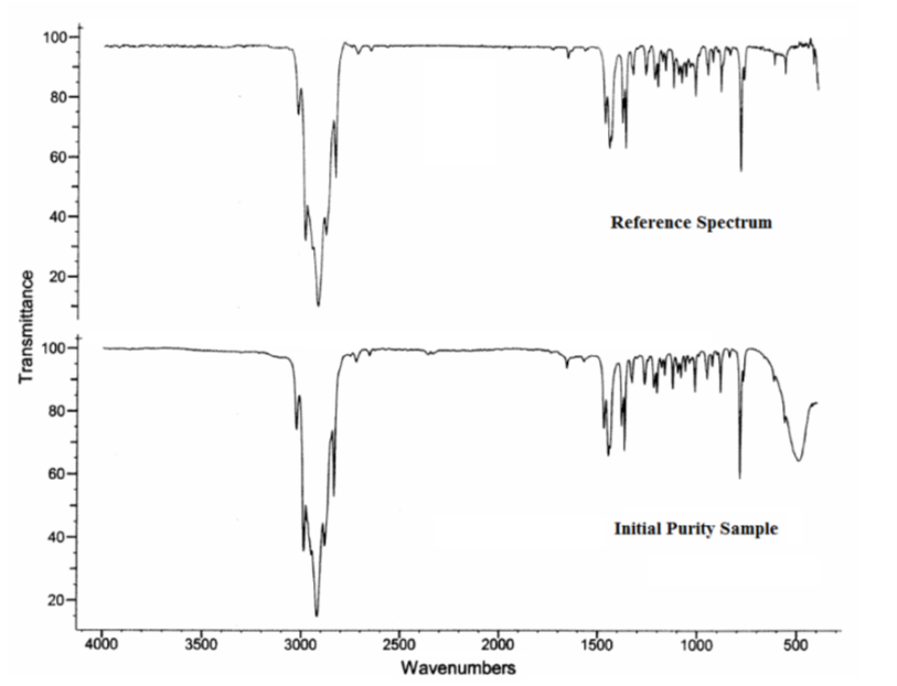
Figure A-2. 1H Nuclear Magnetic Resonance Spectrum of α-Pinene
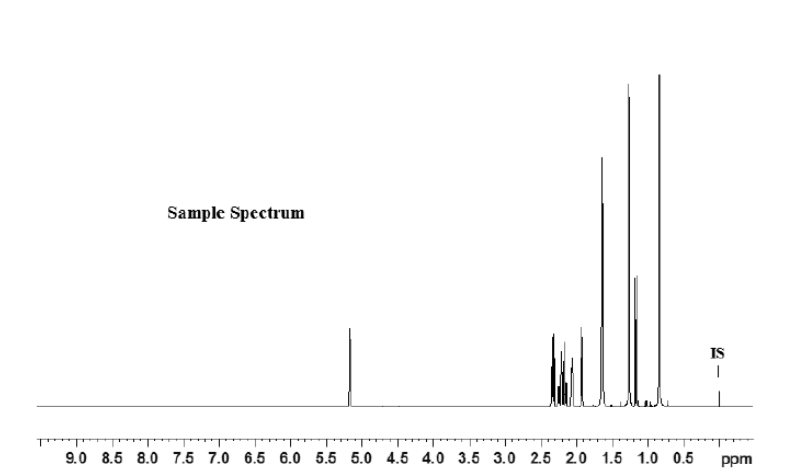
Figure A-3. 13C Nuclear Magnetic Resonance Spectrum of α-Pinene
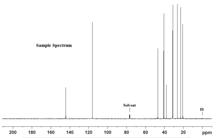
Figure A-4. Representative Chromatogram for Enantiomeric Ratio Determination of α-Pinene
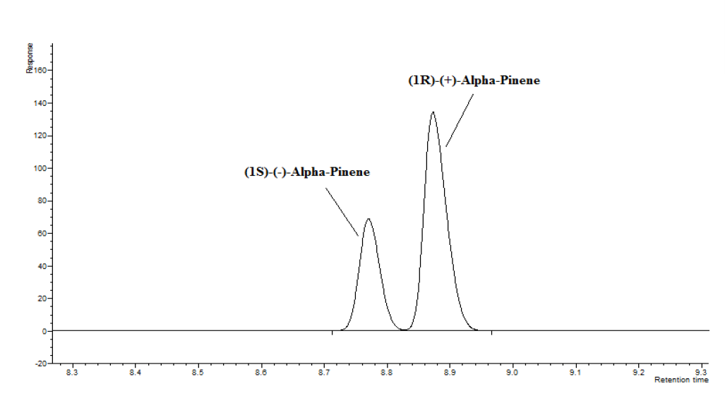
Figure A-5. Schematic of the Vapor Generation and Delivery System in the Inhalation Study of α-Pinene
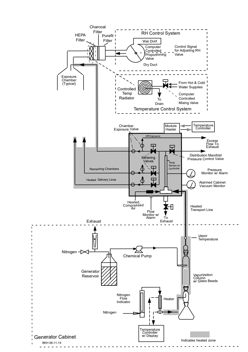
Appendix B. Ingredients, Nutrient Composition, and Contaminant Levels in NIH-07 and NTP-2000 Rat and Mouse Ration
B.1. NIH-07 Feed
Table B-1. Ingredients of NIH-07 Rat and Mouse Ration
| Ingredients | Percent by Weight |
|---|---|
| Ground hard winter wheat | 23.00 |
| Ground #2 yellow shelled corn | 24.25 |
| Wheat middlings | 10.00 |
| Oat hulls | 0.0 |
| Alfalfa meal (dehydrated, 17% protein) | 4.0 |
| Purified cellulose | 0.0 |
| Soybean meal (47% protein) | 12.0 |
| Fish meal (62% protein) | 10.0 |
| Corn oil (without preservatives) | 0.0 |
| Soy oil (without preservatives) | 2.5 |
| Dried brewer’s yeast | 2.0 |
| Calcium carbonate (USP) | 0.5 |
| Vitamin premixa | 0.25 |
| Mineral premixb | 0.15 |
| Calcium phosphate, dibasic (USP) | 1.25 |
| Sodium chloride | 0.5 |
| Choline chloride (70% choline) | 0.10 |
| Dried skim milk | 5.00 |
| Dried molasses | 1.50 |
| Corn gluten meal (60% protein) | 3.00 |
| Methionine | 0.0 |
Table B-2. Vitamins and Minerals in NIH-07 Rat and Mouse Ration
| Amounta | Source | |
|---|---|---|
| Vitamins | ||
| Vitamin A | 6,062 IU | Stabilized vitamin A palmitate or acetate |
| Vitamin D | 5,070 IU | D-activated animal sterol |
| Vitamin K | 3.1 mg | Menadione sodium bisulfite complex |
| Vitamin E | 22 IU | α-Tocopheryl acetate |
| Niacin | 33 mg | – |
| Folic acid | 2.4 mg | – |
| d-Pantothenic acid | 19.8 mg | d-Calcium pantothenate |
| Riboflavin | 3.8 mg | – |
| Thiamine | 11 mg | Thiamine mononitrate |
| B12 | 50 µg | – |
| Pyridoxine | 6.5 mg | Pyridoxine hydrochloride |
| Biotin | 0.15 mg | d-Biotin |
| Minerals | ||
| Iron | 132 mg | Iron sulfate |
| Zinc | 18 mg | Zinc oxide |
| Manganese | 66 mg | Manganese oxide |
| Copper | 4.4 mg | Copper sulfate |
| Iodine | 2.0 mg | Calcium iodate |
| Cobalt | 0.44 mg | Cobalt carbonate |
Table B-3. Nutrient Composition of NIH-07 Rat and Mouse Ration
| Nutrient | Mean ± Standard Deviation | Range | Number of Samples |
|---|---|---|---|
| Protein (% by weight) | 22.7 ± 0.4 | 22.3–23.1 | 3 |
| Crude fat (% by weight) | 5.2 ± 0.2 | 5.0–5.4 | 3 |
| Crude fiber (% by weight) | 3.58 ± 0.144 | 3.46–3.74 | 3 |
| Ash (% by weight) | 6.26 ± 0.384 | 5.85–6.61 | 3 |
| Amino acids (% of total diet) | |||
| Arginine | 1.120 ± 0.425 | 0.258–1.49 | 12 |
| Cystine | 0.515 ± 0.802 | 0.116–3.05 | 12 |
| Glycine | 1.017 ± 0.320 | 0.217–1.31 | 12 |
| Histidine | 0.922 ± 1.458 | 0.125–5.53 | 12 |
| Isoleucine | 0.920 ± 0.223 | 0.214–1.03 | 12 |
| Leucine | 1.879 ± 0.463 | 0.423–2.13 | 12 |
| Lysine | 0.454 ± 0.100 | 0.111–1.32 | 12 |
| Methionine | 0.454 ± 0.112 | 0.102–0.515 | 12 |
| Phenylalanine | 1.023 ± 0.237 | 0.276–1.12 | 12 |
| Threonine | 769.11 ± 2,661.3 | 0.168–9,220 | 12 |
| Tryptophan | 0.312 ± 0.201 | 0.076–0.922 | 12 |
| Tyrosine | 0.804 ± 0.190 | 0.209–0.894 | 12 |
| Valine | 0.971 ± 0.367 | 0.12–1.17 | 12 |
| Essential fatty acids (% of total diet) | |||
| Linoleic | 2.407 ± 0.477 | 1.99–3.77 | 12 |
| Linolenic | 0.188 ± 0.112 | 0.003–0.296 | 12 |
| Vitamins | |||
| Vitamin A (IU/kg) | 5,016 ± 31.84 | 4,780–5,380 | 3 |
| α-Tocopherol (ppm) | 69.08 ± 9.14 | 46.8–78.7 | 12 |
| Thiamine (ppm)a | 11.87 ± 1.46 | 10.7–13.5 | 3 |
| Riboflavin (ppm) | 12.91 ± 4.76 | 4.2–19.8 | 12 |
| Niacin (ppm) | 95.22 ± 15.55 | 51.9–112.0 | 12 |
| Pantothenic acid (ppm) | 42.6 ± 5.70 | 29.4–51.1 | 12 |
| Pyridoxine (ppm)a | 14.92 ± 9.09 | 8.27–42.0 | 12 |
| Folic acid (ppm) | 2.32 ± 0.580 | 1.37–3.09 | 12 |
| Biotin (ppm) | 0.274 ± 0.201 | 0.0–0.638 | 12 |
| Vitamin B12 (ppb) | 48.53 ± 6.74 | 40.0–61.6 | 12 |
| Choline (as chloride) (ppm) | 0.643 ± 0.107 | 0.441–0.8 | 12 |
| Minerals | |||
| Calcium (%) | 1.14 ± 0.11 | 1.06–1.26 | 3 |
| Phosphorus (%) | 4.04 ± 5.43 | 0.87–10.3 | 3 |
| Potassium (%) | 0.838 ± 0.037 | 0.769–0.88 | 12 |
| Chloride (%) | 0.643 ± 0.107 | 0.441–0.8 | 12 |
| Sodium (%) | 0.398 ± 0.114 | 0.274–0.721 | 12 |
| Magnesium (%) | 0.185 ± 0.015 | 0.162–0.218 | 12 |
| Iron (ppm) | 373.25 ± 59.74 | 271.0–469.0 | 12 |
| Manganese (ppm) | 84.225 ± 18.16 | 35.3–104.0 | 12 |
| Zinc (ppm) | 61.91 ± 10.60 | 47.4–89.2 | 12 |
| Copper (ppm) | 12.61 ± 4.63 | 0.683–21.1 | 12 |
| Iodine (ppm) | 1.61 ± 1.05 | 0.0–3.45 | 12 |
| Chromium (ppm) | 2.00 ± 1.324 | 0.277–3.97 | 12 |
| Cobalt (ppm) | 0.432 ± 0.312 | 0.0–0.965 | 12 |
Table B-4. Contaminant Levels in NIH-07 Rat and Mouse Ration
| Mean ± Standard Deviation | Range | Number of Samples | |
|---|---|---|---|
| Contaminants | |||
| Arsenic (ppm) | 0.40 ± 0.018 | 0.391–0.423 | 3 |
| Cadmium (ppm) | 0.09 ± 0.007 | 0.08–0.093 | 3 |
| Lead (ppm) | 0.17 ± 0.02 | 0.142–0.177 | 3 |
| Mercury (ppm) | 0.01 ± 0.00 | 0.01–0.01 | 3 |
| Selenium (ppm) | 0.302 ± 0.03 | 0.278–0.335 | 3 |
| Aflatoxins (ppb)a | <5.0 | – | 3 |
| Nitrate nitrogen (ppm)b | 8.79 ± 1.66 | 7.79–10.7 | 3 |
| Nitrite nitrogen (ppm)b | 0.15 ± 0.001 | 0.15–0.152 | 3 |
| BHA (ppm)a,c | <1.0 | – | 3 |
| BHT (ppm)a,c | <1.0 | – | 3 |
| Aerobic plate count (CFU/g)d | <10.0 | – | 3 |
| Coliform (MPN/g)d | <3.0 | – | 3 |
| Escherichia coli (MPN/g)d | <3.0 | – | 3 |
| Total nitrosamines (ppb)e | 2.93 ± 3.43 | 0.0–6.7 | 3 |
| N-Nitrosodimethylamine (ppb)e | 1.63 ± 1.46 | 0.0–2.8 | 3 |
| N-Nitrosopyrrolidine (ppb)e | 1.95 ± 2.758 | 0.0–3.9 | 2 |
| Pesticides (ppm)f | |||
| Methyl chlorpyrifos | 0.045 ± 0.014 | 0.034–0.06 | 3 |
| Malathion | 0.034 ± 0.013 | 0.022–0.049 | 3 |
B.2. NTP-2000 Feed
Table B-5. Ingredients of NTP-2000 Rat and Mouse Ration
| Ingredients | Percent by Weight |
|---|---|
| Ground hard winter wheat | 23.00 |
| Ground #2 yellow shelled corn | 22.44 |
| Wheat middlings | 15.0 |
| Oat hulls | 8.5 |
| Alfalfa meal (dehydrated, 17% protein) | 7.5 |
| Purified cellulose | 5.5 |
| Soybean meal (49% protein) | 4.0 |
| Fish meal (60% protein) | 4.0 |
| Corn oil (without preservatives) | 3.0 |
| Soy oil (without preservatives) | 3.0 |
| Dried brewer’s yeast | 1.0 |
| Calcium carbonate (USP) | 0.9 |
| Vitamin premixa | 0.5 |
| Mineral premixb | 0.5 |
| Calcium phosphate, dibasic (USP) | 0.4 |
| Sodium chloride | 0.3 |
| Choline chloride (70% choline) | 0.26 |
| Methionine | 0.2 |
Table B-6. Vitamins and Minerals in NTP-2000 Rat and Mouse Ration
| Amounta | Source | |
|---|---|---|
| Vitamins | ||
| Vitamin A | 4,000 IU | Stabilized vitamin A palmitate or acetate |
| Vitamin D | 1,000 IU | D-activated animal sterol |
| Vitamin K | 1.0 mg | Menadione sodium bisulfite complex |
| α-Tocopheryl acetate | 100 IU | – |
| Niacin | 23 mg | – |
| Folic acid | 1.1 mg | – |
| d-Pantothenic acid | 10 mg | d-Calcium pantothenate |
| Riboflavin | 3.3 mg | – |
| Thiamine | 4 mg | Thiamine mononitrate |
| Vitamin B12 | 52 µg | – |
| Pyridoxine | 6.3 mg | Pyridoxine hydrochloride |
| Biotin | 0.2 mg | d-Biotin |
| Minerals | ||
| Magnesium | 514 mg | Magnesium oxide |
| Iron | 35 mg | Iron sulfate |
| Zinc | 12 mg | Zinc oxide |
| Manganese | 10 mg | Manganese oxide |
| Copper | 2.0 mg | Copper sulfate |
| Iodine | 0.2 mg | Calcium iodate |
| Chromium | 0.2 mg | Chromium acetate |
Table B-7. Nutrient Composition of NTP-2000 Rat and Mouse Ration
| Nutrient | Mean ± Standard Deviation | Range | Number of Samples |
|---|---|---|---|
| Protein (% by weight) | 14.72 ± 0.534 | 13.7–15.7 | 27 |
| Crude fat (% by weight) | 8.08 ± 0.626 | 5.1–8.6 | 27 |
| Crude fiber (% by weight) | 9.54 ± 0.511 | 7.7–10.3 | 27 |
| Ash (% by weight) | 4.97 ± 0.164 | 4.41–5.22 | 27 |
| Amino acids (% of total diet) | |||
| Arginine | 0.807 ± 0.070 | 0.67–0.97 | 32 |
| Cystine | 0.220 ± 0.021 | 0.15–0.25 | 32 |
| Glycine | 0.704 ± 0.037 | 0.62–0.8 | 32 |
| Histidine | 0.340 ± 0.067 | 0.27–0.68 | 32 |
| Isoleucine | 0.546 ± 0.037 | 0.43–0.66 | 32 |
| Leucine | 1.095 ± 0.060 | 0.96–1.24 | 32 |
| Lysine | 0.698 ± 0.100 | 0.31–0.86 | 32 |
| Methionine | 0.407 ± 0.039 | 0.26–0.49 | 32 |
| Phenylalanine | 0.628 ± 0.035 | 0.54–0.72 | 32 |
| Threonine | 0.513 ± 0.039 | 0.43–0.61 | 32 |
| Tryptophan | 0.168 ± 0.070 | 0.11–0.525 | 32 |
| Tyrosine | 0.425 ± 0.063 | 0.28–0.54 | 32 |
| Valine | 0.668 ± 0.042 | 0.55–0.77 | 32 |
| Essential fatty acids (% of total diet) | |||
| Linoleic | 3.920 ± 0.242 | 3.49–4.55 | 32 |
| Linolenic | 0.224 ± 0.134 | 0.004–0.35 | 32 |
| Vitamins | |||
| Vitamin A (IU/kg) | 5,406 ± 548.43 | 208–2,430 | 27 |
| α-Tocopherol (ppm) | 74.38 ± 23.62 | 20.3–124.0 | 32 |
| Thiamine (ppm)a | 7.75 ± 1.13 | 6.8–12.5 | 27 |
| Riboflavin (ppm) | 8.02 ± 3.46 | 1.1–17.5 | 32 |
| Niacin (ppm) | 81.46 ± 10.62 | 66.4–107.0 | 32 |
| Pantothenic acid (ppm) | 26.61 ± 10.6 | 17.4–81.0 | 32 |
| Pyridoxine (ppm)a | 9.60 ± 2.45 | 2.3–14.3 | 32 |
| Folic acid (ppm) | 1.65 ± 0.47 | 1.15–3.27 | 32 |
| Biotin (ppm) | 0.323 ± 0.112 | 0.0–0.704 | 32 |
| B12 (ppb) | 50.57 ± 33.14 | 18.3–174.0 | 32 |
| Choline (as chloride) (ppm) | 2,537 ± 628 | 1,160–3,790 | 32 |
| Minerals | |||
| Calcium (%) | 0.905 ± 0.05 | 0.802–0.992 | 27 |
| Phosphorus (%) | 0.57 ± 0.028 | 0.522–0.639 | 27 |
| Potassium (%) | 0.662 ± 0.035 | 0.569–0.733 | 32 |
| Chloride (%) | 0.395 ± 0.068 | 0.3–0.688 | 32 |
| Sodium (%) | 0.192 ± 0.026 | 0.153–0.283 | 32 |
| Magnesium (%) | 0.216 ± 0.052 | 0.185–0.49 | 32 |
| Iron (ppm) | 181.8 ± 46.27 | 21–311 | 32 |
| Manganese (ppm) | 49.87 ± 8.97 | 21.0–73.1 | 32 |
| Zinc (ppm) | 51.13 ± 9.64 | 18.4–78.5 | 32 |
| Copper (ppm) | 7.70 ± 2.42 | 3.21–16.3 | 32 |
| Iodine (ppm) | 0.50 ± 0.236 | 0–1.0 | 32 |
| Chromium (ppm) | 0.72 ± 0.66 | 0.249–3.97 | 31 |
| Cobalt (ppm) | 0.215 ± 0.146 | 0.086–0.864 | 30 |
Table B-8. Contaminant Levels in NTP-2000 Rat and Mouse Ration
| Mean ± Standard Deviation | Range | Number of Samples | |
|---|---|---|---|
| Contaminants | |||
| Arsenic (ppm) | 0.248 ± 0.032 | 0.205–0.314 | 27 |
| Cadmium (ppm) | 0.052 ± 0.004 | 0.045–0.061 | 27 |
| Lead (ppm) | 0.117 ± 0.13 | 0.058–0.621 | 27 |
| Mercury (ppm) | 0.01 ± 0.002 | 0.005–0.015 | 27 |
| Selenium (ppm) | 0.164 ± 0.025 | 0.127–0.251 | 27 |
| Aflatoxins (ppb)a | <5.0 | – | 24 |
| Nitrate nitrogen (ppm)b | 10.67 ± 2.97 | 5.05–17.7 | 27 |
| Nitrite nitrogen (ppm)b | 0.129 ± 0.013 | 0.12–0.152 | 27 |
| BHA (ppm)c | 1.06 ± 0.245 | 1.0–2.24 | 27 |
| BHT (ppm)a,c | <1 | – | 27 |
| Aerobic plate count (CFU/g) | 16.30 ± 32.72 | 10.0–180 | 27 |
| Coliform (MPN/g)d | <3 | – | 27 |
| Escherichia coli (MPN/g)d | <3 | – | 27 |
| Salmonella sp. (MPN/g) | Negative | – | 3 |
| Total nitrosamines (ppb)e | 8.31 ± 3.72 | 2.4–17.1 | 25 |
| N-Nitrosodimethylamine (ppb)e | 2.74 ± 2.19 | 0.0–7.9 | 25 |
| N-Nitrosopyrrolidine (ppb)e | 5.6 ± 2.468 | 1.2–12.0 | 25 |
| Pesticides (ppm)f | |||
| Methyl chlorpyrifos | 0.114 ± 0.08 | 0.026–0.36 | 25 |
| Ethyl chlorpyrifos | 0.025 | 0.025–0.025 | 13 |
| Methyl pirimiphos | 0.027 ± 0.007 | 0.025–0.05 | 13 |
| Malathion | 0.114 ± 0.112 | 0.016–0.54 | 27 |
| Piperonyl butoxide | 0.011 ± 0 | 0.011–0.011 | 1 |
| Deltamethrin | 0.03 ± 0.001 | 0.029–0.03 | 2 |
Appendix C. Sentinel Animal Program
C.1. Methods
Rodents used in the National Toxicology Program are produced in optimally clean facilities to eliminate potential pathogens that might affect study results. The Sentinel Animal Program is part of the periodic monitoring of animal health that occurs during the toxicological evaluation of test compounds. Under this program, the disease state of the rodents is monitored via sera or feces from extra (sentinel) or exposed animals in the study rooms. The sentinel animals and the study animals are subject to identical environmental conditions. Furthermore, the sentinel animals come from the same production source and weanling groups as the animals used for the studies of test compounds.
For these toxicology and carcinogenesis studies, blood samples were collected from each sentinel animal, allowed to clot, and the serum was separated. Additionally, fecal samples were collected and tested for endoparasites and Helicobacter species. All samples were processed appropriately with serology and Helicobacter testing performed by IDEXX BioResearch (formerly Rodent Animal Diagnostic Laboratory [RADIL], University of Missouri), Columbia, MO, for determination of the presence of pathogens. Evaluation for endo- and ectoparasites was performed in-house by the testing laboratory.
The laboratory methods and agents for which testing was performed are tabulated in Table C-1 and Table C-2 below; the times at which samples were collected during the studies are also listed.
C.2. Results
Rats: All test results were negative.
Mice: All test results were negative.
Table C-1. Methods and Results for Sentinel Animal Testing in Male and Female Rats in the Three-month Reproductive and Two-year Inhalation Studies of α-Pinene
| Collection Time Points | Three-month Reproductive Study | Two-year Study | ||||||||||
|---|---|---|---|---|---|---|---|---|---|---|---|---|
| Quarantine | Study Termination | Study Termination (Replacement Animal) | Quarantine | 4 Weeks | 6 Months | 12 Months | 14 Months (Unscheduled) | 15 Months (Unscheduled) | 16 Months (Unscheduled) | 18 Months | Study Termination | |
| Number examined (males/females) | 0/10 | 5/5 | 0/1 | 5/5 | 5/5 | 5/5 | 5/5 | 1/0 | 0/1 | 0/1 | 4/2 | 5/5 |
| Method/test | ||||||||||||
| Multiplex fluorescent immunoassay (MFI) | ||||||||||||
Kilham rat virus (KRV) | − | − | − | − | − | − | − | − | − | − | − | − |
Mycoplasma pulmonis | − | − | − | − | − | − | − | − | − | − | − | − |
Pneumocystis carinii | NT | NT | NT | NT | NT | − | NT | NT | NT | NT | NT | NT |
Pneumonia virus of mice (PVM) | − | − | − | − | − | − | − | − | − | − | − | − |
Rat coronavirus/ | − | − | − | − | − | − | − | − | − | − | − | − |
Rat minute virus (RMV) | − | − | − | − | − | − | − | − | − | − | − | − |
Rat parvo virus (RPV) | − | − | − | − | − | − | − | − | − | − | − | − |
Rat theilovirus (RTV) | − | − | − | − | − | − | − | − | − | − | − | − |
Sendai | − | − | − | − | − | − | − | − | − | − | − | − |
Theiler’s murine encephalomyelitis virus (TMEV) | − | − | − | − | − | − | − | − | − | − | − | − |
Toolan’s H-1 | − | − | − | − | − | − | − | − | − | − | − | − |
| Immunofluorescence assay (IFA) | ||||||||||||
RCV/SDA | NT | NT | NT | NT | NT | NT | NT | NT | NT | NT | − | NT |
| Number examined (males/females) | 0/10 | 0/0 | 0/0 | 5/5 | 5/5 | 5/5 | 5/5 | 1/0 | 0/2 | 0/1 | 4/2 | 0/0 |
| Method/test | ||||||||||||
| In-house evaluation | ||||||||||||
Endoparasites (evaluation of cecal content) | − | NT | NT | − | − | − | − | − | − | − | − | NT |
Ectoparasites (evaluation of perianal surface) | − | NT | NT | − | − | − | − | − | − | − | − | NT |
Table C-2. Methods and Results for Sentinel Animal Testing in Male and Female Mice in the Three-month Reproductive and Two-year Inhalation Studies of α-Pinene
| Collection Time Points | Three-month Reproductive Studya | Two-year Studyb | |||||||
|---|---|---|---|---|---|---|---|---|---|
| Quarantine | Study Termination | Quarantine | 4 Weeks | 2 Months (Unscheduled) | 6 Months | 12 Months | 18 Months | Study Termination | |
| Number examined (males/females) | 0/10 | 5/5 | 10/5 | 10/5 | 0/1 | 5/3 | 5/3 | 4/3 | 5/5 |
| Method/test | |||||||||
| Multiplex fluorescent immunoassay (MFI) | |||||||||
Ectromelia virus | − | − | − | − | − | − | − | − | − |
Epizootic diarrhea of infant mice (EDIM) | − | − | − | − | − | − | − | − | − |
Lymphocytic choriomeningitis virus | − | − | − | − | − | − | − | − | − |
Mycoplasma pulmonis | − | − | − | − | − | − | − | − | − |
Mouse hepatitis virus (MHV) | − | − | − | − | − | − | − | − | − |
Mouse norovirus (MNV) | − | − | − | − | − | − | − | − | − |
Mouse parvovirus (MPV) | − | − | − | − | − | − | − | − | − |
Minute virus of mice (MVM) | − | − | − | − | − | − | − | − | − |
Pneumonia virus of mice (PVM) | − | − | − | − | − | − | − | − | − |
Rat theilovirus (RTV) | − | − | − | − | − | − | − | − | − |
Reovirus (REO3) | − | − | − | − | − | − | − | − | − |
Sendai | − | − | − | − | − | − | − | − | − |
Theiler’s murine encephalomyelitis virus (TMEV) GDVII | − | − | − | − | − | − | − | − | − |
| Immunofluorescence assay (IFA) | |||||||||
Mouse parvovirus (MPV) | NT | NT | NT | NT | NT | NT | − | NT | NT |
Mouse hepatitis virus (MHV) | NT | NT | NT | NT | NT | NT | NT | NT | − |
| Polymerase chain reaction (PCR) | |||||||||
| Helicobacter species | NT | NT | NT | NT | NT | NT | NT | − | NT |
| Number examined (males/females) | 0/10 | 0/0 | 10/5 | 10/5 | 0/1 | 5/3 | 5/3 | 4/3 | 0/0 |
| Method/test | |||||||||
| In-house evaluation | |||||||||
Endoparasites (evaluation of cecal content) | − | NT | − | − | − | − | − | − | NT |
Ectoparasites (evaluation of perianal surface) | − | NT | − | − | − | − | − | − | NT |
Table C-3. Methods and Results for Sentinel Animal Testing in Male and Female Rats in the Three-month Investigative Inhalation Study of α-Pinene
| Collection Time Point | Quarantine | 4 Weeks | Study Termination |
|---|---|---|---|
| Number examined (males/females) | 5/5 | 5/5 | 5/5 |
| Method/test | |||
| Multiplex fluorescent immunoassay (MFI) | |||
Kilham rat virus (KRV) | − | − | − |
Lymphocytic choriomeningitis virus (LCMV) | − | − | − |
Mycoplasma pulmonis | − | − | − |
Pneumocystis carinii | − | − | − |
Pneumonia virus of mice (PVM) | − | − | − |
Rat coronavirus/sialodacryoadenitis virus (RCV/SDA) | − | − | − |
Rat minute virus (RMV) | − | − | − |
Rat parvo virus (RPV) | − | − | − |
Rat theilovirus (RTV) | − | − | − |
Reovirus (REO3) | − | − | − |
Sendai | − | − | − |
Theiler’s murine encephalomyelitis virus (TMEV) | − | − | − |
Toolan’s H-1 | − | − | − |
| Polymerase chain reaction (PCR) – feces | |||
| Helicobacter spp. | − | − | − |
Helicobacter bilis | − | − | − |
Helicobacter ganmani | − | − | − |
Helicobacter hepaticus | − | − | − |
Helicobacter mastomyrinus | − | − | − |
Helicobacter rodentium | − | − | − |
Helicobacter typhlonius | − | − | − |
Aspiculuris tetraptera | − | − | − |
Syphacia muris | − | − | − |
Syphacia obvelata | − | − | − |
| Polymerase chain reaction (PCR) – pelt swabs | − | − | − |
Myocoptes | − | − | − |
Radfordia/myobia | − | − | − |
Appendix D. Internal Concentration Assessment
D.1. Sample Collection
Blood and mammary gland from male and female Sprague Dawley (Hsd:Sprague Dawley SD) rats in the 3-month investigative study and female rats and B6C3F1/N mice from the 2-year inhalation studies of α-pinene and blood from male rats and mice in the 2-year inhalation studies of α-pinene were analyzed to determine the concentrations of α-pinene and α-pinene oxide, a metabolite of α-pinene, present in the matrix at the time of collection. Lipid content was also measured in blood and mammary gland from the 3-month study and in mammary gland from the 2-year study.
Samples were shipped frozen in sealed vials to the analytical chemistry laboratory at RTI International (Research Triangle Park, NC) and stored at approximately −70°C until analysis. Standards were prepared with α-pinene (lot A-9211; The John D. Walsh Company, Inc.; Ringwood, NJ) and α-pinene oxide (lot MKBT1323V; Sigma-Aldrich; St. Louis, MO).
D.1.1. Two-year Studies in Rats and Mice
D.1.1.1. Blood
At study termination, rats and mice were anesthetized with a 70% CO2/30% O2 mixture, and blood was collected from the heart of up to 10 (rats) or 11 (mice) randomly selected animals per sex per exposure group into tubes containing K3EDTA (tripotassium ethylene diamine tetraacetic acid). Blood samples were aliquoted as appropriate (e.g., 100 µL aliquots in 2-mL clear glass crimp-top vials for analysis of α-pinene and α-pinene oxide). Samples were flash frozen and stored between −60°C and −85°C.
D.1.1.2. Mammary Gland
Following blood collection at study termination, mammary glands 1, 2, and 3 were collected from the left and right side of up to 16 female rats or 26 female mice per exposure group. Female animals were wiped down with 70% ethanol prior to tissue collection. Each side was stored separately, allocated as appropriate for analysis of α-pinene and α-pinene oxide, snap frozen in liquid nitrogen, and stored at −60°C to −85°C. Tissue from one side was used for internal concentration assessment, and tissue from the other side was used for molecular pathology.
D.1.2. Three-month Investigative Study in Rats
At the beginning of the 3-month study, five male and female rats per exposure group were randomly assigned to the biosample group and exposed in an identical fashion to the core group. Prior to study sample collection, the stability of α-pinene and α-pinene oxide under these conditions was confirmed by the analytical chemistry laboratory.
D.1.2.1. Blood
At study termination, rats designated for biosampling were anesthetized with a 70% CO2/30% O2 mixture, and blood was collected from the retroorbital plexus of five rats/sex/exposure group into tubes containing K3EDTA and immediately placed on wet ice. Separate aliquots of the samples were allocated for α-pinene and α-pinene oxide analysis. Blood allocated for α-pinene concentration analysis was spiked with internal standard (α-pinene-d3; AromaLAB GmbH; Planegg, Germany). Samples were kept on wet ice during collection and then frozen and stored between −60°C and −85°C.
D.1.2.2. Mammary Gland
Following blood collection at study termination, mammary glands 4 and/or 5 were collected from the left and right side of five female rats per exposure group. At least 100 mg of mammary gland sample was used for internal concentration assessment of α-pinene and α-pinene oxide. Tissue allocated for α-pinene concentration analysis was spiked with internal standard (α-pinene-d3; AromaLAB GmbH; Planegg, Germany), and stainless steel balls used for homogenization (Section D.2.2) were added. Each vial was stored separately, weighed, and snap frozen in liquid nitrogen and stored at −60°C to −85°C.
D.2. Sample Analysis
D.2.1. α-Pinene and α-Pinene Oxide in Blood
D.2.1.1. Two-year Rat and Mouse Studies
α-Pinene and α-pinene oxide concentrations in rat and mouse blood were quantified using qualified methods described in Section D.2.4.1; method qualification data are presented in Table D-2.
For quantitation of α-pinene, study samples (100 µL aliquots) were thawed and analyzed using headspace (HS) gas chromatography (GC)-mass spectrometry (MS) detection. The sample vials were heated for 10 minutes at 60°C before a 250 µL injection was made from the headspace into the HS-GC/MS (Table D-1, System A).
For quantitation of α-pinene oxide, study samples (100 µL) were vortex mixed with 300 µL of 66.7 ng/mL (+)-limonene oxide internal standard in ethyl acetate in a 1.5 mL microcentrifuge tube for 3 minutes. Samples were then centrifuged for 3 minutes at 16,000 g, and the resulting supernatant was transferred to GC autosampler vials. One microliter (1 µL) of each sample was analyzed using GC/MS (Table D-1, System B).
Corresponding matrix calibration standards, blanks, and quality control (QC) standards were prepared with commercially obtained pooled mixed-sexed Sprague Dawley rat blood and male CD-1 mouse blood (BioreclamationIVT, Westbury, NY), both containing K3EDTA anticoagulant. α-Pinene in rat and mouse blood was quantified using calibration curves prepared in Sprague Dawley rat blood. α-Pinene oxide was quantified using calibration curves prepared in respective rat or mouse matrix. Blank and QC standards of α-pinene oxide and α-pinene were prepared in respective rat or mouse matrix. Each standard was prepared by spiking with the appropriate volume of standard solution containing analyte (α-pinene or α-pinene oxide) and respective internal standard (n-nonane or (+)-limonene oxide) to reach the desired target concentrations. (Note: For α-pinene analysis, n-nonane standard was used in calibration standards—and not in study samples—to serve as an internal check of the instrument response only and was not used in quantification of α-pinene concentration.) Six calibration standards were prepared for α-pinene, spanning a range of 10 to 1,000 ng/mL α-pinene for both species. Seven matrix calibration standards were prepared for α-pinene oxide, spanning a range of 5 to 250 ng/mL. α-Pinene QC standards were prepared at 20, 100, and 500 ng/mL. α-Pinene oxide QC standards were prepared at 10, 50, and 175 ng/mL. All standards and blanks were prepared for analysis in the same manner as the study samples.
All samples were analyzed for α-pinene and α-pinene oxide using HS-GC/MS and GC/MS, respectively, as described in Section D.2.4.1. Blood α-pinene and α-pinene oxide concentrations were reported as ng analyte/mL blood.
D.2.1.2. Three-month Investigative Rat Study
α-Pinene and α-pinene oxide concentrations in rat blood were quantified using a validated method; data for the method was published previously.62,63
Matrix calibration standards, blanks, and QC standards were prepared with commercial male Sprague Dawley rat blood containing K3EDTA anticoagulant (BioreclamationIVT, Westbury, NY) and run with study samples. Seven calibration standards for each analyte were prepared, spanning a range of 5 to 500 ng/mL α-pinene and 5 to 250 ng/mL α-pinene oxide. Blanks were mixed with the appropriate internal standard. QC standards were prepared at 10, 100, and 250 ng/mL α-pinene or 10, 50, and 200 ng/mL α-pinene oxide. Blood α-pinene and α-pinene oxide concentrations were reported as both ng analyte/mL blood and ng analyte/g lipid.
D.2.2. α-Pinene and α-Pinene Oxide in Mammary Gland
α-Pinene and α-pinene oxide concentrations in mammary gland were quantified using a validated method for both the 2-year chronic and 3-month investigative studies, and the details were published previously.62,63 For quantitation of α-pinene in mammary gland, calibration curves prepared in mammary gland were used while for quantitation of α-pinene oxide in mammary gland, calibration curves prepared in blood were used, which met the acceptance criteria for accuracy and precision.62
For a small number of samples, dilutions were made to acquire measurements within the calibration curve range.
Matrix calibration standards, blanks, and QC standards were prepared using female Sprague Dawley rat blood or mammary gland (BioreclamationIVT, Westbury, NY) as appropriate and run with study samples. Six calibration standards, spanning a range of 100 to 5,000 ng/mL α-pinene, were prepared in mammary gland, and 25 to 500 ng/mL α-pinene oxide were prepared in blood. QC standards were prepared at 250 and 2,500 ng/mL α-pinene or 50 and 400 ng/mL α-pinene oxide. For quantitation of α-pinene oxide, the mammary gland tissue dilution used to prepare the homogenate was used to convert ng/mL homogenate to ng/g of analyte in mammary gland. Mammary gland α-pinene and α-pinene oxide concentrations were reported as both ng/g mammary and ng/g lipid.
D.2.3. Lipid Content in Blood and Mammary Gland
Lipid content of blood (3-month investigative rat study only) and mammary gland was determined based on the method described in Johnson et al.114 Blood samples used were separate aliquots from blood used for concentration determination of α-pinene and α-pinene oxide. Lipid content was analyzed in the following samples: the same aliquot of mammary gland used for concentration determination of α-pinene and the corresponding lipid value was used to generate the lipid-adjusted α-pinene in mammary gland; an aliquot of mammary gland homogenate used for concentration determination of α-pinene oxide and the corresponding lipid value was used to generate lipid-adjusted α-pinene oxide in mammary gland.
Soybean oil was mixed with 50/50 (v/v) methylene chloride and methanol with the appropriate volumes to create standards for a calibration concentration range of 0.12 to 1.2 mg/mL total lipids and high and low concentration QC standards.
Mammary gland samples following concentration determination of α-pinene were diluted with 800 µL of deionized water and then heated at 90°C for 2 hours, followed by vortex mixing. A 100 µL aliquot of each blood and diluted mammary gland sample was vortex mixed with 4.9 mL of 50/50 (v/v) methylene chloride and methanol and then centrifuged for 5 to 20 minutes at 3,000 rpm. A 250 µL aliquot of the mammary extract or a 1,000 µL aliquot of the blood extract was transferred to the bottom of a 13 × 100 mm glass culture tube for later reaction.
A 200 µL aliquot of the mammary gland homogenate prepared for α-pinene oxide analysis was placed in a 15-mL polypropylene centrifuge tube. Each sample was extracted as above with 50/50 (v/v) methylene chloride and methanol using 2 × 1 mL and 1 × 2 mL, and extracts were combined. The extract was transferred to a 5-mL volumetric flask, with additional extraction solvent added to the mark, and then mixed by inversion. A 100 µL aliquot of the extract was transferred to the bottom of a 13 × 100 mm glass culture tube for later reaction.
All samples, calibrations, and QC standards were evaporated to dryness in their 13 × 100 mm culture tubes in a dry block heater set at 100°C. After cooling, samples were vortex mixed with 200 µL of concentrated sulfuric acid and then dried again at 100°C in the dry block heater for approximately 15 minutes before cooling.
Samples were vortex mixed with 3 mg vanillin in 68% aqueous phosphoric acid and then allowed to react in the dark for at least 30 minutes, which produced colored solutions for analysis. Aliquots were transferred to cuvettes for ultraviolet (UV) spectroscopy analysis at 490 nm using an Acquity 2998 photodiode array detector with cuvette holder (Waters Corporation; Milford, MA). Calibration standards, QC standards, and blanks were subject to the hydrolysis reaction and UV absorption analysis in the same manner as the samples.
Calibration curves relating the response of absorbance to the concentration of the calibration standards were constructed using a 1/X weighted linear regression. The lipid concentration in study samples were calculated using their absorbance, the calibration regression equation, the sample weight, and the dilution factor. The concentration values for the standards and samples were used to calculate the individual and average concentrations and uncertainties. The sample results were calculated for the sample matrix (i.e., the lipid content in mammary gland) using the weight/volume ratio of the initial extract.
D.2.4. Instrumentation and Quantitation
D.2.4.1. Qualified Analytical Methods of Blood for Two-year Studies
α-Pinene and α-pinene oxide in rat and mouse whole blood were detected using System A and System B in Table D-1, respectively. Calibration curves relating the analyte response (α-pinene) or response ratio (α-pinene oxide) to the internal standard and the concentration of α-pinene or α-pinene oxide in blood were constructed using a 1/X weighted linear regression. The concentration of α-pinene and α-pinene oxide in study samples was calculated using the response or response ratio, as appropriate, and the regression equation. Analyte concentrations were reported as ng of α-pinene and α-pinene oxide per mL of whole blood. The performance of the calibration curves for each analyte was evaluated once before study sample analysis and during study sample analysis. Each sample set, method blank, and control group was bracketed by a QC set, which consisted of three concentrations (low, medium, and high).
For the analysis of α-pinene in rat blood samples, the calibration standard curve had a correlation coefficient (r) >0.99 and a relative error (RE) ranging from −15.6% to 16.0%. The low (20.0 ng/mL), mid (100 ng/mL), and high (500 ng/mL) rat blood QC standard results gave RE values ranging from −38.2% to −7.3% and had a relative standard deviation (RSD) ≤20.4%. The higher RE and RSD values were likely attributed to not using an internal standard in the study samples to compensate for any sample preparation or instrumentation variability. The calibration curve of α-pinene mouse blood analysis had an (r) >0.99 and an RE ranging from −16.4% to 12.3%. The triplicate low (20.0 ng/mL), mid (100 ng/mL), and high (500 ng/mL) mouse blood QC standard results gave RE values ranging from −17.0% to 1.5% and an RSD of 4.5%. These data demonstrate that the analytical method is acceptable to quantitate α-pinene in rodent blood.
For the analysis of α-pinene oxide in rat blood samples, the calibration standard curve had an (r) >0.99 and an RE ranging from −9.6% to 12.2%. The triplicate low (10.0 ng/mL), mid (50.0 ng/mL), and high (175 ng/mL) rat blood QC standard results gave RE values ranging from −16.4% to 6.8% and an RSD ≤11.8%. The triplicate low (10.0 ng/mL), mid (50.0 ng/mL), and high (175 ng/mL) mouse blood QC standard results gave RE values ranging from −14.1% to −2.5% and an RSD ≤3.4%. These data demonstrate that the analytical method is acceptable to quantitate α-pinene oxide in rodent blood.
D.2.4.2. Validated Methods
The validated methods for analyzing α-pinene and α-pinene oxide in blood and mammary gland from the 3-month investigative rat study and mammary gland from the 2-year study were published previously.62,63
During blood α-pinene concentration determination in samples from the 3-month investigative study, a drop in internal standard response was observed, which was traced back to the day of sample collection, suggesting a potential error in internal standard addition between the 2 days. Therefore, to allow comparisons between sexes and matrices, quantitation of α-pinene in study sample blood was achieved without using internal standard.
Data from study samples were considered valid if they were bracketed by valid QC sets. In general, each sample set, method blank, and control was bracketed by two QC sets, which consisted of a calibration blank and two concentrations of calibration standards (QC low and QC high). A QC set passed when the measured concentration for QC standards was within 15% of its nominal value for at least 67% of all QC standards. All QC sets met acceptance criteria.
Table D-1. Analytical Systems and Parameters Used in the Internal Concentration Assessment of α-Pinene and α-Pinene Oxide in Blood in the Two-year Studies
| Instrument and Parameter | System A | System B |
|---|---|---|
| System | HP 6890 plus GC/HP 5973 MSD with Agilent MassHunter software (version A.01.02) | Agilent 7890A GC/5975A MSD with MassHunter software (version B.07.00, SP2) |
| Sampling | ||
| Headspace autosampler | CombiPAL Autosampler (CTC Analytics, Zwingen, Switzerland) | NA |
| Sample cycle time | 20 minutesa | NA |
| Vial size | 2 mL | NA |
| Syringe volume and temperature | 2.5 mL at 60°C | NA |
| Sample temperature and equilibrium time | 60°C for 10 minutes | NA |
| Mixer | On | NA |
| Sample volume | 250 µL (headspace injection) | 1 µL (liquid injection) |
| Column and program | ||
| Column | Agilent DB-5MS (30 m × 0.25 mm ID, 0.25 μm film thickness) | Agilent DB-5MS (29 m × 0.25 mm ID, 0.25 μm film thickness) |
| Carrier gas | Helium at 1.2 mL/min | Helium at 1.2 mL/min |
| Oven temperature program | 40°C for 5 minutes, then 5°C/min to 75°C, then 37.5°C/min to 150°C, 1 minute hold | 40°C for 5 minutes, then 20°C/min to 140°C, then 20°C/min to 300°C, 10 minute hold |
| Inlet temperature | 270°C | 200°C |
| Injection mode | Splitless | Splitless |
| Retention time | ~10.6 minutes | α-pinene oxide: 16.0 minutes (+)-limonene oxide: 17.3 minutes |
| Auxiliary temperature | 300°C | 280°C |
| MS detector | ||
| Source temperature | 230°C | 230°C |
| Quadrupole temperature | 150°C | 150°C |
| Ionization mode | EI (70 eV) | EI (70 eV) |
| Acquisition mode | SIM for 10.8 to 15.0 minutesb m/z 93 (quantitation ion) m/z 136 (confirmation ion) | SIM α-pinene oxide: m/z 109 (+)-limonene oxide: m/z 94 |
Table D-2. Qualification and Stability Data for α-Pinene and α-Pinene Oxide in Rat and Mouse Whole Blood Used in the Two-year Studies
| Parameter | α-Pinene | α-Pinene Oxide | ||
|---|---|---|---|---|
| Male SD Rat | Male CD-1 Mouse | Male SD Rat | Male CD-1 Mouse | |
| Matrix concentration range (ng/mL) | 10–1,000a | 10–1,000a | 5–250b | 5–175c |
| Sensitivity | ||||
LLOQ (ng/mL) | 10.0 | 10.0 | 5.0 | 5.0 |
LLOQ %RE | ≤±7.7 | ≤±7.7 | ≤±12.2 | ≤±11.9 |
LLOQ %RSD | 5.9 | 3.8 | ≤6.8 | 7.4 |
LOD (ng/mL) | 1.51 | 1.14 | 1.04 | 1.05 |
| Correlation coefficient (r) | 0.9985 | 0.9939 | 0.9997 | 0.9985d |
| Precision and accuracye | ||||
%RSD Low | 20.7 | 4.5 | 11.8 | 3.4 |
%RSD Medium | 12.6 | 3.4 | 2.0 | 0.8 |
%RSD High | 13.5 | 1.7 | 3.8 | 0.7 |
%RE Low | −38.2 to −7.3 | ≤1.5 | −16.4 to 5.9 | −2.5 to −8.7 |
%RE Medium | −35.4 to −18.9 | ≤−11.2 | −1.8 to 1.8 | −11.5 to −10.2 |
%RE High | −33.3 to −12.5 | ≤−1.7 | 0.0 to 6.8 | −14.1 to −12.9 |
Appendix E. Evaluation of Mutation Burden and Unique Mutation Signatures in Rat and Mouse Tumors Following Chronic Exposure to α-Pinene
E.1. Introduction
Examination of the mutation signatures (mutographs) in tumors can provide potential insights into the mechanistic bases of carcinogenesis. Mutation signatures capture the overall genetic events that lead to distinct phenotypes including carcinogenic outcomes. Several well-known human carcinogens, such as aflatoxin, aristolochic acid, ultraviolet (UV) light, and tobacco, elicit exposure-specific mutation signatures in tumors from these respective exposures, and these signatures often provide a mechanistic understanding of the underlying carcinogenic process.
Chronic exposure to α-pinene has resulted in increases in neoplasia in multiple target organs, including the liver, lung, Harderian gland, urinary bladder, forestomach, mammary gland, and ovary in B6C3F1/N mice and the urinary bladder, uterus, and mammary gland in Sprague Dawley (Hsd:Sprague Dawley SD) rats. Incidentally, there are relatively high background incidences of tumors within these mouse (liver and lung tumors) and rat (mammary and uterine tumors) strains. Upon chronic chemical exposures, there are often statistically significant increases in these tumor types that are histologically indistinguishable from spontaneous tumors. The underlying genomic alterations and the mode of carcinogenesis mechanisms in these tumors that arise either spontaneously or due to chronic chemical exposures are not well understood.
In this study, whole-genome sequencing experiments were performed on hepatocellular and alveolar/bronchiolar tumors from mice and mammary tumors from rats following chronic inhalation exposure to α-pinene, as well as on the corresponding tumors arising spontaneously due to aging in chamber controls. Mutation burden and de novo mutation signatures were determined, along with their decomposition into the respective somatic variant mutational signature within the published Catalogue of Somatic Mutations in Cancer (COSMIC) signatures derived from human tumors. A brief summary of findings is presented here; additional details will be included in a future publication.
E.2. Materials and Methods
Whole-genome sequencing was performed on fresh frozen tissue samples collected from animals euthanized as moribund or at scheduled termination following the 2-year exposure. The samples included mouse hepatocellular carcinomas (mHCCs, n = 38), mouse alveolar/bronchiolar carcinomas (mABCs, n = 12), and rat mammary tumors (rMTs, n = 32), which included adenocarcinomas and fibroadenomas arising spontaneously due to aging or due to chronic inhalation exposure to α-pinene (Table E-1). Age-matched normal tissues for each of the target organs were sourced from various studies, including the current α-pinene studies (mouse liver, n = 36; mouse lung, n = 7; rat mammary gland, n = 5), to serve as genomic controls. Genomic DNA was extracted from the above fresh frozen samples using a Gentra Puregene kit (Qiagen, Germantown, MD) and shipped to the Sanger Institute (Cambridge, UK) for paired-end 150 base-paired (bp) whole-genome sequencing at a read depth of 45X.
Table E-1. Sample Counts of Tumors and Nontumor Age-matched Target Organ Controls for Whole-genome Sequencing in the Two-year Inhalation Study of α-Pinene
| Spontaneous Tumor Controls | 50 ppm | 100 ppm | 200 ppm | 400 ppm | Total Tumors | Nontumor Age-matched Target Organ Controls | |
|---|---|---|---|---|---|---|---|
| mHCCs | 12 | –a | 12 | 1 | 13 | 38 | 36 |
| mABCs | 5 | – | 0 | 4 | 3 | 12 | 7 |
| rMTs | 11 | 7 | 7 | 7 | – | 32 | 5 |
E.2.1. Bioinformatics Analysis
The bioinformatics analysis pipelines for both the mouse and rat tumors are similar, and the description below applies to all the tumors examined in this study.
Binary alignment map (BAM) files, generated from the alignment of 150 bp paired-end reads to the Rn6 or Mm10 reference genome, were provided by the Sanger Institute. Unmapped reads were discarded, and alignments with a quality score >20 were extracted using the samtools v.1.18 view function with flags -F 4 -q 20.115 A panel of normals (PON) was generated from the nontumor mammary glands following Genome Analysis Toolkit (GATK) recommendations.116,117 In summary, the GATK (v.4.2.4) Mutect2 module with -max-mnp-distance 0 flag and the CreateSomaticPanelOfNormals module with --min-sample-count [rat = 1 flag; mouse = 2 flag (default)] were used to create the PON variant call set. Single nucleotide variants (SNVs) and small insertions or deletions (Indels) were called in tumor samples with Mutect2 using the PON and subsequently filtered with FilterMutectCalls to discard mutations derived from reads with median mapping quality <40 and median base quality <30. Clustered SNVs and Indels were discarded with the Bcftools v.1.18 filter function with flags -g 20 and -G 20 for rat only.115,118 The following GATK-recommended hard filters for a PON experimental design without available matched normals were applied: MIN(INFO/QD) > 2.0, MIN(INFO/SOR) < 3.0, MIN(INFO/DP) > 10, MIN(INFO/MQRankSum) > −12.5, MIN(INFO/ReadPosRankSum) > −8.0, and MIN(INFO/FS) < 60.0 (SNPs) or MIN(INFO/FS) < 200.0 (Indels). To reduce potential germline contamination in the remaining high confidence set, mutations with GERMQ score [>30 for mouse; >20 for rat] and allele frequency ≤0.1 and ≥0.9 were discarded. Previously reported mutations in rats and mice were removed to further reduce germline contamination.119,120 Variant call format (vcf) files were provided to generate mutation count matrices from the Rn6 or Mm10 genome with SigProfilerMatrixGenerator v.1.2.4 at default parameters.121 Mutation signatures were extracted from 96-single base substitution trinucleotide count matrices and decomposed into COSMIC signatures (https://cancer.sanger.ac.uk/signatures/) using SigProfilerExtractor v.1.1.4 with flags minimum_signatures = 1, reference_genome = [“rn6”; “mm10”], maximum_signatures = 10, opportunity_genome = [“rn6”; “mm10”].122 All figures were generated with custom R v.4.3.1 scripts.
E.3. Results
E.3.1. Linear Relationship between Mutation Burden and Exposure Concentration
Following chronic inhalation exposure to α-pinene, the mutation burden (single base substitutions and small indels) increased in an exposure concentration-dependent manner in both mHCCs and rMTs (Figure E-1A, Figure E-1B). However, the mutation burden in mABCs, although increased in some α-pinene-exposed mABCs (increased in three out of seven tumors), was not significant when compared to the spontaneous tumor group (Figure E-6). There were no significant differences in mutation burden between male and female mHCCs (Figure E-3).
Figure E-1. Mutation Burden and Exposure Concentration Linear Relationship in Rodent Tumors Following Chronic (Two-year) Inhalation Exposure to α-Pinene
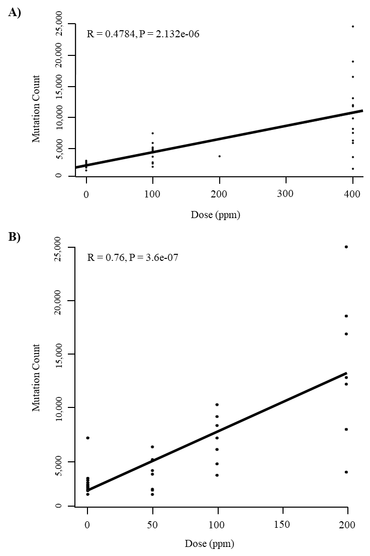
(A) B6C3F1/N mouse hepatocellular carcinoma mutational burden. (B) Sprague Dawley rat mammary tumor mutational burden. R = Pearson’s correlation coefficient; 0 ppm = spontaneous tumor control samples.
E.3.2. Mutation Signatures of Mouse Hepatocellular Carcinomas Arising Spontaneously or Following α-Pinene Exposure
Extraction of the de novo mutation signatures from the mHCCs resulted in three (A, B, and C) signatures (Figure E-2, Figure E-3). Signature A was predominant in the α-pinene-exposed mHCCs compared to the spontaneous mHCCs. When these de novo signatures were decomposed into COSMIC signatures (>0.80 cosine similarity), signature A did not resolve into known COSMIC signatures and was assigned a novel mouse-specific signature, mSig1. Overall, these de novo signatures resolved into mSig1 (61.8%), single base substitution 40 (SBS40) (22.1%), SBS5 (9.6%), SBS58 (2.8%), SBS3 (1.4%), and SBS1 (1.3%) (Figure E-4).
Figure E-2. De novo Mutational Spectra of Hepatocellular Carcinomas in B6C3F1/N Mice Arising Spontaneously or Following Chronic (Two-year) Inhalation Exposure to α-Pinene
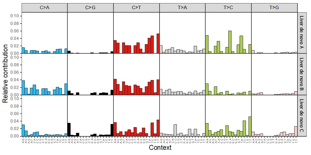
De novo mutational signatures (A, B, and C) extracted from mouse hepatocellular carcinomas. C = cytosine; A = adenine; G = guanine; T = thymine.
Figure E-3. Mutation Burden of Hepatocellular Carcinomas in B6C3F1/N Mice Arising Spontaneously or Following Chronic (Two-year) Inhalation Exposure to α-Pinene
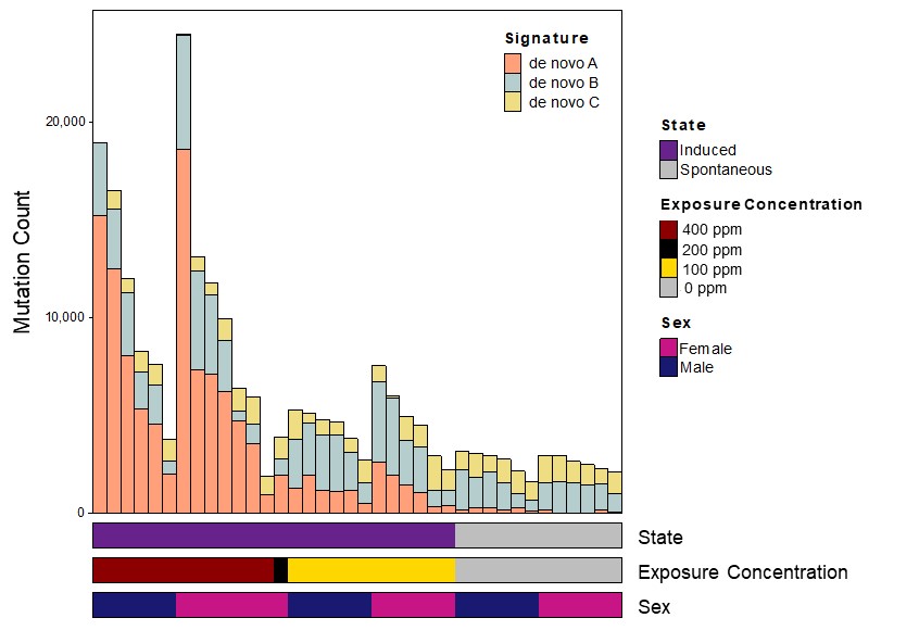
Mutation burden (absolute) with the corresponding constituent de novo signatures with the metadata on exposure level and sex.
Figure E-4. Proportion of COSMIC Signatures Decomposed from the de novo Signatures of Hepatocellular Carcinomas in B6C3F1/N Mice Arising Spontaneously or Following Chronic (Two-year) Inhalation Exposure to α-Pinene
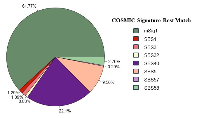
mSig1 did not resolve into known COSMIC signatures at the default cosine similarity (0.8). COSMIC = Catalogue of Somatic Mutations in Cancer; SBS = single base substitutions.
E.3.3. Mutation Signatures of Mouse Alveolar/Bronchiolar Carcinomas Arising Spontaneously or Following α-Pinene Exposure
Extraction of the de novo mutation signatures from the mABCs resulted in two (A and B) signatures (Figure E-5, Figure E-6). Signature A was predominant in some of the α-pinene-exposed mABCs compared to the spontaneous mABCs. These de novo signatures resolved into the following COSMIC signatures: SBS5 (71.3%), SBS58 (17.7%), SBS57 (7.7%), and SBS7a (2.7%) (Figure E-7).
Figure E-5. De novo Mutational Spectra of Alveolar/Bronchiolar Carcinomas in B6C3F1/N Mice Arising Spontaneously or Following Chronic (Two-year) Inhalation Exposure to α-Pinene
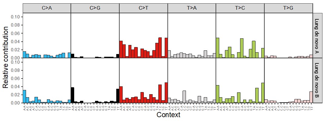
De novo signatures (A and B) extracted from mouse alveolar/bronchiolar carcinomas. C = cytosine; A = adenine; G = guanine; T = thymine.
Figure E-6. Mutation Burden of Alveolar/Bronchiolar Carcinomas in B6C3F1/N Mice Arising Spontaneously or Following Chronic (Two-year) Inhalation Exposure to α-Pinene
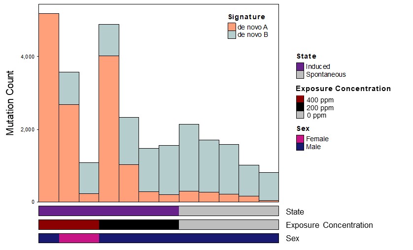
Mutation burden (absolute) with the corresponding constituent de novo signatures with the metadata on exposure level and sex.
Figure E-7. Proportion of COSMIC Signatures Decomposed from the de novo Signatures of Alveolar/Bronchiolar Carcinomas in B6C3F1/N Mice Arising Spontaneously or Following Chronic (Two-year) Inhalation Exposure to α-Pinene
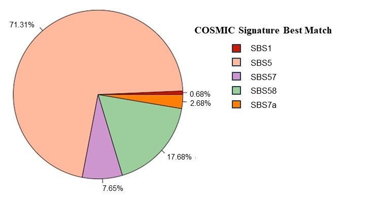
COSMIC = Catalogue of Somatic Mutations in Cancer; SBS = single base substitutions.
E.3.4. Mutation Signatures of Rat Mammary Tumors Arising Spontaneously or Following α-Pinene Exposure
Extraction of the de novo mutation signatures from rMTs resulted in three (A, B, and C) signatures (Figure E-8, Figure E-9). Signature A was unique to only α-pinene-exposed rMTs compared to the spontaneous rMTs. These de novo signatures resolved into SBS5 (63%), SBS21 (16%), SBS40 (11%), SBS12 (8%), and SBS1 (3%) (Figure E-10). SBS21 and SBS12 were unique to α-pinene-exposed rMTs (data not shown).
Figure E-8. De novo Mutational Spectra of Mammary Tumors in Sprague Dawley Rats Arising Spontaneously or Following Chronic (Two-year) Inhalation Exposure to α-Pinene
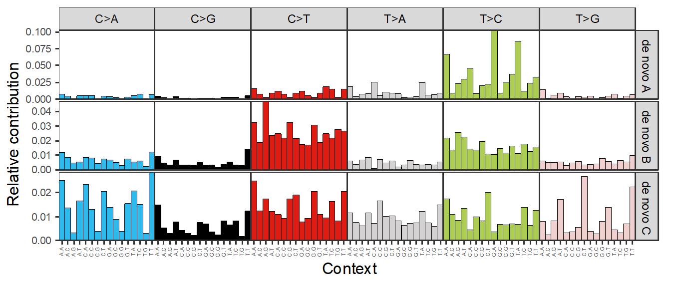
De novo signatures (A, B, and C) extracted from rat mammary tumors. C = cytosine; A = adenine; G = guanine; T = thymine.
Figure E-9. Mutation Burden of Mammary Tumors in Sprague Dawley Rats Arising Spontaneously or Following Chronic (Two-year) Inhalation Exposure to α-Pinene
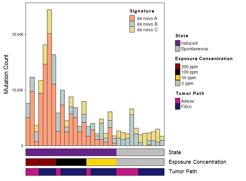
Mutation burden (absolute) with the corresponding constituent de novo signatures with the metadata on exposure level and tumor origination. Adeno = adenocarcinoma; Fibro = fibroadenoma.
Figure E-10. Proportion of COSMIC Signatures Decomposed from the de novo Signatures of Mammary Tumors in Sprague Dawley Rats Arising Spontaneously or Following Chronic (Two-year) Inhalation Exposure to α-Pinene
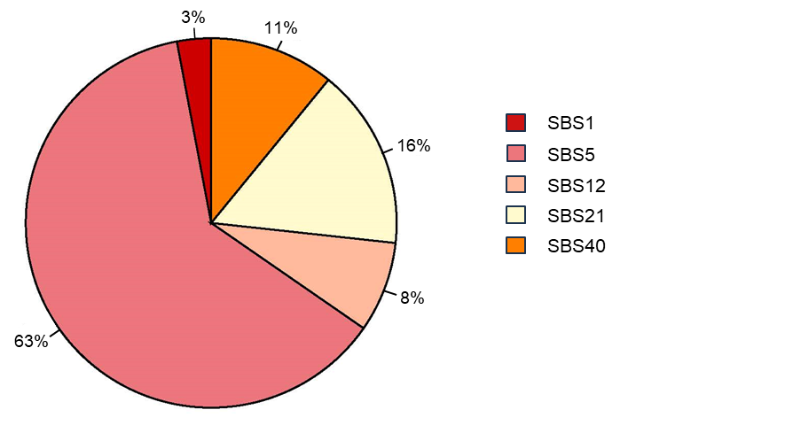
COSMIC = Catalogue of Somatic Mutations in Cancer; SBS = single base substitutions.
E.4. Discussion
Chronic α-pinene exposure resulted in a multisite carcinogenic response in mice and rats of both sexes. A recent study by Waidyanatha et al.5 indicated that α-pinene oxide, a metabolite of α-pinene, is mutagenic at ≥25 µg/plate in vitro. There was a significant exposure concentration-dependent increase in mutation burden in mHCCs but not in mABCs even with inhalation as the route of exposure. These data support the findings by Waidyanatha et al.,5 wherein inhalation and subsequent metabolism of α-pinene to α-pinene oxide in the liver may lead to higher mutagenicity at the principal site of metabolism and thus, a higher mutation burden in the liver compared to the lung in mice. While similar data in rats are not available, the exposure concentration-dependent mutation burden in the rMTs supports the findings by Waidyanatha et al.,6 which reported a higher blood maximum concentration (Cmax) and area under the curve (AUC) in the mammary gland than in blood for both α-pinene and α-pinene oxide. Overall, the weight of evidence suggests α-pinene’s carcinogenic activity is operating through a mutagenic mode of action following metabolic activation to α-pinene oxide.
While there were significant age/clock-like signatures (SBS1, SBS5, SBS40) in all tumors assessed, some tumors following α-pinene exposure showed distinct signatures that were likely reflective of the exposure. The novel mutation signature mSig1 was not readily resolved into known COSMIC signatures at the default setting (>0.8 cosine similarity) and was predominant in the mHCCs following α-pinene exposure but not in those arising spontaneously, suggesting that it may be related to the exposure. Similarly, the presence of SBS21 and SBS12 COSMIC signatures in the rMTs following α-pinene exposure, but not in those arising spontaneously, suggests these signatures may also be related to the exposure. These tumor-specific differences in mutation signatures may be related to the different mutagenic and DNA repair processes in the respective target organs. Further adductomic studies on the tumor target organs in rats and mice exposed to α-pinene may provide more specific information on the exogenous biochemical modification of the nucleotides leading to mutagenicity and subsequent carcinogenicity. In addition, the adductomic and multiomic studies on in vitro studies using relevant human cells/organoids will provide translationally relevant information and better inform public health risk assessments.
Appendix F. Peer Review Comments
F.1. Peer Reviewers
External peer review of the draft NTP Technical Report on the Toxicology and Carcinogenesis Studies of α-Pinene (CASRN 80-56-8) Administered by Inhalation to Sprague Dawley (Hsd:Sprague Dawley SD) Rats, B6C3F1/N Mice, and CD-1 Mice was conducted by seven subject matter experts who individually provided comments via letter. Individuals outside the federal government were invited to serve as peer reviewers because of their expertise and then vetted, following established National Toxicology Program (NTP) practices, to ensure no conflicts of interest. For peer review of this draft NTP report, the peer reviewers had expertise in carcinogenicity, anatomical pathology, reproductive toxicology, genetic toxicology, monoterpenes, and occupational inhalation.
The peer reviewers were charged to peer review the draft NTP report and provide recommended revisions that would strengthen the scientific analyses or conclusions.
The report preparation team carefully considered the peer reviewers’ comments in revising the report. The peer reviewers’ anonymized comments, presented in random order, are provided verbatim in this appendix, other than for the correction of minor typographical or grammatical errors.
Reviewers:
Javed A. Bhalli, Ph.D., M.Phil
Vice President and Site Head, Safety and Toxicology
Frontage Laboratories, Inc.
Chicago, Illinois, USA
Daniel J. Conklin, Ph.D.
Director, Exposure Studies Shared Resource, Omics & Exposure Facility Core
Co-Director, Research Engagement and Training Coordination Core
University of Louisville
Louisville, Kentucky, USA
Terry Gordon, Ph.D.
Research Professor, Department of Medicine
New York University School of Medicine
New York City, New York, USA
Wendy G. Halpern, D.V.M., Ph.D.
Senior Fellow – Pathologist
Genentech, Inc.
San Francisco, California, USA
Stephen S. Hecht, Ph.D.
Professor, Department of Laboratory Medicine and Pathology
Masonic Cancer Center, University of Minnesota
Minneapolis, Minnesota, USA
Lisa A. Miller, Ph.D.
Professor, Department of Anatomy, Physiology, and Cell Biology
University of California, Davis School of Veterinary Medicine
Davis, California, USA
Wanying Zhang, M.D.
Laboratory Director, Clinical Consultant
Variantyx, Inc.
Fort Lauderdale, Florida, USA
F.2. Peer Review Charge and Instructions
Charge:
Peer review the draft NTP Technical Report on the Toxicology and Carcinogenesis Studies of α-Pinene (CASRN 80-56-8) Administered by Inhalation to Sprague Dawley (Hsd:Sprague Dawley SD) Rats, B6C3F1/N Mice, and CD-1 Mice.
Instructions:
For the numbered charge questions, provide any recommended revisions necessary for strengthening the scientific analyses or conclusions of the draft NTP Technical Report. As available, please provide the page and line number(s), table number, and/or figure number to which the specific comment applies.
F.3. Peer Review Comments
F.3.1. Reviewer 1
1. Information presentation:
Please comment on whether the information presented in the draft NTP Technical Report, including presentation of data in any tables and figures, is technically correct, clearly stated, and objectively presented.
Reviewer Comments:
Two-year Study in Rats
Line 43, page 101 “Whole-genome sequencing of fresh frozen rat mammary tumors revealed an exposure concentration-dependent increase in mutation burden and an exposure-specific mutation signature (SBS21 COSMIC signature) that is enriched for T>C transitions (data not shown).” It is odd to include this statement in the Discussion as there is no mention of conducting this analysis in the Results section. There are data shown in the Appendix, but it is presented as a completely separate results section.
Two-year Study in Mice
Whole-genome sequencing appears to have been conducted in mice, but these data are limited to the Appendix section. As with the rat study, there is no mention of the sequencing analysis in the Results.
Reproductive Performance in Rats and Mice
The results of the study were limited to males, despite the observation that females appear more sensitive to chronic exposure.
Three-month Investigative Study in Rats
Why is this study listed separately from the other rat studies in the Materials and Methods?
The Results for this are confusing as it only describes deposition, weight, and survival.
Line 17, page xix: The abstract states “Early mortalities in the chronic study, attributed to mammary masses or nodules, prompted the addition of a follow-up 3-month investigative study in male and female Sprague Dawley rats to evaluate early biomarkers of carcinogenicity in mammary gland and collect definitive internal concentration data for α-pinene and α-pinene oxide in blood and mammary gland. Mammary gland was collected for future analysis, and sperm parameters were also evaluated.” This was not mentioned in the Results. What early markers were evaluated?
Line 21, page xxvii: “The early signs of mammary tumors in the chronic study prompted the addition of a follow-up investigative 3-month study in male and female Sprague Dawley rats to evaluate early biomarkers of carcinogenicity in the mammary gland using error-corrected duplex sequencing and to perform a definitive evaluation of internal concentrations of α-pinene and α-pinene oxide in the blood and mammary gland using validated methods.” I could not find these data from the 3-month investigative study.
Please suggest any improvements to the information presented and provide your rationale or scientific support for proposed improvements where applicable.
Reviewer Comments:
In general, the report was challenging to review given the different species, strains, exposure periods and assessments (e.g., reproductive versus investigative). Perhaps consider the organization and order in which the information is presented? For example, the Materials and Methods do not align with the Results section. The Materials and Methods starts with the two-year rat study, but the Results starts with the 3-month reproductive study, etc.
What was the rationale for using different mouse and rat strains? And how might this have affected the interpretation of the data?
This is minor, but for consistency it might help to start with male outcomes, followed by female outcomes for both rats and mice.
In some sections the 3-month reproduction study is combined with the 2-year study. Again, it makes it difficult to follow and correlate outcomes with conclusions (see lines 23-26 page xix).
It is very confusing to have the mutation data presented as a separate appendix and only superficially addressed in the main body of the report.
While this was a comprehensive study (acute vs chronic, two species), there are potential limitations to the exposure conditions that warrant mention in the discussion. Specifically, there appeared to be some variability in particle counts (Table A-1) and less than proportional blood concentration (Tables 27, 28), which could have influenced the severity of the outcomes (although not the overall interpretation).
Please identify any information that should be added or deleted and provide your rationale behind the additions or deletions.
Reviewer Comments:
I don't have any deletions to suggest beyond my comments on various sections.
For the Material and Methods, I would suggest including a sentence that clearly states that inhalation exposure was for the entire duration of the study period (at least that is how I'm interpreting the writing). In other words, for a 3-month study, animals are exposed to alpha pinene for 24 hours/day, 7 days/week for a total of 90 days. Presumably, the animal husbandry would require periodic access to the exposure chambers, which would require evacuation of the alpha pinene to reduce occupational exposures?
Line 28 page xxvii has a typo (Appendix E).
2. Study design, conduct, and findings:
Please comment on the adequacy of the experimental design and methods used for execution of the studies. Provide your rationale or scientific support for proposed changes where the approach is deemed insufficient.
Reviewer Comments:
Is it possible that the different sources [for] rat and mouse strains could influence interpretation of the findings between exposure periods? Particularly since the 3-month investigative study in rats showed the highest blood concentration of alpha pinene?
Line 35, page 9: “Male and female Sprague Dawley rats were obtained from Envigo (formerly Harlan Laboratories, Inc.; Indianapolis, IN) for the 2-year and 3-month reproductive studies. Male and female Sprague Dawley rats were obtained from Envigo (Haslett, MI) for the 3-month investigative study.” This is confusing - did the company move or is there a typographical error? Vendor sources of animals can certainly influence study outcomes.
It is notable that the stability of the compound was tested through the duration of the studies to determine if degradation occurred (no evidence).
Line 23, page 8: ”Particle counts were collected from all chambers at regular intervals during all studies, and particle counts were frequently >200 particles/cm3 except for the 400 ppm chamber for Sprague Dawley (Hsd:Sprague Dawley SD) rats in the 3-month reproductive study. The source and identity of the particles were undetermined.” Could this be addressed in the discussion?
Please comment on whether the statistical analyses have been appropriately applied and scientifically justified. Provide your rationale or scientific support for any proposed changes to the statistical analyses.
Reviewer Comments: The statistical analyses appears to be appropriately applied and scientifically justified.
Please comment on whether the scientific interpretations of the data are objectively presented and scientifically justified. Please specify any study conclusions that are not justified and provide alternate interpretation of the data.
Reviewer Comments:
Is it possible that greater death in female rats is due to higher dose/body weight?
The concentration assessment measures alpha pinene and alpha pinene oxide in blood with no apparent differences between males and females. Are there differences in blood volume between males and females?
The reproductive assessments were exclusively done in males, but females (particularly for rats) appear to be more sensitive. This is a gap in knowledge and should be pointed out in the discussion.
Although the concentration of blood alpha pinene and alpha pinene oxide with exposure does seem to increase with dose, the lack of proportional concentration is concerning with regard to interpretation of findings as it may have been influenced by exposure methods. At a minimum this should be addressed in the discussion.
Line 9, page 99: “α-Pinene and α-pinene oxide concentrations in the 3-month investigative rat study were higher than those in the 2-year rat study, and the difference was not consistent across matrices and analytes. Up to 4-fold and 5- to 15-fold higher concentrations were observed in male and female blood, respectively, and 2- to 43-fold higher concentrations were observed in female mammary gland.” This should be of concern, as it may be due to variability in exposure conditions, which would influence outcomes. In this case, it might suggest that the observed effects in the 2-year study are mild relative to what would be expected.
3. Evaluation of mutation burden and unique mutation signatures:
Please comment on the adequacy of the experimental design and methods used for execution of the studies. Provide your rationale or scientific support for proposed changes where the approach is deemed insufficient:
Reviewer Comments: There appeared to be variability in exposure concentrations. Particle counts (Table A-1) seemed widely different between studies, particularly mice versus rats.
Please comment on whether the bioinformatic analyses were appropriately applied and scientifically justified. Provide your rationale or scientific support for any proposed changes to the statistical analyses.
Reviewer Comments: The bioinformatic analysis appears to be appropriately applied and scientifically justified.
Please comment on whether the scientific interpretations of the data are objectively presented and scientifically justified. Please specify any study conclusions that are not scientifically justified and provide an alternate interpretation of the data.
Reviewer Comments:
I agree that the overall conclusions of alpha pinene toxicity and carcinogenicity for the 2-year study (both rats and mice) are justified by the evidence of blood concentration and histopathologic assessments.
Less than proportional blood deposition might suggest variability introduced by exposure conditions, however this observation does not discount the overall conclusions of toxicity and carcinogenicity.
4. Levels of Evidence of Carcinogenic Activity categories:
Additional information on the NTP Levels of Evidence of Carcinogenic Activity is available at https://ntp.niehs.nih.gov/sites/default/files/ntp/test_info/cartox_loe_508.pdf and in the draft Technical Report.
Indicate your agreement or disagreement with each draft NTP Level of Evidence conclusion, taking into consideration the strength of the toxicology and carcinogenicity results in Hsd:Sprague Dawley rats and B6C3F1/N mice exposed to α-pinene.
Male Hsd:Sprague Dawley SD rats:
Some evidence of carcinogenic activity
Higher incidence of urinary bladder papilloma
☒ Agree
☐ Agree in principle with the exceptions listed below:
☐ Do not agree with interpretations because:
Female Hsd:Sprague Dawley SD rats:
Clear evidence of carcinogenic activity
Increased incidences of mammary gland adenocarcinoma and adenoma or adenocarcinoma (combined)
Increased incidences of stromal polyp; adenocarcinoma; squamous cell carcinoma; and squamous cell papilloma, squamous cell carcinoma, adenoma, or adenocarcinoma (combined) in the uterus were also considered to be related to exposure
☒ Agree
☐ Agree in principle with the exceptions listed below:
☐ Do not agree with interpretations because:
Male B6C3F1/N mice:
Clear evidence of carcinogenic activity
Increased incidences of Harderian glad adenoma, adenocarcinoma, and adenoma or adenocarcinoma (combined); hepatocellular adenoma, carcinoma, and adenoma or carcinoma (combined); and alveolar/bronchiolar adenoma and adenoma or carcinoma (combined)
Higher incidences of urinary bladder papilloma and forestomach papilloma were also considered to be related to exposure
☒ Agree
☐ Agree in principle with the exceptions listed below:
☐ Do not agree with interpretations because:
Female B6C3F1/N mice:
Clear evidence of carcinogenic activity
Increased incidences of Harderian gland adenoma and adenoma or adenocarcinoma (combined); hepatocellular adenoma, carcinoma, and adenoma or carcinoma (combined); alveolar/bronchiolar adenoma, carcinoma, and adenoma or carcinoma (combined); and mammary gland adenocarcinoma and adenoma or adenocarcinoma (combined)
Increased incidences of granulosa cell tumor, benign, malignant (combined) and tubulostromal adenoma in the ovary and a higher incidence of squamous cell carcinoma in the forestomach were also considered to be related to exposure
☒ Agree
☐ Agree in principle with the exceptions listed below:
☐ Do not agree with interpretations because:
5. Please provide any additional comments, suggestions, or recommendations for improving the report and provide rationale or scientific support for proposed improvements where applicable.
Reviewer Comments: This study provides a comprehensive histopathologic assessment of the toxicity and carcinogenicity of alpha pinene using two in vivo models of exposure. It is possible that some of the observed findings regarding alpha pinene blood deposition may be attributed to variability in exposure conditions, but ancillary data from prior peer-reviewed studies suggest that the metabolites of alpha pinene could also play a role in toxicity when less than proportional deposition concentrations of the inhalant were observed. Regardless of the gaps in understanding the mechanisms of alpha pinene metabolism, the outcome of the study in both rats and mice provides scientific evidence of toxicity and carcinogenicity following two years of chronic exposure.
F.3.2. Reviewer 2
1. Information presentation:
Please comment on whether the information presented in the draft NTP Technical Report, including presentation of data in any tables and figures, is technically correct, clearly stated, and objectively presented.
Reviewer Comments: The information provided in this report, including tables and figures, is clear and objectively presented. However, summary can be improved to make it clear to the readers without going into the main sections of the report. For example,
Summary
Two-year Study in Rats
Summary states that animals were exposed to α-pinene for two years, but it is not clear if this was whole body or nose only exposure. This is also not clear if exposure was continuous during this duration or was for certain days per week etc.
It is stated that survival of male rats was comparable to that of control rats, whereas female survival was significantly decreased compared to control rats. However, it is not clear survival of male rats was decreased in any particular dose group or this statement talks about the cumulative decrease.
Same is for body weight, should be clarified which dose groups are significantly lower or if this statement is for cumulative decrease.
Two-year Study in Mice:
This title is confusing. There are two studies in this section.
3 months exposure in CD-1 males and B6C3F1/N males and females for reproductive assessment.
2 years toxicity and carcinogenicity study in B6C3F1/N male and female mice.
However, the title is “Two-year Study in Mice.” It is suggested either to clearly state the title or make Subsets A and B for the two types of studies. Also, the summary of the two study types is mixed. It is suggested to summarize the results of the 3-month study in the first paragraph and then start a separate paragraph to summarize the results of the carcinogenicity study.
Paragraph 2, line 3: It states that α-pinene exposure did not significantly affect survival. However, it is not stated which sex.
Paragraph 2, last sentence: It is stated that clinical observations were sporadic and not attributed to α-pinene exposure, but this does not state which sex, male, females, or both.
Paragraph 4, first sentence: Urinary bladder papilloma was observed in the 400 ppm male mice, but it is not stated which male mice, CD1 or B6C3F1/N.
Please suggest any improvements to the information presented and provide your rationale or scientific support for proposed improvements where applicable.
Reviewer Comments: The studies presented in the report are well designed and well conducted. Data supports the conclusion.
Please identify any information that should be added or deleted and provide your rationale behind the additions or deletions.
Reviewer Comments: None.
2. Study design, conduct, and findings:
Please comment on the adequacy of the experimental design and methods used for execution of the studies. Provide your rationale or scientific support for proposed changes where the approach is deemed insufficient.
Reviewer Comments: In the Genetic Toxicity section of Overview, studies conducted reported in literature are stated. However, it does not look like an Ames study is conducted in the past where all five strains (TA98, TA100, TA1535, TA1537, and WP2uvrA) were used where α-pinene and α-pinene oxide were tested up to the dose levels recommended by the OECD and ICH guidelines, 5000 µg/plate to determine if parent compound or one of its metabolite can cause mutation induction.
Because now data from 2-year carcinogenicities are available to support the carcinogenic potential of these compounds, not having data for the Ames assay up to most relevant doses or in all recommended strains is not critical.
Please comment on whether the statistical analyses have been appropriately applied and scientifically justified. Provide your rationale or scientific support for any proposed changes to the statistical analyses.
Reviewer Comments: Statistical analysis conducted is appropriate.
Please comment on whether the scientific interpretations of the data are objectively presented and scientifically justified. Please specify any study conclusions that are not justified and provide alternate interpretation of the data.
Reviewer Comments: Yes.
3. Evaluation of mutation burden and unique mutation signatures:
Please comment on the adequacy of the experimental design and methods used for execution of the studies. Provide your rationale or scientific support for proposed changes where the approach is deemed insufficient.
Reviewer Comments: Mutational spectrum as conducted using tissues from treated animals. Tissues were shipped to Sanger Institute where these were analyzed with a read depth of 45X. However, a new approach of error corrected next generation sequencing (ecNGS) also called duplex sequencing is available with multiple versions. This could have been more relevant with very low percentage of error which could have provided better results. It is recommended that this technique should be considered in future for similar studies.
Please comment on whether the bioinformatic analyses were appropriately applied and scientifically justified. Provide your rationale or scientific support for any proposed changes to the statistical analyses.
Reviewer Comments: Bioinformatic analyses used in this report is appropriate for the mutation method used here.
Please comment on whether the scientific interpretations of the data are objectively presented and scientifically justified. Please specify any study conclusions that are not scientifically justified and provide an alternate interpretation of the data.
Reviewer Comments: Yes.
4. Levels of Evidence of Carcinogenic Activity categories:
Additional information on the NTP Levels of Evidence of Carcinogenic Activity is available at https://ntp.niehs.nih.gov/sites/default/files/ntp/test_info/cartox_loe_508.pdf and in the draft Technical Report.
Indicate your agreement or disagreement with each draft NTP Level of Evidence conclusion, taking into consideration the strength of the toxicology and carcinogenicity results in Hsd:Sprague Dawley rats and B6C3F1/N mice exposed to α-pinene.
Male Hsd:Sprague Dawley SD rats:
Some evidence of carcinogenic activity
Higher incidence of urinary bladder papilloma
☒ Agree
☐ Agree in principle with the exceptions listed below:
☐ Do not agree with interpretations because:
Female Hsd:Sprague Dawley SD rats:
Clear evidence of carcinogenic activity
Increased incidences of mammary gland adenocarcinoma and adenoma or adenocarcinoma (combined)
Increased incidences of stromal polyp; adenocarcinoma; squamous cell carcinoma; and squamous cell papilloma, squamous cell carcinoma, adenoma, or adenocarcinoma (combined) in the uterus were also considered to be related to exposure
☒ Agree
☐ Agree in principle with the exceptions listed below:
☐ Do not agree with interpretations because:
Male B6C3F1/N mice:
Clear evidence of carcinogenic activity
Increased incidences of Harderian glad adenoma, adenocarcinoma, and adenoma or adenocarcinoma (combined); hepatocellular adenoma, carcinoma, and adenoma or carcinoma (combined); and alveolar/bronchiolar adenoma and adenoma or carcinoma (combined)
Higher incidences of urinary bladder papilloma and forestomach papilloma were also considered to be related to exposure
☒ Agree
☐ Agree in principle with the exceptions listed below:
☐ Do not agree with interpretations because:
Female B6C3F1/N mice:
Clear evidence of carcinogenic activity
Increased incidences of Harderian gland adenoma and adenoma or adenocarcinoma (combined); hepatocellular adenoma, carcinoma, and adenoma or carcinoma (combined); alveolar/bronchiolar adenoma, carcinoma, and adenoma or carcinoma (combined); and mammary gland adenocarcinoma and adenoma or adenocarcinoma (combined)
Increased incidences of granulosa cell tumor, benign, malignant (combined) and tubulostromal adenoma in the ovary and a higher incidence of squamous cell carcinoma in the forestomach were also considered to be related to exposure
☒ Agree
☐ Agree in principle with the exceptions listed below:
☐ Do not agree with interpretations because:
5. Please provide any additional comments, suggestions, or recommendations for improving the report and provide rationale or scientific support for proposed improvements where applicable.
Reviewer Comments: Studies presented in this report were appropriately conducted, data was interpreted with scientific vigor, and the report is written and presented well. Report can be accepted in its original form with some more clarity in the summary section.
F.3.3. Reviewer 3
1. Information presentation:
Please comment on whether the information presented in the draft NTP Technical Report, including presentation of data in any tables and figures, is technically correct, clearly stated, and objectively presented.
Reviewer Comments: The information follows a well structured and long used pattern of presentation that is technically great, clear, and objective. Minor issues are noted in the last question below.
Please suggest any improvements to the information presented and provide your rationale or scientific support for proposed improvements where applicable.
Reviewer Comments: Given the decades of successful formatting and the proven rationale and science that is supported by the data presentation in these NTP reports, this question no longer needs to be asked and could be removed/eliminated.
Please identify any information that should be added or deleted and provide your rationale behind the additions or deletions.
Reviewer Comments: No suggestions for additions or deletion other than the inclusion of the histopathology in the report. Yes, they are important and catch my inquisitive eye as a toxicologist, but they could be eliminated or relegated to an appendix as while they do serve the purpose of backing up the data upon which the determinations are made, they don’t add much to the report.
2. Study design, conduct, and findings:
Please comment on the adequacy of the experimental design and methods used for execution of the studies. Provide your rationale or scientific support for proposed changes where the approach is deemed insufficient.
Reviewer Comments: The study design is excellent (as all NTP chronic studies generally are). The choice of the inhalation route, the exposure concentrations, and the subchronic add-on studies to examine the potential adverse effects was logical and well warranted. Similarly, the mechanistic studies (e.g., mutations) were appropriately designed for inclusion. Only one minor comment – The generation reservoir and a nitrogen exhaust vent/rotameter are unclear in Figure A-5.
Please comment on whether the statistical analyses have been appropriately applied and scientifically justified. Provide your rationale or scientific support for any proposed changes to the statistical analyses.
Reviewer Comments: The statistical analyses were appropriately applied and justified. The ‘newer’ trend analyses are quite valuable. As I believe I’ve noted on previous reviews, the inclusion of the trend’s statistical results under the control column still appears odd or confusing to me until I carefully read the footnotes. Perhaps there is a simpler or clearer way to present the trend results (or I’m just from an older generation that likes clearly understandable tables).
Please comment on whether the scientific interpretations of the data are objectively presented and scientifically justified. Please specify any study conclusions that are not justified and provide alternate interpretation of the data.
Reviewer Comments: The data interpretations are clearly presented and justified. One small suggestion would be to consistently present the most significant/important findings first. For example, in the Abstract, page xxii, lines 2-4, the ‘some evidence’ finding is presented first followed by the ‘clear evidence.’
3. Evaluation of mutation burden and unique mutation signatures:
Please comment on the adequacy of the experimental design and methods used for execution of the studies. Provide your rationale or scientific support for proposed changes where the approach is deemed insufficient.
Reviewer Comments: The mutation burden and signature data were well documented and I agree that it was logical to assess the endpoints in three major tumor types for comparison. Mutational spectrum research is not my field of expertise, but I do know that it is quite complicated. Thus, to this reviewer, it is not clear if the approach would be improved by the more precise (and costly) gene by gene analysis.
In Figure E-10, SBS and such are not defined.
Please comment on whether the bioinformatic analyses were appropriately applied and scientifically justified. Provide your rationale or scientific support for any proposed changes to the statistical analyses.
Reviewer Comments: To the best knowledge of this reviewer (which is limited), these analyses were appropriate.
Please comment on whether the scientific interpretations of the data are objectively presented and scientifically justified. Please specify any study conclusions that are not scientifically justified and provide an alternate interpretation of the data.
Reviewer Comments: The interpretations of the data are objectively and well presented in the report. The writing leads to clear justifications for the proposed levels of evidence of carcinogenic activity for alpha-pinene. This was particularly true for the well written Discussion section that clearly linked the findings to the exposures.
4. Levels of Evidence of Carcinogenic Activity categories:
Additional information on the NTP Levels of Evidence of Carcinogenic Activity is available at https://ntp.niehs.nih.gov/sites/default/files/ntp/test_info/cartox_loe_508.pdf and in the draft Technical Report.
Indicate your agreement or disagreement with each draft NTP Level of Evidence conclusion, taking into consideration the strength of the toxicology and carcinogenicity results in Hsd:Sprague Dawley rats and B6C3F1/N mice exposed to α-pinene.
Male Hsd:Sprague Dawley SD rats:
Some evidence of carcinogenic activity
Higher incidence of urinary bladder papilloma
☒ Agree
☐ Agree in principle with the exceptions listed below:
☐ Do not agree with interpretations because:
Female Hsd:Sprague Dawley SD rats:
Clear evidence of carcinogenic activity
Increased incidences of mammary gland adenocarcinoma and adenoma or adenocarcinoma (combined)
Increased incidences of stromal polyp; adenocarcinoma; squamous cell carcinoma; and squamous cell papilloma, squamous cell carcinoma, adenoma, or adenocarcinoma (combined) in the uterus were also considered to be related to exposure
☒ Agree
☐ Agree in principle with the exceptions listed below:
☐ Do not agree with interpretations because:
Male B6C3F1/N mice:
Clear evidence of carcinogenic activity
Increased incidences of Harderian glad adenoma, adenocarcinoma, and adenoma or adenocarcinoma (combined); hepatocellular adenoma, carcinoma, and adenoma or carcinoma (combined); and alveolar/bronchiolar adenoma and adenoma or carcinoma (combined)
Higher incidences of urinary bladder papilloma and forestomach papilloma were also considered to be related to exposure
☒ Agree
☐ Agree in principle with the exceptions listed below:
☐ Do not agree with interpretations because:
Female B6C3F1/N mice:
Clear evidence of carcinogenic activity
Increased incidences of Harderian gland adenoma and adenoma or adenocarcinoma (combined); hepatocellular adenoma, carcinoma, and adenoma or carcinoma (combined); alveolar/bronchiolar adenoma, carcinoma, and adenoma or carcinoma (combined); and mammary gland adenocarcinoma and adenoma or adenocarcinoma (combined)
Increased incidences of granulosa cell tumor, benign, malignant (combined) and tubulostromal adenoma in the ovary and a higher incidence of squamous cell carcinoma in the forestomach were also considered to be related to exposure
☒ Agree
☐ Agree in principle with the exceptions listed below:
☐ Do not agree with interpretations because:
5. Please provide any additional comments, suggestions, or recommendations for improving the report and provide rationale or scientific support for proposed improvements where applicable.
Reviewer Comments:
Abstract – page xx, line 15 – it’s not clear what this % decrease refers to as it’s male mice and one concentration.
Abstract – page xx, lines 26-31 – here and elsewhere, why are there redundant sentences where the first sentence states the general finding and the second sentence is nearly identical but just adds the data (e.g., “There were exposure-related significant increases in the incidences of hepatocellular adenoma, carcinoma, and adenoma or carcinoma (combined) in male and female mice. The incidences of hepatocellular adenoma, carcinoma, and adenoma or carcinoma (combined) were significantly increased at concentrations of ≥200, 400, and ≥100 ppm α-pinene, respectively, in male mice, and ≥100, 400, and ≥100 ppm, respectively, in female mice.”)
Abstract – page xxi, line 15-16 – I suggest that this be a new paragraph as it’s unrelated to the preceding.
Abstract – page xxii, line 27 – should ‘these’ be ‘the’?
Abstract – page xxv – Unclear why ‘Equivocal Findings’ is noted here. [Does]n’t ‘equivocal’ come after ‘Some evidence’? It just seems odd to see it pop up here as a subcategory when Some and Clear are presented in the next line of Level of Evidence.
Abstract – page xxviii – Shouldn’t ‘NTP’ be ‘alpha-pinene’?
Page 2, lines 17-31 – To show clearer real-world relevance, perhaps add some comparative calculations for an oral dose/day vs. an inhaled dose/day for occupational and environmental/indoor exposures?
Page 5, line 39 – Is it correct to state “No in vivo studies…’ if Reference 81 is a mouse micronucleus study?
Page 24, lines 9-12 – First, to the non-initiated, ‘censored’ and ‘uncensored’ are unclear terms (i.e., this inhalation toxicologist with over 40 years of experience had to read this 3 or 4 times to figure out what ‘censored’ meant (i.e., as in it means counted or eliminated as in ‘this thesis was censored’). Second, for the less knowledgeable, like myself, it would help if ‘natural’ and ‘unnatural’ causes are defined. Bottom line, this ‘survival analyses’ terminology can be confusing.
Page 87, line 12 – ‘the presence of artifacts’ needs further explanation for the decision to reject data output. Was it technical error or the machine broke or there was contamination or the samples were not stored properly or analyzed in time?
Page 90, line 13 – confusing as the sentence first states that the decrease in lutea occurred in all groups compared to controls, then talks about natural variability. Is it implying the decreases were within historical variability? Were there statistics compared to control?
Page 90, line 15 – the subheading title could be improved – “Investigative” is very broad.
Page 90, lines 16-19 – Unclear – this starts with biomarkers being investigated for mammary gland carcinogenesis and then male rat moribund data are reported next. Then, male repro data are presented on page 94. Thus, the given reasoning for the 3-month investigative study doesn’t match the endpoints or the inclusion of male rats.
Page 90, lines 18-19 – Unclear why this is mentioned under this subheading when the next subheading is all about the internal concentrations.
The Discussion section is particularly clear and links everything nicely.
F.3.4. Reviewer 4
1. Information presentation:
Please comment on whether the information presented in the draft NTP Technical Report, including presentation of data in any tables and figures, is technically correct, clearly stated, and objectively presented.
Reviewer Comments: In general, the data appear internally consistent across narrative descriptions, figures, and tables. Analytical and histopathological findings are clearly linked to exposure concentrations. Tables and figures are generally well-constructed with informative titles, footnotes, and unites of measurement. Statistical trends and significance are clearly annotated. The narrative maintains scientific neutrality. Interpretations of findings are well-supported by data without overreaching conclusion. Minor changes will clarify some of the confusions including:
In some of the tables, for example Table 5 (page 67, line 5), the legends of “Average severity grade of lesions in affected animals: 1=minimal, 2=mild, 3=moderate, 4=marked” should have a clarification of how was this graded. If it is a grade system for hyperplasia or adenoma, it can be based on proliferative indices like Ki-67, size of the lesions, or purely morphological evaluation.
Figure 4 on the page of 68 shows an area where only tumor tissue presents. It will be beneficial to include some uninvolved mammary gland tissue from control group. This can help identify species-related, strain-related, age-related changes, and other normal variability that occur naturally or spontaneously vs. those potentially induced by the chemical.
Figure 9 on the page of 79, legend B is not accurately stated. The ratio of hematopoietic cells and marrow fat is age related in healthy individuals. The right comparison of the ratio should be in between control group animals and experimental group animals of the same age.
Please suggest any improvements to the information presented and provide your rationale or scientific support for proposed improvements where applicable.
Reviewer Comments: The information is clearly presented, technically accurate, and appropriately supported by the data. However, there is information suggested to be added. Please review the next section.
Please identify any information that should be added or deleted and provide your rationale behind the additions or deletions.
Reviewer Comments: Two areas need to be taken into consideration or if additional data is available to clarify:
Given the reported tumors in the uterus and mammary glands of female animals, the report would benefit from a discussion on the potential hormonal relevance of these findings. Specifically, the authors should clarify whether endocrine mechanisms were considered and if hormone measurements (e.g., estrogen, prolactin) were taken or could be analyzed from archived samples. This would help explain sex-specific findings, particularly the broader systemic impact observed in male animals at high doses. For example, on page 22, line 36, female rats showed higher concentrations of both α-pinene and α-pinene oxide, suggesting possible sex-specific pharmacokinetics. This may be due to differences in metabolism, body fat distribution (supporting mammary gland as a primary target), or hormone-regulated enzyme activity. Supporting evidence includes the presence of stromal polyps, which are often hormone responsive. It is suggested to discuss whether specific enzyme systems (e.g., cytochrome P450s) may be involved in the bioactivation of α-pinene and whether any known sex-specific activity in these pathways could explain the observed differences.
On page 76, line 7 where bone marrow changes were described, to enhance the interpretation of the observed bone marrow changes, the report should compare histological findings with peripheral blood parameters (e.g., reticulocyte counts, red and white blood cell indices). This would help clarify whether the changes primarily affect a single lineage or are multilineage, and whether the response appears reactive, regenerative, or stress induced.
2. Study design, conduct, and findings:
Please comment on the adequacy of the experimental design and methods used for execution of the studies. Provide your rationale or scientific support for proposed changes where the approach is deemed insufficient.
Reviewer Comments: The selected exposure concentrations (50 and 100 ppm) for toxicokinetic studies in rodents are relevant for investigating potential mechanistic pathways and internal dose relationships. However, these concentrations may exceed typical human occupational exposures—particularly in non-industrial environments. For context, the maximum reported workplace exposure to α-pinene is approximately 27 ppm, which is considerably lower than the high-dose test condition of 100 ppm. While the highest dose (400 ppm) is appropriate for hazard identification purposes, the report should acknowledge its limited relevance to human exposure scenarios, especially in consumer or indoor settings where α-pinene levels are typically in the sub-ppm range. To strengthen the rationale for dose selection, the report should:
State whether selected doses approach or exceed the maximum tolerated dose (MTD).
Clarify if these were based on prior studies indicating target organ toxicity.
Explain how these doses support hazard identification or margin-of-exposure assessments for humans.
Justify the use of 100 ppm as the lowest dose in the chronic study—was this based on ambient human exposures or extrapolated from the 3-month study where 25 ppm was used as the lowest dose?
The report identifies changes in the Harderian gland and forestomach. These organs have limited relevance to human health:
Harderian gland is rudimentary or absent in humans. This limitation should be acknowledged, although it does not invalidate the importance of mechanistic insights.
Page 115, line 9: Clarify whether similar changes occurred in the glandular stomach, or if findings were strictly localized to the forestomach which has mucosal squamous epithelium. This is important because forestomach is a rodent specific structure. Human stomach tissue is mainly glandular.
Please comment on whether the statistical analyses have been appropriately applied and scientifically justified. Provide your rationale or scientific support for any proposed changes to the statistical analyses.
Reviewer Comments: The statistical analyses appear to be appropriately applied and scientifically justified. A threshold of p ≤ 0.05 was used to determine statistical significance across analyses. Importantly, the determination of biological relevance or potential carcinogenicity of α-pinene was not based solely on statistical significance. Instead, the findings were evaluated using a comprehensive weight-of-evidence approach as described in the Explanation of Levels of Evidence of Carcinogenic Activity section. This multifactorial assessment appropriately considers dose-response relationships, biological plausibility, consistency across studies, and concordance with related endpoints (e.g., histopathological changes, organ weights, and mechanistic data), providing a scientifically balanced interpretation of the results.
One area to be taken into consideration is Table 15 on page 97 includes the notations “multiple” or “including multiple.” While formal statistical analyses were not conducted on multiplicity data, the inclusion of a brief discussion on quantitative tumor burden (e.g., total mass or volume of neoplasms per dose group) would improve interpretability. This addition would help contextualize the biological significance of findings, especially in cases where multiple tumor types or co-occurring lesions were observed.
Please comment on whether the scientific interpretations of the data are objectively presented and scientifically justified. Please specify any study conclusions that are not justified and provide alternate interpretation of the data.
Reviewer Comments: Overall, the interpretations presented in the draft report appear to be scientifically justified, objective, and grounded in the data. The narrative accurately reflects the findings and maintains a balanced tone when drawing conclusions, particularly in areas where statistical significance was not achieved but biological plausibility or relevance warranted discussion. However, several points would benefit from clarification to further strengthen the objectivity and rigor of the interpretations:
Page 96, line 28. A more detailed discussion of quantitative tumor burden (e.g., total mass or volume of neoplasms) would enhance the interpretation of biological significance, particularly in cases where tumor multiplicity or co-occurrence is noted but not statistically analyzed.
Page 101, line 26. Please clarify whether squamous differentiation within carcinomas was observed incidentally or occurred more frequently in high-dose animals. This may indicate divergent differentiation pathways and could have implications for understanding tumor heterogeneity and chemical-specific effects.
Page 110, figure 25. Clarify whether the hyperplastic and papillomatous regions occurred in the same animals or within the same anatomical locations. Figure 25 appears to show a papilloma adjacent to hyperplasia, which may suggest progression or spatial correlation worthy of further discussion.
Page 74, line 9. Although not statistically significant, the observed incidence of papilloma should still be noted, especially given their rarity. It would be valuable to include data on whether inflammatory or degenerative changes were observed in the bladder epithelium or submucosa across dose groups, as this may support a local irritative mechanism.
Page 97, line 9. Further characterization of the severity and histological grading of basophilic foci and necrosis is recommended. This would help differentiate between spontaneous background lesions and those potentially induced by α-pinene exposure.
3. Evaluation of mutation burden and unique mutation signatures:
Please comment on the adequacy of the experimental design and methods used for execution of the studies. Provide your rationale or scientific support for proposed changes where the approach is deemed insufficient.
Reviewer Comments: The study utilized whole-genome sequencing (WGS) to characterize mutational profiles across three tumor types—mouse hepatocellular carcinomas (mHCCs), mouse alveolar/bronchiolar carcinomas (mABCs), and rat mammary tumors (rMTs)—using fresh frozen samples obtained from animals exposed to α-pinene over a 2-year period. The general design, which includes both tumor and age-matched normal tissues for comparison, is scientifically appropriate and well aligned with current genomic profiling standards. The use of recognized bioinformatics tools (e.g., GATK, Mutect2, SigProfiler) and COSMIC mutational signature databases, especially given the solid tumor types, reflects current best practices. However, several elements merit further clarification or enhancement to improve transparency and interpretability. Please review the next sections.
Please comment on whether the bioinformatic analyses were appropriately applied and scientifically justified. Provide your rationale or scientific support for any proposed changes to the statistical analyses.
Reviewer Comments: The bioinformatic analyses appear to be generally appropriate and aligned with standard practices for tumor genomic profiling. However, critical quality control (QC) metrics were not clearly reported, which limits the ability to fully assess the reliability and interpretability of the findings.
Inclusion of key QC metric is recommended.
DNA input amount: To assess whether sufficient material was used for reliable sequencing.
Tumor cellularity: Low tumor purity can obscure true variants and introduce false negatives. This should be estimated (e.g., via histopathology, computational tools) and discussed.
Analytical sensitivity and limit of detection (LOD): Particularly relevant for low-frequency variants or samples with low cellularity. These should be clearly stated to contextualize mutation burden results.
Reporting QC metrics and assay sensitivity is standard practice in genomic studies (e.g., The Cancer Genome Atlas, clinical sequencing guidelines). These metrics are necessary to ensure reproducibility and transparency, determine the adequacy of variant calling thresholds (e.g., VAF cutoffs), as well as justify the inclusion or exclusion of samples with borderline quality. In the absence of tumor purity estimates and LOD, conclusions regarding differences in mutation burden (e.g., lower burden in certain subgroups) should be interpreted with caution, as they may reflect technical artifacts rather than biological signal.
Please comment on whether the scientific interpretations of the data are objectively presented and scientifically justified. Please specify any study conclusions that are not scientifically justified and provide an alternate interpretation of the data.
Reviewer Comments: Tumor cellularity (also referred to as tumor burden or purity) is a critical factor in genomic analysis, particularly when using whole genome sequencing (WGS). Reliable detection of somatic variants generally requires a minimum tumor content of approximately 30%. If cellularity is low, it may result in: false negatives due to insufficient variant allele frequency (VAF) to surpass the detection threshold; increased background noise, potentially skewing mutational signature analysis; underestimation of mutational burden, especially in morphologically low-grade or mixed tumors. The lack of a clear mutational burden signal in mABCs may indeed reflect biological phenomena; however, it could also result from technical limitations imposed by low tumor cellularity. This concern is particularly relevant because ABCs often exhibit diffuse growth patterns and low tumor cell density, which are known challenges in genomic profiling of low-grade lung cancers.
To support the interpretations and strengthen the scientific conclusions, the following clarifications and additions are recommended:
Indicate whether tumor cellularity was assessed, either through histopathologic evaluation or morphologically assessment by a pathologist. If not performed, this should be explicitly stated as a limitation of the analysis.
Provide key analytical parameters, such as:
Minimum and median tumor cellularity across all sequenced samples;
Minimum sequencing depth and VAF thresholds used in variant calling;
Criteria used to determine sample quality or inclusion.
Clarify the intersection between sequencing sensitivity and tumor purity, including whether a minimum sensitivity threshold was applied to ensure reliability in mutation detection.
Discuss tumor cellularity as a potential confounder in mutational burden analysis—particularly in mABCs—given the known architectural and cytological features that may influence the interpretation.
Consider including power calculations or sensitivity analyses to demonstrate that the absence of detectable mutations in certain tumor types is unlikely to be due to methodological limitation.
4. Levels of Evidence of Carcinogenic Activity categories:
Additional information on the NTP Levels of Evidence of Carcinogenic Activity is available at https://ntp.niehs.nih.gov/sites/default/files/ntp/test_info/cartox_loe_508.pdf and in the draft Technical Report.
Indicate your agreement or disagreement with each draft NTP Level of Evidence conclusion, taking into consideration the strength of the toxicology and carcinogenicity results in Hsd:Sprague Dawley rats and B6C3F1/N mice exposed to α-pinene.
Male Hsd:Sprague Dawley SD rats:
Some evidence of carcinogenic activity
Higher incidence of urinary bladder papilloma
☒ Agree
☐ Agree in principle with the exceptions listed below:
☐ Do not agree with interpretations because:
Female Hsd:Sprague Dawley SD rats:
Clear evidence of carcinogenic activity
Increased incidences of mammary gland adenocarcinoma and adenoma or adenocarcinoma (combined)
Increased incidences of stromal polyp; adenocarcinoma; squamous cell carcinoma; and squamous cell papilloma, squamous cell carcinoma, adenoma, or adenocarcinoma (combined) in the uterus were also considered to be related to exposure
☒ Agree
☐ Agree in principle with the exceptions listed below:
☐ Do not agree with interpretations because:
Male B6C3F1/N mice:
Clear evidence of carcinogenic activity
Increased incidences of Harderian glad adenoma, adenocarcinoma, and adenoma or adenocarcinoma (combined); hepatocellular adenoma, carcinoma, and adenoma or carcinoma (combined); and alveolar/bronchiolar adenoma and adenoma or carcinoma (combined)
Higher incidences of urinary bladder papilloma and forestomach papilloma were also considered to be related to exposure
☒ Agree
☐ Agree in principle with the exceptions listed below:
☐ Do not agree with interpretations because:
Female B6C3F1/N mice:
Clear evidence of carcinogenic activity
Increased incidences of Harderian gland adenoma and adenoma or adenocarcinoma (combined); hepatocellular adenoma, carcinoma, and adenoma or carcinoma (combined); alveolar/bronchiolar adenoma, carcinoma, and adenoma or carcinoma (combined); and mammary gland adenocarcinoma and adenoma or adenocarcinoma (combined)
Increased incidences of granulosa cell tumor, benign, malignant (combined) and tubulostromal adenoma in the ovary and a higher incidence of squamous cell carcinoma in the forestomach were also considered to be related to exposure
☒ Agree
☐ Agree in principle with the exceptions listed below:
☐ Do not agree with interpretations because:
5. Please provide any additional comments, suggestions, or recommendations for improving the report and provide rationale or scientific support for proposed improvements where applicable.
Reviewer Comments: The draft report presents a scientifically robust and comprehensive toxicologic evaluation of α-pinene. However, several areas require clarification or expanded discussion to enhance transparency, contextual relevance, and the scientific defensibility of genomic and histopathological conclusions. Addressing tumor purity, dose relevance, and species-specific anatomical differences will strengthen the reliability and translational value of the findings.
F.3.5. Reviewer 5
1. Information presentation:
Please comment on whether the information presented in the draft NTP Technical Report, including presentation of data in any tables and figures, is technically correct, clearly stated, and objectively presented.
Reviewer Comments: Presentation of data in tables and figures is technically correct, clearly stated, and objectively presented.
Please suggest any improvements to the information presented and provide your rationale or scientific support for proposed improvements where applicable.
Reviewer Comments: In image presentations, there could be two improvements: 1) images could include scale bars especially as magnifications are changed; and, 2) it could be more appropriate to include representative images from all treatment groups (e.g., male and female with air and alpha-pinene; or, 0, 50, 100, 200 alpha-pinene) arranged in a 4-cluster frame. Both changes will improve the utility and clarity of information contained in these images, which are excellent in terms of staining and clarity.
Please identify any information that should be added or deleted and provide your rationale behind the additions or deletions.
Reviewer Comments: It would have been highly useful to have images of nasal/nasopharynx histopathology. As rodents are obligate nose breathers, the nasal epithelium gets dosed with each breath. Others have shown that exposures short-term to aldehydes or chronically to cigarette/e-cigarette aerosols stimulate nasal epithelium remodeling/hyperplasia. Similarly, there were no special stains performed to identify select changes such as fibrosis or immune cell infiltration. Perhaps, these additional outcomes may have been deemed secondary or resource intensive.
2. Study design, conduct, and findings:
Please comment on the adequacy of the experimental design and methods used for execution of the studies. Provide your rationale or scientific support for proposed changes where the approach is deemed insufficient.
Reviewer Comments: Experimental design and methods were excellent, well detailed, and well rationalized. These were challenging exposure studies to carry out and they were well executed and well documented.
Please comment on whether the statistical analyses have been appropriately applied and scientifically justified. Provide your rationale or scientific support for any proposed changes to the statistical analyses.
Reviewer Comments: The statistical analyses were appropriately applied and scientifically justified especially the use of the Poly 3 k-test. The table legends are incredibly helpful in explanation of specific statistical findings.
Please comment on whether the scientific interpretations of the data are objectively presented and scientifically justified. Please specify any study conclusions that are not justified and provide alternate interpretation of the data.
Reviewer Comments: Based on the statistical testing outcomes, the interpretations of the data are objectively presented and scientifically justified. There was a sense of conservative interpretation, but it is hard to objectively criticize any of the interpretations.
However, the data in Table 27 on the presence of alpha-pinene in female rat mammary glands but not in blood were concerning because of:
Measurable values in control (unexposed) rats; and,
Loss of level dependence at 200 ppm.
Notably, there were also alpha-pinene levels measured in female mammary gland but not in blood in control (unexposed) mice (Table 28). Explanation of these findings is warranted.
3. Evaluation of mutation burden and unique mutation signatures:
Please comment on the adequacy of the experimental design and methods used for execution of the studies. Provide your rationale or scientific support for proposed changes where the approach is deemed insufficient.
Reviewer Comments: The experimental design and methods used for execution of these studies appears adequate although this is an area out of my expertise.
Please comment on whether the bioinformatic analyses were appropriately applied and scientifically justified. Provide your rationale or scientific support for any proposed changes to the statistical analyses.
Reviewer Comments: The presence of “data not shown” in the Discussion in reference to the SBS21 COSMIC was abrupt. These data could be included in the Results section and more fully elaborated there. However, these studies appear adequate although this is an area out of my expertise.
Please comment on whether the scientific interpretations of the data are objectively presented and scientifically justified. Please specify any study conclusions that are not scientifically justified and provide an alternate interpretation of the data.
Reviewer Comments: These data in Appendix E could be included in the Results section and more fully elaborated there. However, these studies appear adequate although this is an area out of my expertise.
4. Levels of Evidence of Carcinogenic Activity categories:
Additional information on the NTP Levels of Evidence of Carcinogenic Activity is available at https://ntp.niehs.nih.gov/sites/default/files/ntp/test_info/cartox_loe_508.pdf and in the draft Technical Report.
Indicate your agreement or disagreement with each draft NTP Level of Evidence conclusion, taking into consideration the strength of the toxicology and carcinogenicity results in Hsd:Sprague Dawley rats and B6C3F1/N mice exposed to α-pinene.
Male Hsd:Sprague Dawley SD rats:
Some evidence of carcinogenic activity
Higher incidence of urinary bladder papilloma
☒ Agree
☐ Agree in principle with the exceptions listed below:
☐ Do not agree with interpretations because:
Female Hsd:Sprague Dawley SD rats:
Clear evidence of carcinogenic activity
Increased incidences of mammary gland adenocarcinoma and adenoma or adenocarcinoma (combined)
Increased incidences of stromal polyp; adenocarcinoma; squamous cell carcinoma; and squamous cell papilloma, squamous cell carcinoma, adenoma, or adenocarcinoma (combined) in the uterus were also considered to be related to exposure
☒ Agree
☐ Agree in principle with the exceptions listed below:
☐ Do not agree with interpretations because:
Male B6C3F1/N mice:
Clear evidence of carcinogenic activity
Increased incidences of Harderian glad adenoma, adenocarcinoma, and adenoma or adenocarcinoma (combined); hepatocellular adenoma, carcinoma, and adenoma or carcinoma (combined); and alveolar/bronchiolar adenoma and adenoma or carcinoma (combined)
Higher incidences of urinary bladder papilloma and forestomach papilloma were also considered to be related to exposure
☒ Agree
☐ Agree in principle with the exceptions listed below:
☐ Do not agree with interpretations because:
Female B6C3F1/N mice:
Clear evidence of carcinogenic activity
Increased incidences of Harderian gland adenoma and adenoma or adenocarcinoma (combined); hepatocellular adenoma, carcinoma, and adenoma or carcinoma (combined); alveolar/bronchiolar adenoma, carcinoma, and adenoma or carcinoma (combined); and mammary gland adenocarcinoma and adenoma or adenocarcinoma (combined)
Increased incidences of granulosa cell tumor, benign, malignant (combined) and tubulostromal adenoma in the ovary and a higher incidence of squamous cell carcinoma in the forestomach were also considered to be related to exposure
☒ Agree
☐ Agree in principle with the exceptions listed below:
☐ Do not agree with interpretations because:
5. Please provide any additional comments, suggestions, or recommendations for improving the report and provide rationale or scientific support for proposed improvements where applicable.
Reviewer Comments: Editorial:
Please use “compare with” or “compared with” rather than “compared to” throughout the narrative. “With” is better for comparing like things whereas “to” is for metaphorical comparison (e.g., poems).
Pg xxi, l. 6: The first use of “suppurative” could include in parentheses “(pus forming)” to define this term.
Pg 64 (92), l. 7-9: Is it “focus” or “foci”?
Pg 78 (106), l. 9-11: Statement is contradictory: “…higher in the 400 ppm group…not statistically significant.” This is repeated in other places as well, but is a number higher than another number if it is not statistically different?
Pg 90 (118), l. 11: Please change “Since” to “Because” as there is no temporal reference here.
Discussion would benefit from the use of sub-headers to organize topics, e.g., Urinary Bladder:.
A section on Limitations could be included in the Discussion, e.g., Nasopharynx/Nasal Epithelium was not evaluated.
The presence of “data not shown” in the Discussion in reference to the SBS21 COSMIC was abrupt. These data could be included in the Results section and more fully elaborated there.
The “Conclusions” section reads more like a “Summary” than being inferential. For example, there were no inferences provided that linked mutational data with mechanisms of carcinogenesis, which may be expected. For example, some of the inferences present in Appendix E could be included. It appears that Appendix E is perhaps being “held back” for inclusion in a separate publication.
Document could include “Future Directions/Follow-up/Regulatory Implications” section(s).
It was slightly surprising that urinary metabolites of alpha-pinene were not measured as more stable biomarkers of exposure (see https://pubmed.ncbi.nlm.nih.gov/37889254/). Given the non-invasive collection of urine, it may be more useful for tracking occupational exposures to alpha-pinene.
F.3.6. Reviewer 6
1. Information presentation:
Please comment on whether the information presented in the draft NTP Technical Report, including presentation of data in any tables and figures, is technically correct, clearly stated, and objectively presented.
Reviewer Comments: Overall, the information is technically correct, clearly stated, and objectively presented. I have some relatively minor comments for consideration, as noted below.
Please suggest any improvements to the information presented and provide your rationale or scientific support for proposed improvements where applicable.
Reviewer Comments:
Abstract, line 5. What is the meaning of exposure to 99 ppm “total terpenes” as presumably some of these are compounds other than α-pinene. Suggest deleting this phrase.
Page xxi, lines 38-40. Abstract should include the method used to quantify blood α-pinene levels.
Page xxvii, lines 30-32. It is not clear why this result is “surprising” and why it would implicate α-pinene oxide specifically in the liver rather than the lung. Suggest revising this sentence.
Page 3, lines 39-40. Structures of cis and trans verbenol and myrtenol should be shown.
Page 6, lines 21-25. Since α-pinene oxide is a metabolite of α-pinene, data on its genotoxicity should be summarized.
Page 7, line 21. Not clear what is meant by “bicyclo[3.1.0]hexane…didehydro deriv (MMEDD).”
Page 94 forward. Studies of α-pinene oxide in blood are reported, but there is limited or no information regarding stability of this metabolite or possible further transformations that would indicate its presence, such as dihydrodiols or glutathione or cysteine conjugates.
Table 27. Why do the α-pinene oxide concentrations in blood and tissues decrease at the highest dose (200 ppm) in female rats?
It would be helpful to see a representative chromatogram on which the quantitation of α-pinene oxide is based.
Page 99, lines 15-17. What is meant by “inadequate sample collection and storage procedures during the 2-year study? In a GLP study?
Page 100, line 26. Remove the word modestly.
Page 101, lines 23-25. Does this statement take cigarette smoking into account?
Please identify any information that should be added or deleted and provide your rationale behind the additions or deletions.
Reviewer Comments: More information on α-pinene oxide as noted above would be helpful in understanding its potential role in the carcinogenicity of α-pinene.
2. Study design, conduct, and findings:
Please comment on the adequacy of the experimental design and methods used for execution of the studies. Provide your rationale or scientific support for proposed changes where the approach is deemed insufficient.
Reviewer Comments: The study design and methods are comprehensive and outstanding.
Please comment on whether the statistical analyses have been appropriately applied and scientifically justified. Provide your rationale or scientific support for any proposed changes to the statistical analyses.
Reviewer Comments: The methods appear to be standard and acceptable, although this is outside of my field of expertise.
Please comment on whether the scientific interpretations of the data are objectively presented and scientifically justified. Please specify any study conclusions that are not justified and provide alternate interpretation of the data.
Reviewer Comments: The interpretation of the bioassay data are objectively presented and scientifically justified. Some of the α-pinene oxide metabolism data require more detail for clarity, as noted above.
3. Evaluation of mutation burden and unique mutation signatures:
Please comment on the adequacy of the experimental design and methods used for execution of the studies. Provide your rationale or scientific support for proposed changes where the approach is deemed insufficient.
Reviewer Comments: The design and methods appear to be acceptable for execution of the mutation studies.
Please comment on whether the bioinformatic analyses were appropriately applied and scientifically justified. Provide your rationale or scientific support for any proposed changes to the statistical analyses.
Reviewer Comments: This is outside of my field of expertise.
Please comment on whether the scientific interpretations of the data are objectively presented and scientifically justified. Please specify any study conclusions that are not scientifically justified and provide an alternate interpretation of the data.
Reviewer Comments: The scientific interpretations of the data appear to be acceptable.
4. Levels of Evidence of Carcinogenic Activity categories:
Additional information on the NTP Levels of Evidence of Carcinogenic Activity is available at https://ntp.niehs.nih.gov/sites/default/files/ntp/test_info/cartox_loe_508.pdf and in the draft Technical Report.
Indicate your agreement or disagreement with each draft NTP Level of Evidence conclusion, taking into consideration the strength of the toxicology and carcinogenicity results in Hsd:Sprague Dawley rats and B6C3F1/N mice exposed to α-pinene.
Male Hsd:Sprague Dawley SD rats:
Some evidence of carcinogenic activity
Higher incidence of urinary bladder papilloma
☒ Agree
☐ Agree in principle with the exceptions listed below:
☐ Do not agree with interpretations because:
Female Hsd:Sprague Dawley SD rats:
Clear evidence of carcinogenic activity
Increased incidences of mammary gland adenocarcinoma and adenoma or adenocarcinoma (combined)
Increased incidences of stromal polyp; adenocarcinoma; squamous cell carcinoma; and squamous cell papilloma, squamous cell carcinoma, adenoma, or adenocarcinoma (combined) in the uterus were also considered to be related to exposure
☒ Agree
☐ Agree in principle with the exceptions listed below:
☐ Do not agree with interpretations because:
Male B6C3F1/N mice:
Clear evidence of carcinogenic activity
Increased incidences of Harderian glad adenoma, adenocarcinoma, and adenoma or adenocarcinoma (combined); hepatocellular adenoma, carcinoma, and adenoma or carcinoma (combined); and alveolar/bronchiolar adenoma and adenoma or carcinoma (combined)
Higher incidences of urinary bladder papilloma and forestomach papilloma were also considered to be related to exposure
☒ Agree
☐ Agree in principle with the exceptions listed below:
☐ Do not agree with interpretations because:
Female B6C3F1/N mice:
Clear evidence of carcinogenic activity
Increased incidences of Harderian gland adenoma and adenoma or adenocarcinoma (combined); hepatocellular adenoma, carcinoma, and adenoma or carcinoma (combined); alveolar/bronchiolar adenoma, carcinoma, and adenoma or carcinoma (combined); and mammary gland adenocarcinoma and adenoma or adenocarcinoma (combined)
Increased incidences of granulosa cell tumor, benign, malignant (combined) and tubulostromal adenoma in the ovary and a higher incidence of squamous cell carcinoma in the forestomach were also considered to be related to exposure
☒ Agree
☐ Agree in principle with the exceptions listed below:
☐ Do not agree with interpretations because:
5. Please provide any additional comments, suggestions, or recommendations for improving the report and provide rationale or scientific support for proposed improvements where applicable.
Reviewer Comments: Overall, the report is outstanding. I have no further suggestions.
F.3.7. Reviewer 7
1. Information presentation:
Please comment on whether the information presented in the draft NTP Technical Report, including presentation of data in any tables and figures, is technically correct, clearly stated, and objectively presented.
Reviewer Comments: Generally clear and straightforward. A suggestion for Table 1, page 23, for both the 3-month reproductive study in male SD rats and the 3-month study in B6C3F1/N mice, suggest to remove the following sentence “The following parameters were evaluated: cauda epididymal sperm motility, cauda epididymal and testicular sperm concentration, cauda epididymal and testicular sperm head counts, and testicular sperm production rate,” as these parameters were NOT evaluated in these studies. Also, for the 3-month reproductive study in CD1 mice, it would be good to repeat the content for clarity (especially that artifacts also precluded evaluation of sperm parameters in this study).
Please suggest any improvements to the information presented and provide your rationale or scientific support for proposed improvements where applicable.
Reviewer Comments: Generally clear and straightforward. A few suggestions to consider: 1) on page 16, lines 25-38, suggest reorganizing the sentences to reflect the procedures more accurately, if correct. That is, describe the vaginal cytology sampling, then the limited necropsy and organ weights, then the cauda epididymis sperm sampling, the mammary gland wet mounts, and finally the tissue collection, preservation and processing through microscopic evaluation. The reference to Table 1 can come at the end, as it applies to all. The current wording suggests, for example, that the sperm sampling was conducted *after* tissue preservation.
Please identify any information that should be added or deleted and provide your rationale behind the additions or deletions.
Reviewer Comments: Generally clear and straightforward. A few suggestions to consider: 1) On page 8, lines 19-27, the high particle counts are briefly described, but with little discussion of potential impact; adding a statement that the overall exposure of the particle contaminants was <1%, and therefore within accepted limits—as stated in Appendix A, page A4, lines 35-37—would benefit the reader. 2) On page 101, line 13, suggest to add …male B6C3F1/N mice…, and line 22 …sex, strain and species… 3) the paragraph starting on page 101 line 26 and ending on page 102 line 3 clearly describes the mammary gland effects of a-pinene in rats, but never explicitly states that this is considered ‘clear evidence of carcinogenic activity’ (in contrast to all other tumor types where a carcinogenicity conclusion is included).
2. Study design, conduct, and findings:
Please comment on the adequacy of the experimental design and methods used for execution of the studies. Provide your rationale or scientific support for proposed changes where the approach is deemed insufficient.
Reviewer Comments: Overall, these studies advance our knowledge of the effects and carcinogenic potential of inhaled a-pinene in rodents. It was disappointing to me that so many of the typically routine reproductive parameter endpoints (sperm analysis, vaginal cytology, mammary wet mounts) were unable to be included due to technical challenges, but these deficiencies do not, on their own, warrant further investigation.
Please comment on whether the statistical analyses have been appropriately applied and scientifically justified. Provide your rationale or scientific support for any proposed changes to the statistical analyses.
Reviewer Comments: No comments or specific concerns.
Please comment on whether the scientific interpretations of the data are objectively presented and scientifically justified. Please specify any study conclusions that are not justified and provide alternate interpretation of the data.
Reviewer Comments: In general, no comments or concerns.
3. Evaluation of mutation burden and unique mutation signatures:
Please comment on the adequacy of the experimental design and methods used for execution of the studies. Provide your rationale or scientific support for proposed changes where the approach is deemed insufficient.
Reviewer Comments: No comments or specific concerns; hopefully, these data will be published.
Please comment on whether the bioinformatic analyses were appropriately applied and scientifically justified. Provide your rationale or scientific support for any proposed changes to the statistical analyses.
Reviewer Comments: No comments or specific concerns.
Please comment on whether the scientific interpretations of the data are objectively presented and scientifically justified. Please specify any study conclusions that are not scientifically justified and provide an alternate interpretation of the data.
Reviewer Comments: In general, the data and conclusions are well supported. My only specific concern is with regard to the male reproductive assessments. I agree that there are no notable effects of a-pinene exposure on reproductive performance in mice or rats, but also think that the nonneoplastic effects on organ weights and histopathology of male reproductive tissues (testes, epididymides, seminal vesicles) warrant a conclusion of a potential male reproductive liability with a-pinene exposure. This could possibly be made more clear in the conclusions on page 106.
4. Levels of Evidence of Carcinogenic Activity categories:
Additional information on the NTP Levels of Evidence of Carcinogenic Activity is available at https://ntp.niehs.nih.gov/sites/default/files/ntp/test_info/cartox_loe_508.pdf and in the draft Technical Report.
Indicate your agreement or disagreement with each draft NTP Level of Evidence conclusion, taking into consideration the strength of the toxicology and carcinogenicity results in Hsd:Sprague Dawley rats and B6C3F1/N mice exposed to α-pinene.
Male Hsd:Sprague Dawley SD rats:
Some evidence of carcinogenic activity
Higher incidence of urinary bladder papilloma
☒ Agree
☐ Agree in principle with the exceptions listed below:
☐ Do not agree with interpretations because:
Female Hsd:Sprague Dawley SD rats:
Clear evidence of carcinogenic activity
Increased incidences of mammary gland adenocarcinoma and adenoma or adenocarcinoma (combined)
Increased incidences of stromal polyp; adenocarcinoma; squamous cell carcinoma; and squamous cell papilloma, squamous cell carcinoma, adenoma, or adenocarcinoma (combined) in the uterus were also considered to be related to exposure
☒ Agree
☐ Agree in principle with the exceptions listed below:
☐ Do not agree with interpretations because:
Male B6C3F1/N mice:
Clear evidence of carcinogenic activity
Increased incidences of Harderian glad adenoma, adenocarcinoma, and adenoma or adenocarcinoma (combined); hepatocellular adenoma, carcinoma, and adenoma or carcinoma (combined); and alveolar/bronchiolar adenoma and adenoma or carcinoma (combined)
Higher incidences of urinary bladder papilloma and forestomach papilloma were also considered to be related to exposure
☒ Agree
☐ Agree in principle with the exceptions listed below:
☐ Do not agree with interpretations because:
Female B6C3F1/N mice:
Clear evidence of carcinogenic activity
Increased incidences of Harderian gland adenoma and adenoma or adenocarcinoma (combined); hepatocellular adenoma, carcinoma, and adenoma or carcinoma (combined); alveolar/bronchiolar adenoma, carcinoma, and adenoma or carcinoma (combined); and mammary gland adenocarcinoma and adenoma or adenocarcinoma (combined)
Increased incidences of granulosa cell tumor, benign, malignant (combined) and tubulostromal adenoma in the ovary and a higher incidence of squamous cell carcinoma in the forestomach were also considered to be related to exposure
☒ Agree
☐ Agree in principle with the exceptions listed below:
☐ Do not agree with interpretations because:
5. Please provide any additional comments, suggestions, or recommendations for improving the report and provide rationale or scientific support for proposed improvements where applicable.
Reviewer Comments:
Given the sex and species differences in exposure and sensitivity, it should be clarified on page 3 that the human volunteers were all men (references 39 and 40). Also, although not specifically stated in references 41 and 42, these studies are also likely to be in men—it would be good to clarify that TK data from women exposed to alpha-pinene have not been specifically evaluated.
Minor typographic clarification on page 104, line 45 (‘Appendix E’).
Appendix G. Supplemental Data
Tables with supplemental data can be found here: https://doi.org/10.22427/NTP-DATA-TR-606.
About This Report
Foreword
The National Toxicology Program (NTP), established in 1978, is an interagency program within the Public Health Service of the U.S. Department of Health and Human Services. Its activities are executed through a partnership of the National Institute for Occupational Safety and Health (NIOSH, part of the Centers for Disease Control and Prevention), the Food and Drug Administration (FDA, primarily at the National Center for Toxicological Research), and the National Institute of Environmental Health Sciences (NIEHS, part of the National Institutes of Health), where the program is administratively located. NTP offers a unique venue for the testing, research, and analysis of agents of concern to identify toxic and biological effects, provide information that strengthens the science base, and inform decisions by health regulatory and research agencies to safeguard public health. NTP also works to develop and apply new and improved methods and approaches that advance toxicology and better assess health effects from environmental exposures.
The Technical Report series began in 1976 with carcinogenesis studies conducted by the National Cancer Institute. In 1981, this bioassay program was transferred to NTP. The studies described in the NTP Technical Report series are designed and conducted to characterize and evaluate the toxicological potential, including carcinogenic activity, of selected substances in laboratory animals (usually two species, rats and mice). Substances (e.g., chemicals, physical agents, and mixtures) selected for NTP toxicity and carcinogenicity studies are chosen primarily on the basis of human exposure, level of commercial production, and chemical structure. The interpretive conclusions presented in NTP Technical Reports are derived solely from the results of these NTP studies and should not be misconstrued to represent an official policy of the individual agencies that participate in the NTP partnership (NIEHS, NIOSH, or FDA). Extrapolation of the results to other species, including characterization of hazards and risks to humans, requires analyses beyond the intent of these reports. Selection for study per se is not an indicator of a substance’s carcinogenic potential.
NTP conducts its studies in compliance with its laboratory health and safety guidelines and the FDA Good Laboratory Practice Regulations and meets or exceeds all applicable federal, state, and local health and safety regulations. Animal care and use are in accordance with the Public Health Service Policy on Humane Care and Use of Laboratory Animals. Studies are subjected to retrospective quality assurance audits before they are presented for public review. Draft reports undergo external peer review before they are finalized and published.
NTP Technical Reports are available free of charge on the NTP website and cataloged in PubMed, a free resource developed and maintained by the National Library of Medicine (part of the National Institutes of Health). Data for these studies are included in NTP’s Chemical Effects in Biological Systems database.
For questions about the reports and studies, please email NTP or call 984-287-3211.
Collaborators and Contributors
Collaborators
Division of Translational Toxicology, National Institute of Environmental Health Sciences, Research Triangle Park, North Carolina, USA
Cynthia V. Rider, Ph.D., Lead Toxicologist1,9,10
Ronald A. Herbert, D.V.M., Ph.D., Lead Pathologist1,3,5,9,10
Chad R. Blystone, Ph.D.1,10
Mark F. Cesta, D.V.M., Ph.D.5,10
Angela P. King-Herbert, D.V.M.5,9,10
Barry S. McIntyre, Ph.D. (Retired)1,9,10
Arun R. Pandiri, B.V.Sc. & A.H., Ph.D.1,3,9,10
Georgia K. Roberts, Ph.D.1,5,10
Kelly A. Shipkowski, Ph.D.5,10
Suramya Waidyanatha, Ph.D.1,5,9,10
Social & Scientific Systems, a DLH Holdings Corp Company, Research Triangle Park, North Carolina, USA
Contracts GS-00F-173CA/75N96022F00055 and HHSN273201600011C
Shawn F. Harris, M.S.2,10
Integrated Laboratory Systems, LLC, an Inotiv Company, Research Triangle Park, North Carolina, USA
Contract HHSN273201500013C
Cynthia J. Willson, D.V.M., Ph.D.3,10
ICF, Reston, Virginia, USA
Contracts 75N96025C00003 and GS00Q14OADU417 (Order No. HHSN273201600015U)
Jeanne Luh, Ph.D.9,10
Contributors
Division of Translational Toxicology, National Institute of Environmental Health Sciences, Research Triangle Park, North Carolina, USA
Danica Andrews, B.S.5
Milene L. Brownlow, Ph.D. (currently at NIH Center for Scientific Review)12
Michelle C. Cora, D.V.M.1
Helen C. Cunny, Ph.D.5
June K. Dunnick, Ph.D.1
Sue E. Fenton, Ph.D. (currently at North Carolina State University)1
Jennifer M. Fostel, Ph.D.2,5
Paul M. Foster, Ph.D. (Retired)1
William M. Gwinn, Ph.D.1
Michelle J. Hooth, Ph.D.10
Grace E. Kissling, Ph.D. (Retired)1
Jian-Liang Li, Ph.D.2,3
Ruth M. Lunn, Dr.P.H.3
Daniel L. Morgan, Ph.D. (Retired)1
Veronica G. Robinson, M.S. (Retired)5
Keith R. Shockley, Ph.D.5
Robert C. Sills, D.V.M., Ph.D.1,3
Stephanie L. Smith-Roe, Ph.D.1
Jason P. Stanko, Ph.D.5
Matthew D. Stout, Ph.D.1,5
Thai-Vu T. Ton, B.S.3
Nigel J. Walker, Ph.D.10
Kristine L. Witt, M.S. (Retired)9
Mary S. Wolfe, Ph.D. (Retired)12
Battelle, Columbus, Ohio, USA
Contract HHSN273201400015C
Milton R. Hejtmancik, Ph.D., Principal Investigator5,6
Barney R. Sparrow, Ph.D., Principal Investigator5,6
Jessica Bailey, D.V.M.3
Brian L. Burback, Ph.D.5,6
P. Scott Clemons, Ph.D.3
Amit Gupta, M.S.3
Barry K. Hayden3
Eve Mylchreest, Ph.D.3
Mark Perry, M.S.3
Jamie S. Richey, M.S.5,6
Roger Renne, D.V.M.3
Kemla Siddoway, B.S.3
Anthony J. Skowronek, D.V.M., Ph.D.3
AmplifyBio, West Jefferson, Ohio, USA
Subcontract to HHSN273201400015C
Karen E. Elsass, B.S.3
Heather Toy, B.S.3
RTI International, Research Triangle Park, North Carolina, USA
Contracts HHSN273201400004C, HHSN273201400022C, HHSN273201100003C
Reshan A. Fernando, Ph.D., Principal Investigator5,6
James C. Blake, B.A. 5,6
Timothy R. Fennell, Ph.D.3
Jennifer Gilliam, B.S.3
Melanie Silinski, Ph.D.3
Taconic BioSciences, Inc., Germantown, New York, USA
Contract HHSN273201600020C
Jeffrey Lohmiller, D.V.M., Principal Investigator5,6
Jami Southard, A.G.S.3
Instem, Staffordshire, United Kingdom
Contract HHSN273201300004C
Mark Handley, Computing H.N.C., Program Manager2,5,6
Pam Reese, B.S.2
Martin Tyszka, M.S.2
Charles River Laboratories, Inc., Durham, North Carolina, USA
Contract HHSN273201500012C
Vivian S. Chen, D.V.M., Ph.D.3
Sheba R. Churchill, D.V.M.10
Crystal L. Johnson, D.V.M.3
Ashley Talley, D.V.M.3
Pathology Working Group on Two-year Study in Mice (March 8, 2011), National Institute of Environmental Health Sciences, Research Triangle Park, North Carolina, USA
Ronald A. Herbert, D.V.M., Ph.D., National Institute of Environmental Health Sciences3
Arun R. Pandiri, B.V.Sc. & A.H., Ph.D., National Institute of Environmental Health Sciences3
Contract HHSN273201500012C
Michael R. Elwell, D.V.M., Ph.D., Apex ToxPath, LLC.3
Contract HHSN273201500014C
Amy E. Brix, D.V.M., Ph.D., Experimental Pathology Laboratories, Inc.3
Torrie A. Crabbs, D.V.M., Experimental Pathology Laboratories, Inc.3
Contract HHSN273201500013C
Kristen R. Hobbie, D.V.M., Ph.D., Integrated Laboratory Systems, LLC, an Inotiv Company3
Cynthia J. Willson, D.V.M., Ph.D., Integrated Laboratory Systems, LLC, an Inotiv Company3
Contract HHSN273201400015C
Anthony J. Skowronek, D.V.M., Ph.D., Battelle3
Pathology Working Group on Two-year Study in Rats (March 8–9, 2011), National Institute of Environmental Health Sciences, Research Triangle Park, North Carolina, USA
Ronald A. Herbert, D.V.M., Ph.D., National Institute of Environmental Health Sciences3
Arun R. Pandiri, B.V.Sc. & A.H., Ph.D., National Institute of Environmental Health Sciences3
Contract HHSN273201500014C
Amy E. Brix, D.V.M., Ph.D., Experimental Pathology Laboratories, Inc.3
Torrie A. Crabbs, D.V.M., Experimental Pathology Laboratories, Inc.3
Contract HHSN273201500013C
Kristen R. Hobbie, D.V.M., Ph.D., Integrated Laboratory Systems, LLC, an Inotiv Company3
Cynthia J. Willson, D.V.M., Ph.D., Integrated Laboratory Systems, LLC, an Inotiv Company3
Contract HHSN273201500012C
Vivian S. Chen, D.V.M., Ph.D., Charles River Laboratories, Inc.3
Michael R. Elwell, D.V.M., Ph.D., Apex ToxPath, LLC.3
Contract HHSN273201400015C
Anthony J. Skowronek, D.V.M., Ph.D., Battelle3
Integrated Laboratory Systems, LLC, an Inotiv Company, Research Triangle Park, North Carolina, USA
Contract HHSN273201500013C
Georgette D. Hill, D.V.M, Ph.D.3
Experimental Pathology Laboratories, Inc., Research Triangle Park, North Carolina, USA
Contracts 75N96024C00002 and HHSN273201800006C
Emily Singletary, A.S., B.S., Manager5,6
Michael J. Carden3
Leslie C. Couch, B.S.3
Lorri N. Ezedin, B.S.3
Jami L. Heller, A.A.S., B.S.3
Alexis Mihaltian, B.S.3
Contract HHSN273201500014C
Karen Y. Cimon, D.V.M., M.S.10
Kelly Government Solutions, Research Triangle Park, North Carolina, USA
Contract 75N95021D00012
Ashley M. Brooks, Ph.D.2,3
Jianying Li, M.S.2,3
Princeton University, Princeton, New Jersey, USA
Ella Gunady, B.S.2,3
Wellcome Sanger Institute, Cambridge, United Kingdom
David Adams, Ph.D.1,2,3
ASRC Federal Data Solutions, Beltsville, Maryland, USA
Contracts 75N96023A00001 and HHSN316201200054W
Bhawana Bariar, Ph.D.2
Julie Berke, B.S.2
Phyllis B. Brown, B.S.2
Karen S. Gilbert, B.S.2
Courtney R. Goslowsky, B.S.2
Marcus A. Jackson, B.S.2
Kelsey J. Oeler, Ph.D.2
Satya S. Uppuganti, M.S.2
CSS Corporation, Research Triangle Park, North Carolina, USA
Contract HHSN273201500006C
Steven Brecher, Ph.D., Principal Investigator5,6,11
Sudha Iyer, B.S.11
Varghese S. Tharakan, D.V.M.11
Social & Scientific Systems, a DLH Holdings Corp Company, Research Triangle Park, North Carolina, USA
Contracts GS-00F-173CA/75N96022F00055 and HHSN273201600011C
Katherine N. Allen, Ph.D., Principal Investigator5,6
Laura J. Betz, M.S.2
Angela Jeffers, B.S.2
Guanhua Xie, Ph.D.2
ICF, Reston, Virginia, USA
Contracts 75N96025C00003 and GS00Q14OADU417 (Order No. HHSN273201600015U)
David Burch, M.E.M., Principal Investigator5,6
Kezia A. Addo, Ph.D.10
Tamara Dawson12
Katherine S. Duke, Ph.D.10
Hannah J. Eglinton, B.A.10
Lindsey M. Green, M.P.H.12
Tara Hamilton, M.S.10
Cary E. Haver, M.P.H.5
Rachel C. McGill, B.S.10
Kevin T. O’Donovan, B.A.10
Jennifer I. Powers, M.A.P.10
Lisa M. Prince, Ph.D.10
Courtney Rosenthal, M.S.10
Samantha J. Snow, Ph.D.10
Swati Sriram, M.P.H.10
J. Wren Tracy, M.H.S.10
Nkoli Ukpabi, M.S.10
Jessica A. Wignall, M.S.P.H.5,6
Integrated Laboratory Systems, LLC, an Inotiv Company, Research Triangle Park, North Carolina, USA
Subcontract to 75N96025C00003
Whitney D. Arroyave, Ph.D.3,5,6
Alton F. Peters, M.S.3
Collaborator and Contributor Roles and Definitionsa
| No. | Role | Definition |
|---|---|---|
| 1 | Conceptualization | Ideas; formulation or evolution of overarching research goals and aims |
| 2 | Data Curation, Formal Analysis, and Software | Management activities to annotate (produce metadata), scrub, and maintain research data (including software code, when it is necessary for interpreting the data) for initial use and later reuse or Application of statistical, mathematical, computational, or other formal techniques to analyze or synthesize study data or Programming and software development; design of computer programs; implementation of computer code and supporting algorithms; testing of existing code components |
| 3 | Investigation | Conduct of the research/investigation process, specifically the performance of experiments or the collection of data/evidence |
| 4 | Methodology | Development or design of methodology; creation of models |
| 5 | Project Administration | Management and coordination responsibility for research planning and execution |
| 6 | Resources for Study Conduct | Provision of study materials, reagents, patients, laboratory samples, animals, instrumentation, computing resources, or other analysis tools |
| 7 | Validation | Verification, whether as a part of the activity or separately, of the overall replication/reproducibility of results/experiments and other research outputs |
| 8 | Visualization | Preparation, creation, and/or presentation of the published work, specifically visualization/data presentation |
| 9 | Writing: Original | Preparation, creation, and/or presentation of the published work, specifically the writing of the initial draft (including substantive translation) |
| 10 | Writing: Review and Editing | Preparation, creation, and/or presentation of the published work by those from the original research group, specifically provision of substantive critical review, commentary, or revision—including pre- or post-publication stages |
| 11 | Quality Assessment | Conduct of independent assessments of accuracy, consistency, and completeness of various aspects of research products and their components, including data; identification of areas in the conduct and documentation of studies that merit correction or improvement of the description of methodologies |
| 12 | Peer Review and Production | Coordination and management of external peer review and publication, including identification of experts, conflict-of-interest screening, correspondence with reviewers, preparation of review documents, and publication activities |
Explanation of Levels of Evidence of Carcinogenic Activity
The National Toxicology Program (NTP) describes the results of individual experiments on a chemical agent and notes the strength of the evidence for conclusions regarding each study. Negative results, in which the study animals do not have a greater incidence of neoplasia than control animals, do not necessarily mean that a chemical is not a carcinogen, in as much as the experiments are conducted under a limited set of conditions. Positive results demonstrate that a chemical is carcinogenic for laboratory animals under the conditions of the study and indicate that exposure to the chemical has the potential for hazard to humans. Other organizations, such as the International Agency for Research on Cancer, assign a strength of evidence for conclusions based on an examination of all available evidence, including animal studies such as those conducted by NTP, epidemiologic studies, and estimates of exposure. Thus, the actual determination of risk to humans from chemicals found to be carcinogenic in laboratory animals requires a wider analysis that extends beyond the purview of these studies.
Five categories of evidence of carcinogenic activity are used in the Technical Report series to summarize the strength of evidence observed in each experiment: two categories for positive results (clear evidence and some evidence); one category for uncertain findings (equivocal evidence); one category for no observable effects (no evidence); and one category for experiments that cannot be evaluated because of major flaws (inadequate study). These categories of interpretative conclusions were first adopted in June 1983 and then revised in March 1986 for use in the Technical Report series to incorporate more specifically the concept of actual weight of evidence of carcinogenic activity. For each separate experiment (male rats, female rats, male mice, female mice), one of the following five categories is selected to describe the findings. These categories refer to the strength of the experimental evidence and not to potency or mechanism.
Clear evidence of carcinogenic activity is demonstrated by studies that are interpreted as showing a dose-related (i) increase of malignant neoplasms, (ii) increase of a combination of malignant and benign neoplasms, or (iii) marked increase of benign neoplasms if there is an indication from this or other studies of the ability of such tumors to progress to malignancy.
Some evidence of carcinogenic activity is demonstrated by studies that are interpreted as showing a chemical-related increased incidence of neoplasms (malignant, benign, or combined) in which the strength of the response is less than that required for clear evidence.
Equivocal evidence of carcinogenic activity is demonstrated by studies that are interpreted as showing a marginal increase of neoplasms that may be chemical related.
No evidence of carcinogenic activity is demonstrated by studies that are interpreted as showing no chemical-related increases in malignant or benign neoplasms.
Inadequate study of carcinogenic activity is demonstrated by studies that, because of major qualitative or quantitative limitations, cannot be interpreted as valid for showing either the presence or absence of carcinogenic activity.
For studies showing multiple chemical-related neoplastic effects that if considered individually would be assigned to different levels of evidence categories, the following convention has been adopted to convey completely the study results. In a study with clear evidence of carcinogenic activity at some tissue sites, other responses that alone might be deemed some evidence are indicated as “were also related” to chemical exposure. In studies with clear or some evidence of carcinogenic activity, other responses that alone might be termed equivocal evidence are indicated as “may have been” related to chemical exposure.
When a conclusion statement for a particular experiment is selected, consideration must be given to key factors that would extend the actual boundary of an individual category of evidence. Such consideration should allow for incorporation of scientific experience and current understanding of long-term carcinogenesis studies in laboratory animals, especially for those evaluations that may be on the borderline between two adjacent levels. These considerations should include:
adequacy of the experimental design and conduct;
occurrence of common versus uncommon neoplasia;
progression (or lack thereof) from benign to malignant neoplasia as well as from preneoplastic to neoplastic lesions;
some benign neoplasms have the capacity to regress but others (of the same morphologic type) progress. At present, it is impossible to identify the difference. Therefore, where progression is known to be a possibility, the most prudent course is to assume that benign neoplasms of those types have the potential to become malignant;
combining benign and malignant tumor incidence known or thought to represent stages of progression in the same organ or tissue;
latency in tumor induction;
multiplicity in site-specific neoplasia;
metastases;
supporting information from proliferative lesions (hyperplasia) in the same site of neoplasia or other experiments (same lesion in another sex or species);
presence or absence of dose relationships;
statistical significance of the observed tumor increase;
concurrent control tumor incidence as well as the historical control rate and variability for a specific neoplasm;
survival-adjusted analyses and false positive or false negative concerns;
structure-activity correlations; and
in some cases, genetic toxicology.
Peer Review
The National Toxicology Program (NTP) conducted a peer review of the draft NTP Technical Report on the Toxicology and Carcinogenesis Studies of α-Pinene (CASRN 80-56-8) Administered by Inhalation to Sprague Dawley (Hsd:Sprague Dawley SD) Rats, B6C3F1/N Mice, and CD-1 Mice by letter in June 2025 by the experts listed below. Reviewer selection and document review followed established NTP practices. The reviewers were charged to:
Peer review the draft NTP Technical Report on the Toxicology and Carcinogenesis Studies of α-Pinene (CASRN 80-56-8) Administered by Inhalation to Sprague Dawley (Hsd:Sprague Dawley SD) Rats, B6C3F1/N Mice, and CD-1 Mice.
NTP carefully considered reviewer comments when finalizing this report. The anonymized peer review comments are provided in Appendix F.
Peer Reviewers
Javed A. Bhalli, Ph.D., M.Phil
Vice President and Site Head, Safety and Toxicology
Frontage Laboratories, Inc.
Chicago, Illinois, USA
Daniel J. Conklin, Ph.D.
Director, Exposure Studies Shared Resource, Omics & Exposure Facility Core
Co-Director, Research Engagement and Training Coordination Core
University of Louisville
Louisville, Kentucky, USA
Terry Gordon, Ph.D.
Research Professor, Department of Medicine
New York University School of Medicine
New York City, New York, USA
Wendy G. Halpern, D.V.M., Ph.D.
Senior Fellow – Pathologist
Genentech, Inc.
San Francisco, California, USA
Stephen S. Hecht, Ph.D.
Professor, Department of Laboratory Medicine and Pathology
Masonic Cancer Center, University of Minnesota
Minneapolis, Minnesota, USA
Lisa A. Miller, Ph.D.
Professor, Department of Anatomy, Physiology, and Cell Biology
University of California, Davis School of Veterinary Medicine
Davis, California, USA
Wanying Zhang, M.D.
Laboratory Director, Clinical Consultant
Variantyx, Inc.
Fort Lauderdale, Florida, USA
Publication Details
Publisher: National Toxicology Program
Publishing Location: Research Triangle Park, NC
ISSN: 2378-8925
DOI: https://doi.org/10.22427/NTP-TR-606
Report Series: NTP Technical Report Series
Report Series Number: 606
Official citation: National Toxicology Program (NTP). 2026. NTP technical report on the toxicology and carcinogenesis studies of α-pinene (CASRN 80-56-8) administered by inhalation to Sprague Dawley (Hsd:Sprague Dawley SD) rats, B6C3F1/N mice, and CD-1 mice. Research Triangle Park, NC: National Toxicology Program. Technical Report 606.
Overview
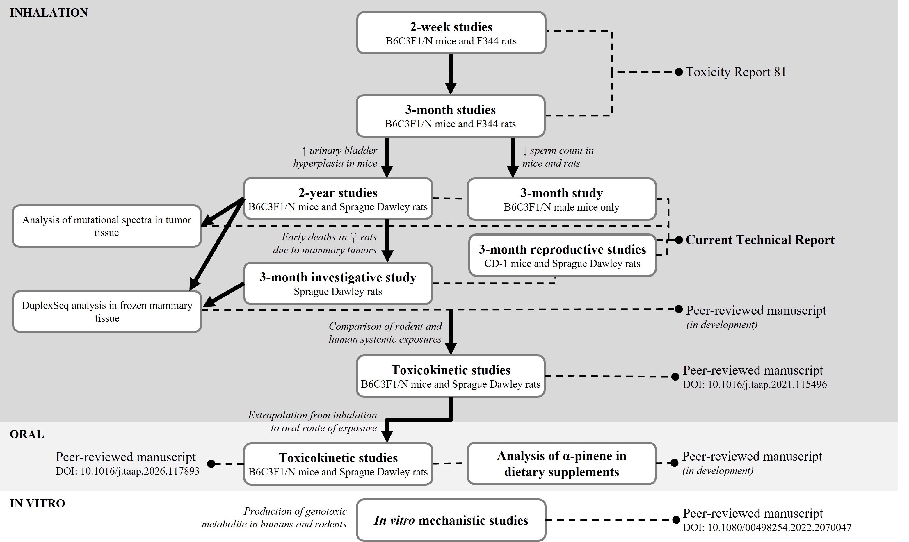
Summary of National Toxicology Program Studies of α-Pinene
The International Union of the United Auto Workers originally nominated turpentine to the National Toxicology Program (NTP) for comprehensive assessment because of widespread exposure potential and a lack of chronic toxicity data. α-Pinene was selected for study because it is a major constituent of turpentine and has a wider exposure profile from its use as a fragrance and flavoring ingredient. An inhalation route was chosen to reflect both occupational and consumer product exposure.
NTP assessment of α-pinene was initiated with 2-week and 3-month studies in Fischer 344 (F344/N) rats and B6C3F1/N mice.2 In light of the findings of hyperplasia in the urinary bladder of male and female mice and the concern expressed in the original nomination for turpentine exposure and chronic nephrotoxicity in humans, the chronic toxicity and carcinogenicity of α-pinene were evaluated. Between the 3-month studies and the beginning of the 2-year studies, a programmatic decision was made to switch from F344/N rats to Sprague Dawley (Hsd:Sprague Dawley SD) rats because of inherent health issues (e.g., high rates of testicular cancer and mononuclear cell leukemia) and decreased fecundity in the F344/N rat strain.3,4 The 3-month F344/N and B6C3F1/N studies observed significant decreases in sperm counts, and thus, reproductive evaluations were added to the chronic study to investigate whether this effect translated into a functional deficit in reproduction; however, B6C3F1/N mice cannot be bred to maintain the same genetic background because they are an F1 hybrid, so the CD-1 mouse strain was selected. A 3-month evaluation of B6C3F1/N mice was included to confirm urinary bladder findings and to conduct histopathological assessment of male reproductive tissues that could not be evaluated in the previous study.
During the chronic rat study, exposure-related early deaths in females were attributed to mammary masses or nodules. At the end of the chronic studies, preliminary data were collected on the internal concentrations of α-pinene and its purported active metabolite, α-pinene oxide. The early signs of mammary tumors in the chronic study prompted the addition of a follow-up investigative 3-month study in male and female Sprague Dawley rats to evaluate early biomarkers of carcinogenicity in the mammary gland using error-corrected duplex sequencing (data are not reported here and will be published separately) and to perform a definitive evaluation of internal concentrations of α-pinene and α-pinene oxide in the blood and mammary gland using validated methods. Whole-genome sequencing of the rat mammary tumors and mouse liver tumors demonstrated an exposure concentration-dependent increase in mutation burden. Surprisingly, in the mouse inhalation study, the mutation burden was higher in the liver tumors compared to the lung tumors, implicating a mutagenic intermediate formed in the liver, likely α-pinene oxide, and thereby supporting a mutagenic mode of carcinogenesis. In addition, exposure-related de novo mutation signatures were observed in each of the respective tumor targets (summary results of these data are provided in Appendix E). Further immunohistochemical studies using human breast cancer-relevant biomarkers are in progress to compare the α-pinene-induced rat mammary tumors with those arising spontaneously due to aging. Results from the evaluation of biomarkers of carcinogenicity will be published in peer-reviewed manuscripts.
Multiple translational research efforts are ongoing, aimed at investigating the relevance of animal toxicity and carcinogenicity findings to human exposure scenarios. As the first step in this process, α-pinene oxide was identified as a possible active metabolite of α-pinene. In vitro assays confirmed the production of α-pinene oxide in rats and humans, and furthermore, α-pinene oxide was found to be mutagenic in the Ames assay.5 The toxicokinetic (TK) behavior of α-pinene and α-pinene oxide was investigated in blood and mammary gland following whole-body inhalation exposure in adult male and female Sprague Dawley rats and B6C3F1/N mice. Rodent systemic exposure has been compared to human systemic exposure data.6
Further collaborations with the National Institute for Occupational Safety and Health are measuring α-pinene and other monoterpene concentrations in various workplaces with potential exposures.
All NTP α-pinene toxicity and carcinogenicity studies used an inhalation route of exposure. However, oral exposure is also relevant for humans given α-pinene’s use as a flavoring ingredient and its possible presence in pine bark extract dietary supplements. Because of a lack of information on the α-pinene content in these products, a survey of monoterpenes in commercially available pine bark supplements was undertaken. Findings from this study will be published in peer-reviewed manuscripts. Additional studies were conducted to investigate the TK behavior of α-pinene and α-pinene oxide following a 7-day oral exposure in male and female Sprague Dawley rats and B6C3F1/N mice and to bridge inhalation and oral exposures.7
Acknowledgments
This work was supported by the Intramural Research Program (ES103374, ES103376, ES103377, and ES103380) at the National Institute of Environmental Health Sciences, National Institutes of Health and performed for the National Toxicology Program, Public Health Service, U.S. Department of Health and Human Services under contracts 75N96025C00003, 75N96024C00002, 75N96023A00001, GS-00F-173CA/75N96022F00055, 75N95021D00012, HHSN273201800006C, HHSN273201600020C, GS00Q14OADU417 (Order No. HHSN273201600015U), HHSN273201600011C, HHSN273201500014C, HHSN273201500013C, HHSN273201500012C, HHSN273201500006C, HHSN273201400015C, HHSN273201400004C, HHSN273201400022C, HHSN273201300004C, HHSN316201200054W, and HHSN273201100003C.