

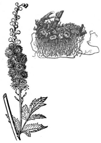
Black cohosh (Actaea racemose L.) is widely used as a botanical dietary supplement to alleviate female gynecological symptoms, such as premenstrual syndrome and changes associated with menopause, and to stimulate labor. Despite its popularity, limited data are available on the long‑term safety of black cohosh products. To address this knowledge gap, 2-year National Toxicology Program (NTP) carcinogenicity studies were conducted in Sprague Dawley (Hsd:Sprague Dawley SD) rats and B6C3F1/N mice. To emulate a potential human exposure scenario in which a woman might use black cohosh throughout pregnancy and lactation, perinatal exposure was included for the rat study.
Time-mated female rats were administered 0, 75, 250, or 750 mg/kg body weight/day (mg/kg/day) of black cohosh root extract (BCE) in 0.5% methylcellulose by gavage starting on gestation day 6 and continuing through lactation. After weaning, female offspring were administered the same doses as their respective dam for 2 years; male offspring were not administered BCE or the methylcellulose vehicle but were maintained for the remainder of the study. Female mice, approximately 5–6 weeks of age, were administered 0, 30, 100, 300, or 1,000 mg/kg/day BCE for 2 years. Interim evaluations of micronucleated peripheral blood erythrocytes, hematology, and bone marrow cytology in these mice were conducted at 3 and 12 months to evaluate the persistence of hematological effects observed in previous studies of BCE.
Two-year study in rats
Mean body weights of BCE-dosed dams were lower (within 10%) than that of vehicle control animals throughout gestation and lactation. Total and live litter sizes were significantly decreased on postnatal day (PND) 1 and PND 4 in the highest dosed group, 750 mg/kg/day, but there was no difference in pup survival throughout the lactation period. Pup mean body weights exhibited a negative trend with increasing dose and remained within 8%–12% of vehicle control mean body weights throughout lactation; this pattern continued into the postweaning phase.
After 2 years, dosed females and perinatally only-exposed males had mean body weights approximately 13% and 11% less, respectively, than vehicle control animals, and survival did not differ significantly among groups. There was a marginal increase in the incidence of rare uterine squamous cell papillomas in the 250 mg/kg/day female rat group. The incidence of squamous metaplasia of the uterus was significantly increased in the 750 mg/kg/day female group. There also were dose-related increases in the incidences of dilation, hemorrhage, thrombus, and ulcers in the uterus. Additionally, ovarian atrophy was considered a dose-related nonneoplastic lesion.
Two-year study in mice
Female mice dosed with BCE for 2 years had survival rates similar to that of the vehicle control group. Mean body weights were lower with increasing BCE dose, indicative of lower body weight gains. Terminal mean body weights of the 30, 100, 300, and 1,000 mg/kg/day groups were approximately 9%, 18%, 25%, and 40% less, respectively, than that of the vehicle control group. No clinical observations were considered related to BCE administration at any point during the study.
At 3 and 12 months, changes in the erythron were observed; these consisted of a decrease in erythrocyte count and hemoglobin concentration (12 months only) and an increase in mean cell volume with increasing dose. In addition, abnormal metarubricytes were observed in the bone marrow at both 3 and 12 months. These hematological changes indicated ineffective erythropoiesis and were consistent with a condition known as megaloblastic anemia. Significant, dose-related increases in peripheral blood micronucleated erythrocytes were observed with no significant changes in the percentage of immature erythrocytes at both 3 and 12 months. These effects were consistent with previously reported NTP studies.
No neoplasms in mice were considered related to BCE administration. Dose-related nonneoplastic lesions included necrosis in the liver and follicle dilation in the thyroid gland.
Genetic toxicology
In addition to interim micronuclei evaluations of peripheral erythrocytes in female mice, two different lots of BCE were tested in two independent bacterial mutagenicity assays in multiple strains of bacteria, with and without induced rodent liver S9 mix. Results were negative for the first BCE lot tested, when both hamster and rat liver S9 was used in seven strains of Salmonella typhimurium; the lot of BCE used in the NTP in vivo rodent studies was negative in two bacterial strains (S. typhimurium TA100 and Escherichia coli WP2 uvrA [pKM101]) but was judged to be equivocal in S. typhimurium TA98 in the presence of induced rat liver S9.
Conclusions
Under the conditions of these 2-year gavage studies, there was equivocal evidence of carcinogenic activity of black cohosh root extract (BCE) in female Hsd:Sprague Dawley SD rats based on marginal increases in the incidence of uterine squamous cell papillomas. There was no evidence of carcinogenic activity of perinatal BCE exposure in male Hsd:Sprague Dawley SD rats at maternal doses of 75, 250, or 750 mg/kg/day.
There was no evidence of carcinogenic activity of BCE in female B6C3F1/N mice at doses of 30, 100, 300, or 1,000 mg/kg/day.
Dose-related nonneoplastic lesions were observed in the uterus and ovary in rats and in the liver and thyroid gland in female mice. A significant decrease in litter size was observed in rats.
Interim hematological evaluations and micronucleus assays in female mice showed disruption of normal erythropoiesis and increased frequency of micronucleated erythrocytes at 3 months; at 12 months, the same effects were observed with similar severity and frequency.
National Toxicology Program (NTP). 2023. NTP technical report on the toxicology and carcinogenesis studies of black cohosh root extract (CASRN 84776-26-1) administered by gavage to Sprague Dawley (Hsd:Sprague Dawley SD) rats and female B6C3F1/N mice. Research Triangle Park, NC: National Toxicology Program. Technical Report 603. https://doi.org/10.22427/NTP-TR-603