

Alimentary System
Esophagus
Narrative
The esophagus lumen is lined by mucosa, which is composed of a keratinized stratified squamous epithelium, with a thin lamina propria composed of connective tissue below, and then a thin muscularis mucosae. Below that is the submucosa, which consists of a collagenous matrix with elastin fibers and then the tunica muscularis, which is composed of striated muscle fibers. Smooth muscle is present only proximal to the lower esophageal sphincter. The outermost layer is the tunica adventitia.
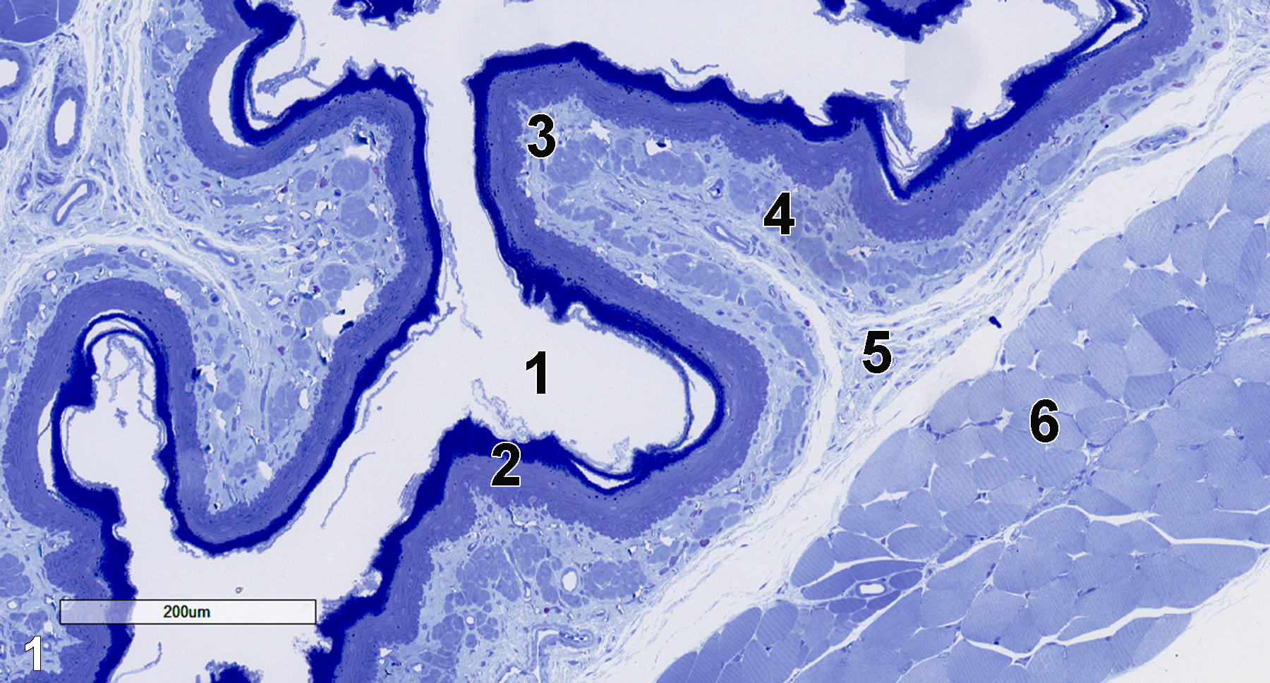
Figure 1. A semithin section (0.5 micrometer thick) of a toluidine blue O-stained esophagus. The esophageal lumen (1) is lined with a stratified squamous epithelium (2), followed by a lamina propria (3) underlaid by the muscularis mucosae (4). Below that layer is the submucosa (5), followed by the inner layer of tunica muscularis (6). 11x.
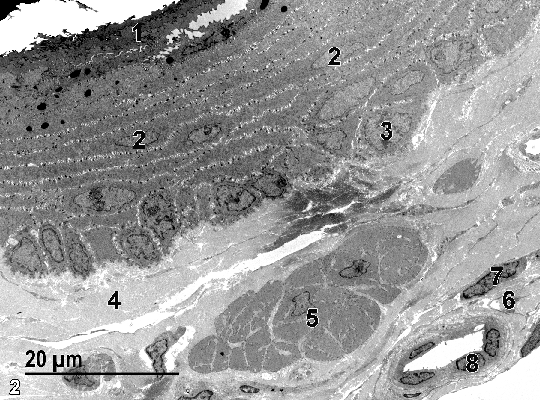
Figure 2. A low magnification electron micrograph of the esophagus, showing the sloughing keratinized epithelial surface (1), the elongated nuclei of squamous epithelial cells (2), a rounded nucleus (3) of a basal epithelial cell, and collagen bundles of the lamina propria (4). Below the lamina propria is the muscularis mucosae, which contains a layer of smooth muscle cells (5) with adjacent collagen fibrils. The submucosal layer (6) beneath that is composed of more connective tissue that contains fibroblasts and small blood vessels. An elongated fibroblast nucleus (7) and an endothelial nucleus of a small vessel (8) are shown. 1900x.
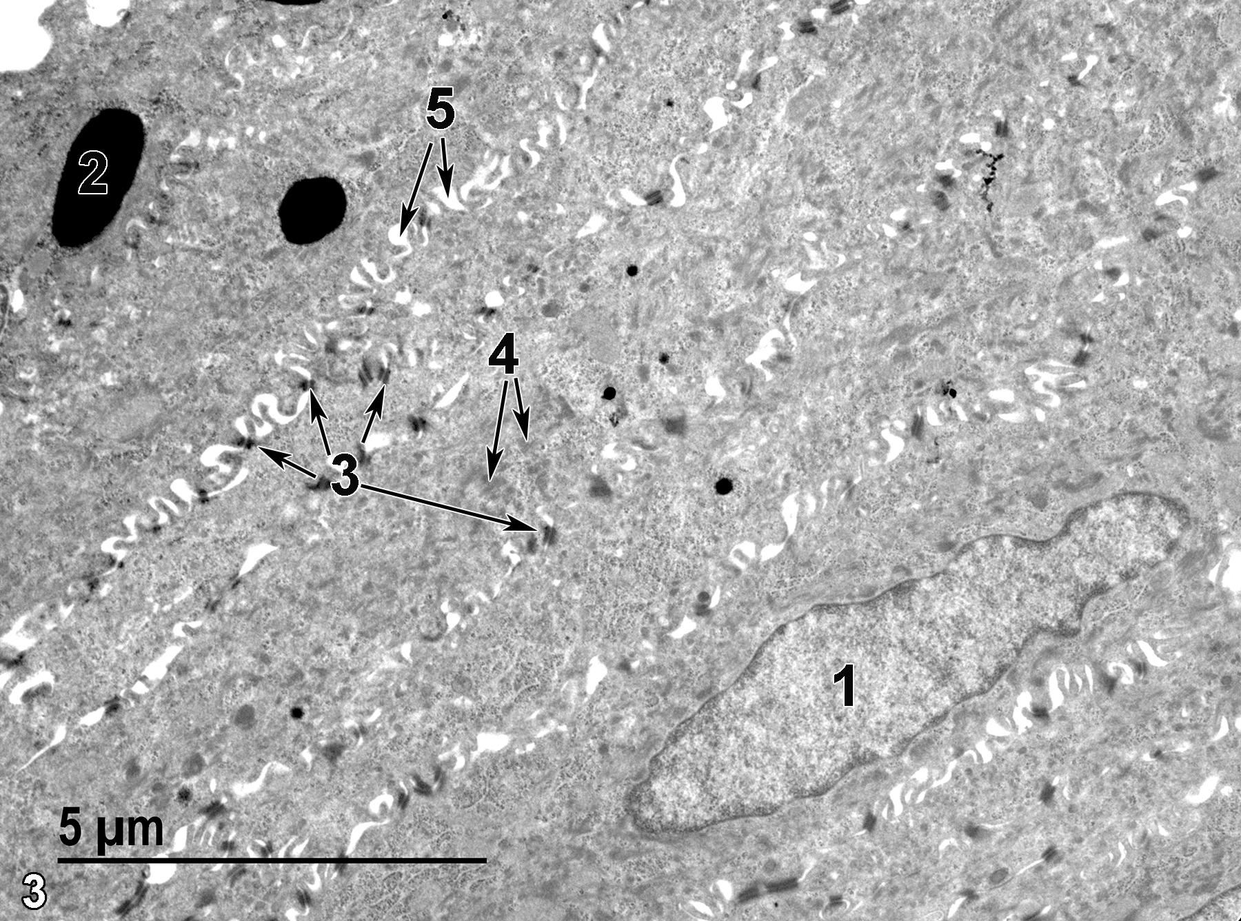
Figure 3. A portion of the stratified squamous epithelium. An elongated nucleus of an epithelial cell (1) is shown. An electron-dense melanosome (2) is present in an epithelial cell. Desmosomes (3) are numerous, primarily binding apical portions of adjacent epithelial cells (arrows). Bundles of tonofilaments (4) are seen in the cytoplasm, often associated with desmosomes (double arrows). Numerous intercellular spaces (5) are visible between the cellular projections (double arrows). 9300x.
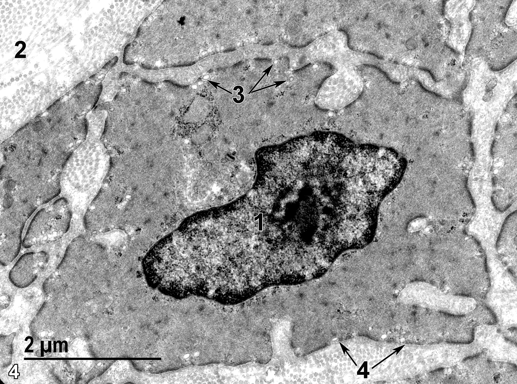
Figure 4. Some details of smooth muscle cells from the muscularis mucosae layer. The smooth muscle cells contain a single nucleus (1) and are surrounded by collagenous fibrils (2).Micropinocytotic vesicles (3) are visible at the surface of the cell (arrows). Each cell is surrounded by a basal lamina (4, double arrows). 18500x.
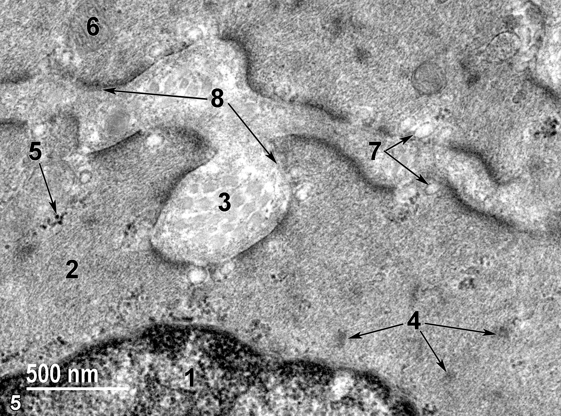
Figure 5. A higher magnification view of the features of a smooth muscle cell in the muscularis mucosae. A portion of the nucleus (1) is visible at the bottom of the image. The cytoplasm is filled with actin and myosin microfilaments (2), as well as fusiform densities (4, arrows). Collagen fibrils (3) can be seen between two adjacent cells. Small clusters of ribosomes (5) are present in the cytoplasm (arrow). Sparse mitochondria (6) are shown. Micropinocytotic vesicles (7) are located adjacent to the plasma membrane (double arrows), and a thin basal lamina (8) surrounds the smooth muscle cells (double arrows). 49000x.
| Boorman GA, Eustis SL, Elwell MR, Montgomery CA, Jr., MacKenzie WF, eds. 1990. Pathology of the Fischer Rat: Reference and Atlas. New York: Academic Press. |
| Cross PC, Mercer KL. 1993. Cell and Tissue Ultrastructure: A Functional Perspective. New York: W.H. Freeman and Company. |
| Dellmann HD, Eurell J, eds. 1998. Textbook of Veterinary Histology. 5th ed. Philadelphia: Lippincott Williams & Wilkins. |
| Rhodin JAG. 1974. Histology: A Text and Atlas. New York: Oxford University Press. |
| Ross MH, Kaye GI, Pawlina W. 2003. Histology: A Text and Atlas. 4th ed. Philadelphia: Lippincott Williams & Wilkins. |
| Uehara T, Elmore SA, K.A. Szabo KA. 2017. Chapter 6: Esophagus and stomach. In Boorman’s Pathology of the Rat (Suttie AW, ed). 2nd ed. London: Academic Press, 35-50. |
| Weiss L, ed. 1988. Cell and Tissue Biology: A Textbook of Histology. 6th ed. Baltimore: Urban & Schwarzenberg. |
All Images


