

Special Senses System
Lacrimal Gland
Narrative
Lacrimal glands are similar in structure to salivary glands and are composed of acini and ducts. The acinar cells contain serous glandular cells associated with basally located myoepithelial cells. The acinar cells contain mucous granules, abundant rough endoplasmic reticulum, and sparse lipid bodies.
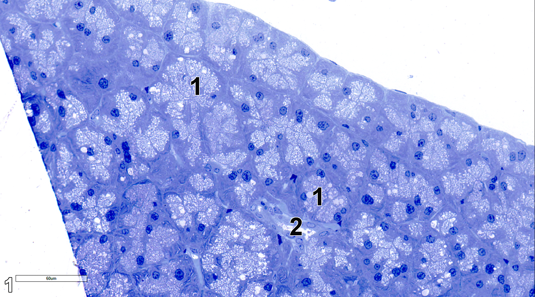
Figure 1. A semithin section (0.5 micrometer thick) of a toluidine blue O-stained portion of a lacrimal gland showing acini (1) and a single secretory duct (2). 38x.
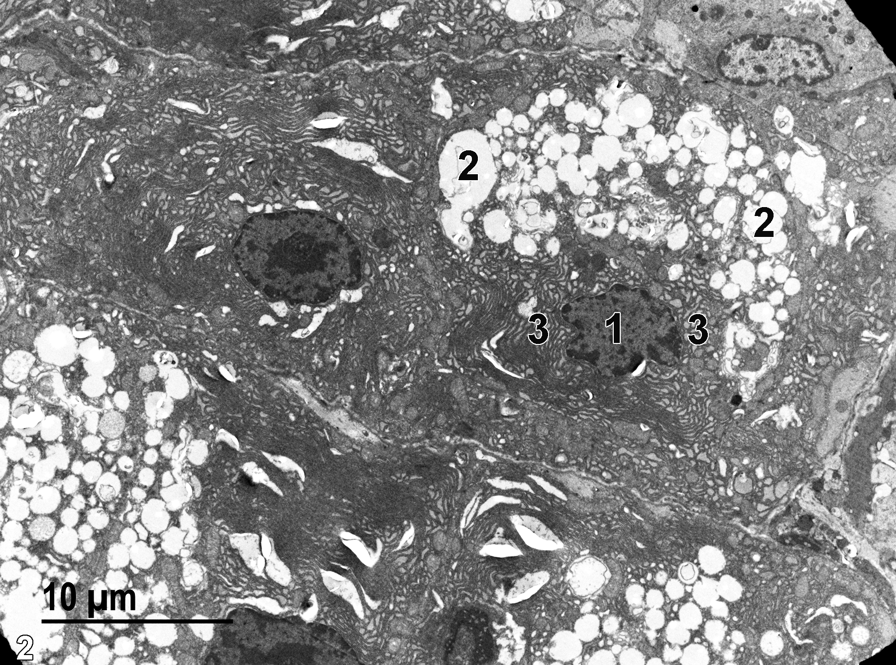
Figure 2. An electron micrograph of a lacrimal gland acinus. The nucleus of an acinar cell (1) is surrounded by stacks of rough endoplasmic reticulum (3). Numerous mucous granules (2) are present. 2900x.
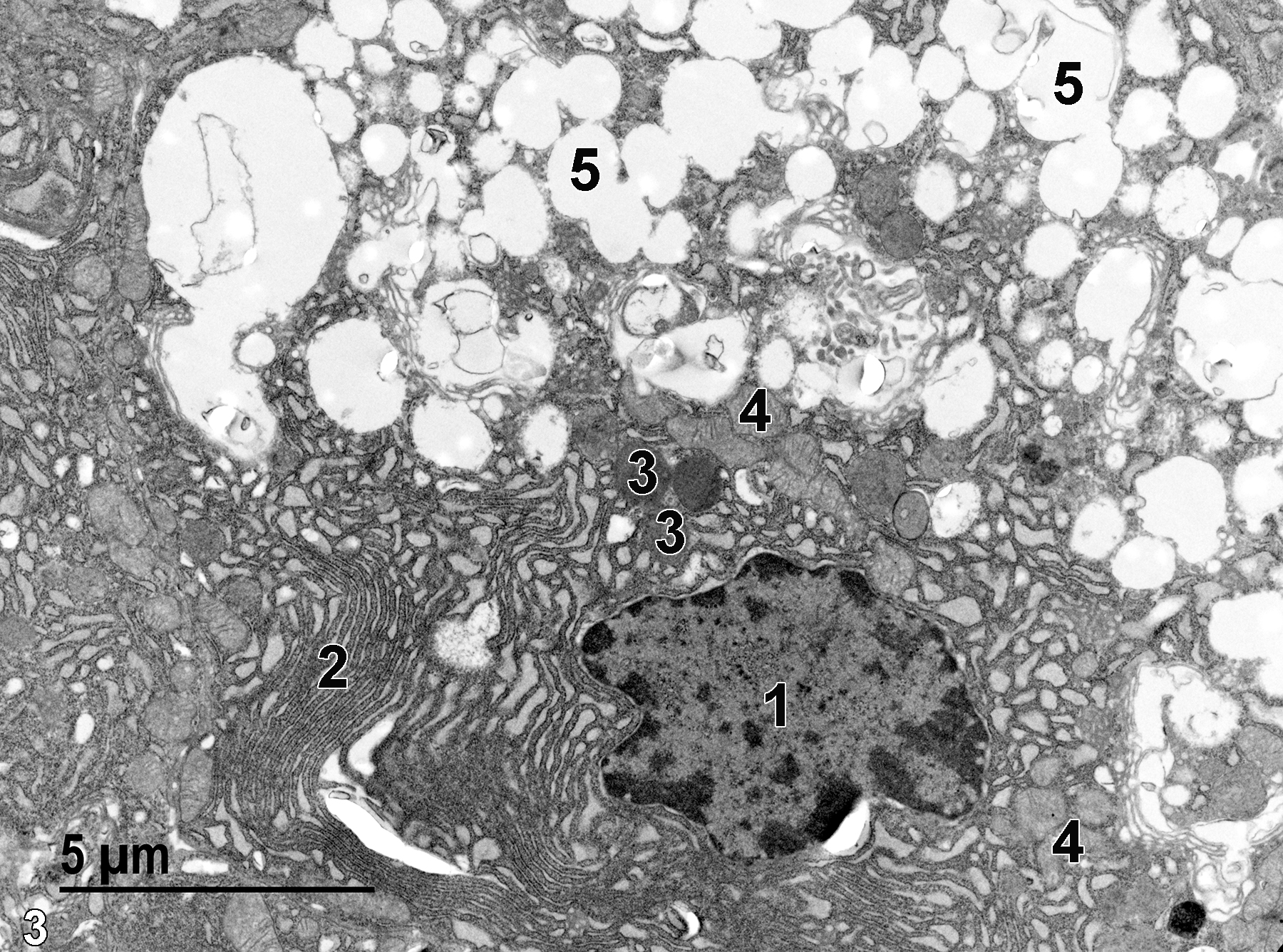
Figure 3. A higher magnification view of the contents of an acinar cell. The nucleus (1) has a large amount of perinuclear rough endoplasmic reticulum (2). Near the nucleus are several lipid droplets (3) and mitochondria (4). The rest of the cell is filled with mucous granules (5). 6800x.
| Dellmann HD, Eurell J, eds. 1998. Textbook of Veterinary Histology. 5th ed. Philadelphia: Lippincott Williams & Wilkins. |
| Weiss L, ed. 1988. Cell and Tissue Biology: A Textbook of Histology. 6th ed. Baltimore: Urban & Schwarzenberg. |
All Images


