

Respiratory System
Lung
Narrative
Two lungs arise from the splitting of the trachea. Each lung, in turn, is subdivided into lobes, consisting of a single left lobe and four right lobes in the rat. The lobes are further divided into bronchopulmonary segments, subsegments, and lobules. These segments are more visibly distinct in other species than those in the rat (Weisz 1988; Herbert et al. 2018; Peake and Pinkerton 2015). Extrapulmonary bronchi arise from the trachea and are structurally similar to the trachea, except that they have diminishing cartilage plates, and they develop a complete ring of smooth muscle between the cartilage and submucosa. When the airway reaches a diameter of approximately 1 mm, they are called bronchioles. They no longer have cartilage plates, glands, or continuous muscularis tissue. The pulmonary acini consist of terminal bronchioles and respiratory bronchioles (not well-developed in the rat), alveolar ducts, and then alveoli (Herbert et al. 2018).
Bronchioles are lined with an epithelium composed of ciliated cells with long microvilli and club cells (formerly known as Clara cells), which are non-ciliated, with short microvilli, and contain secretory granules that produce lipoproteins, smooth endoplasmic reticulum, and stacks of rough endoplasmic reticulum, and Golgi bodies.
Alveolar ducts are lined with alveoli and have some surrounding smooth muscle. The alveolar epithelium consists of type I cells that are thin squamous cells that line most of the alveolar surface (approximately 95%). Mixed in with them are type II cells that tend to aggregate at septal junctions. They cover approximately 5% of the alveolar air surface and contain lamellar bodies with surfactant composed of phospholipids, proteins, and neutral lipids (Ross et al. 2003). Lipofibroblasts (McGowan and Torday 1997) that contain lipid bodies are located in close proximity to the type II cells in the alveolar septa. Occasional brush cells with short, blunt microvilli may be present. Numerous capillaries are located in alveolar septa, and macrophages are present in adjacent airways.
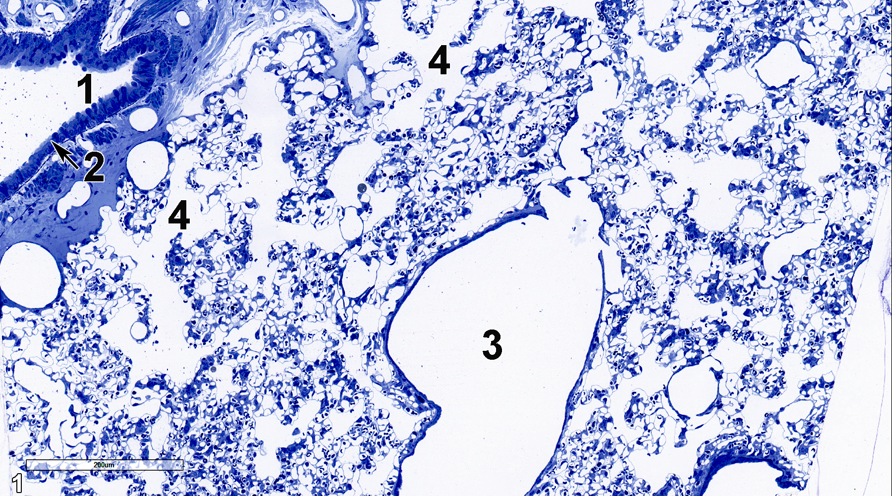
Figure 1. A semithin section (0.5 micrometer thick) of a toluidine blue O-stained portion of the lung, showing a small bronchiole (1) lined with a cuboidal epithelium (2, arrow). A terminal bronchiole (3) is lined with a thin epithelial layer and has an airway (4) leading into alveolar ducts. 15x.
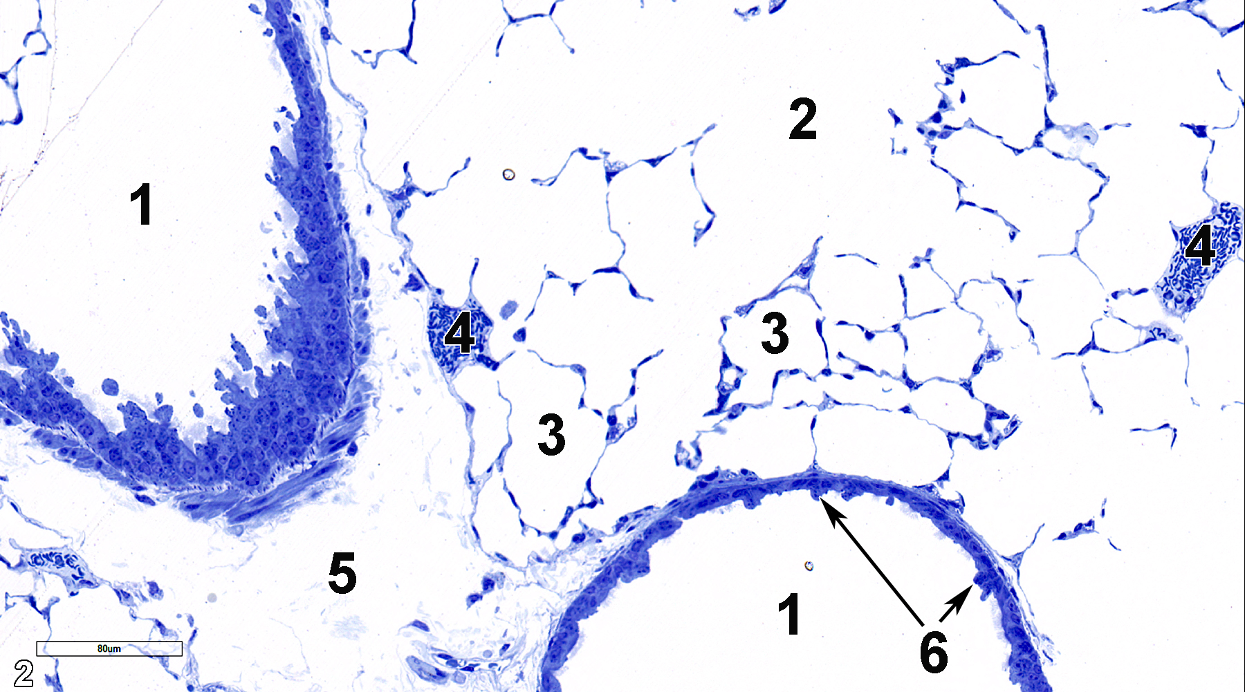
Figure 2. Another semithin section at a slightly higher magnification showing terminal bronchioles (1) with protuberant club cells (6, arrows), alveolar ducts (2), alveoli (3), and blood vessels (4). A connective tissue septum (5) is located between the two bronchiolar segments. 25x.
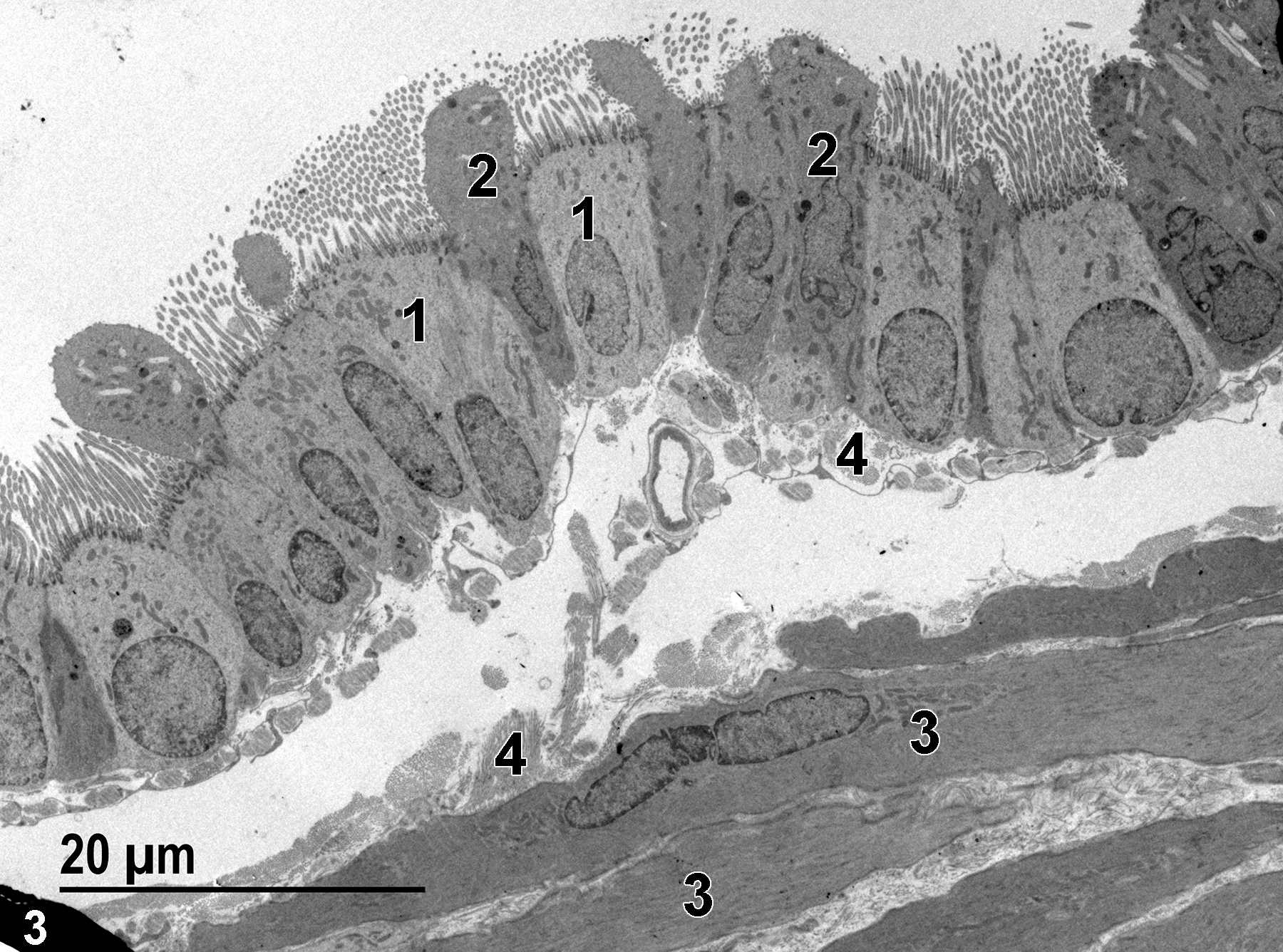
Figure 3. An image of a terminal bronchiole showing ciliated epithelial cells (1), club cells (2), bundles of collagen beneath the epithelial layer (4), and several layers of smooth muscle cells (3) that make up the muscularis. 1900x.
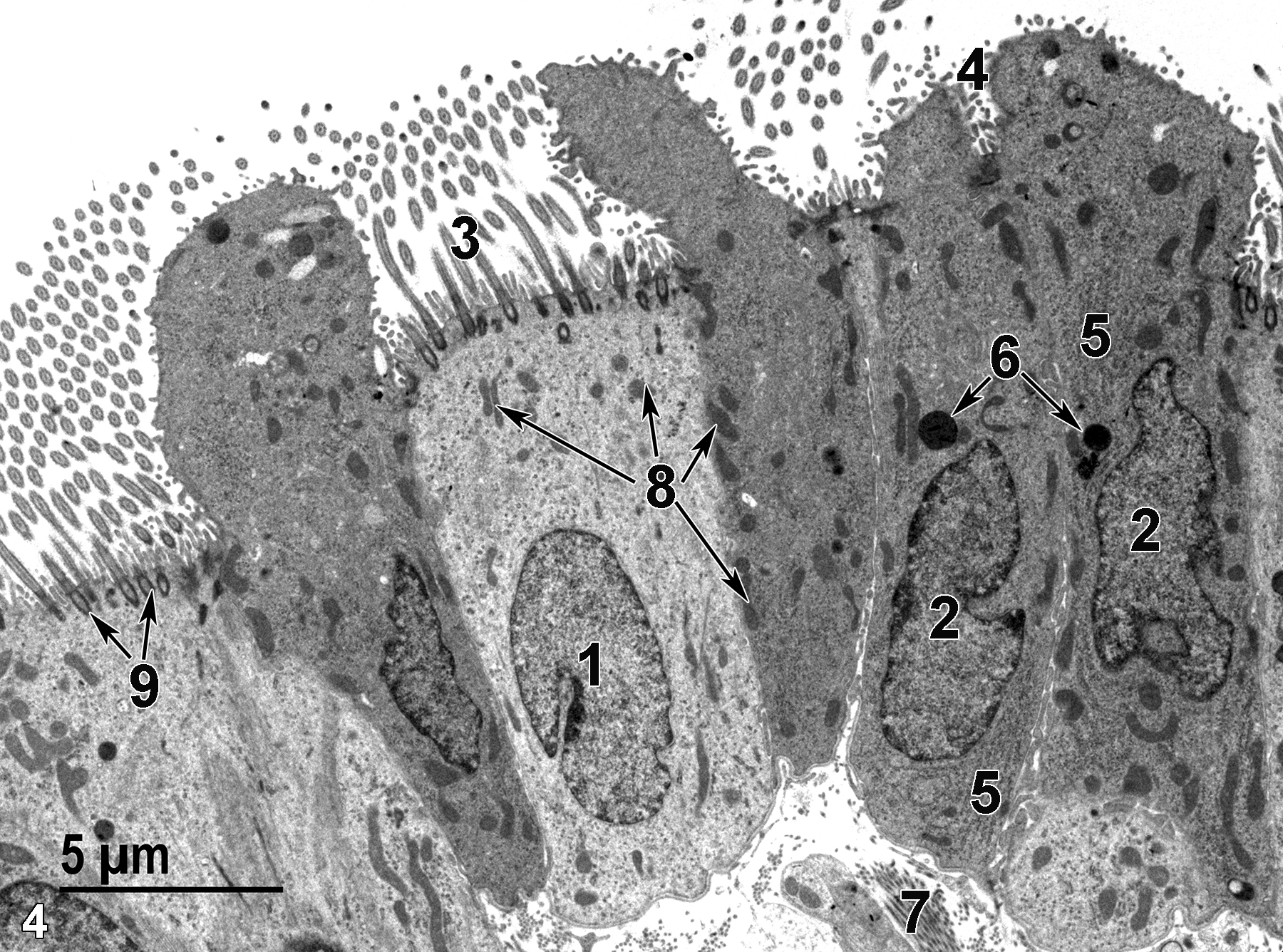
Figure 4. A higher magnification view of the bronchiolar epithelium. The nuclei of a ciliated cell (1) and of a club cell (2) are both located basally in the respective cells. Cilia (3) extend into the airway and are subtended by basal bodies at the apex of the ciliated cells (9, arrows). Microvilli are located between the cilia. The microvilli of club cells (4) are considerably shorter than those seen on the ciliated cells. Stacks of rough endoplasmic reticulum (5) and lysosomes (6, arrows) are visible in the club cells. Bundles of collagen (7) are directly below the epithelial cells. Mitochondria are scattered throughout the cytoplasm of both epithelial cell types (8, arrows). 4800x.
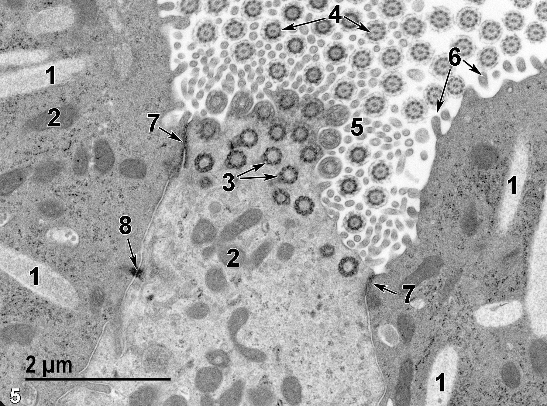
Figure 5. An even higher magnification of the bronchiolar epithelial cells. Secretory granules (1) of the club cells are elongated and have fine granular contents. Mitochondria (2) are relatively electron dense and variable in size and shape. Ciliary basal bodies are seen in cross section (3, arrows), as are the cilia in the airway (4, arrows). Cross sections of ciliary cell microvilli (5) are present as well. Only microvilli are present on the surface of club cells (6, arrows). Junctional complexes are located between adjacent epithelial cells extending to the luminal surface of the airway (7, arrow). At the base of a junctional complex is a desmosome with associated proteinaceous filaments (8, arrow). 18500x.
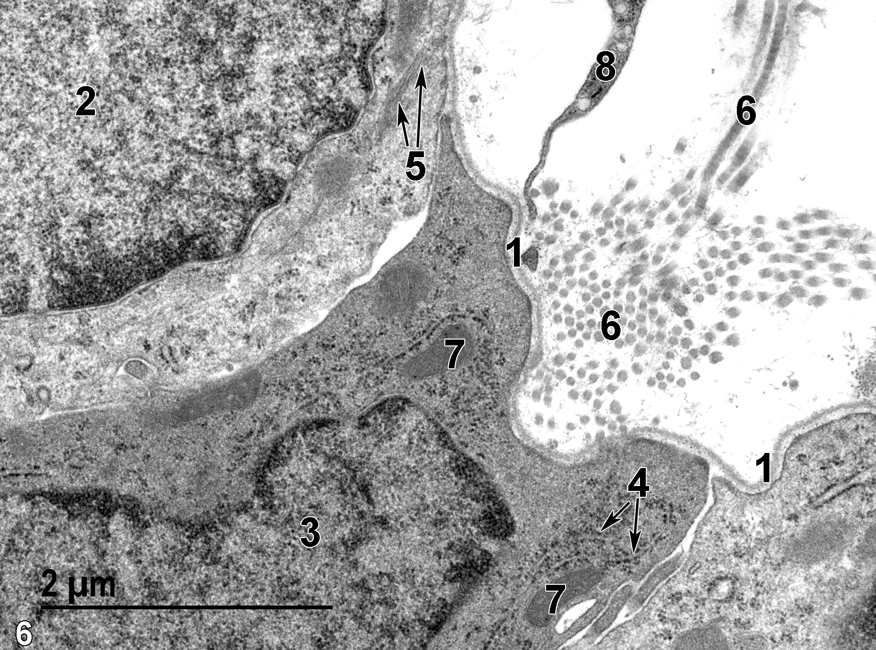
Figure 6. The base of the bronchiolar epithelium delimited by the basal lamina (1). Nuclei of a ciliated epithelial cell (2) and a club cell (3) are shown. The club cell contains rough endoplasmic reticulum cisternae (4, arrows) and mitochondria (7). The ciliated cell contains bundles of cytoplasmic filaments (5, arrows). Below the basal lamina, connective tissue composed of collagen fibrils (6) and a segment of a fibroblast cell (8) can be seen. 23000x.
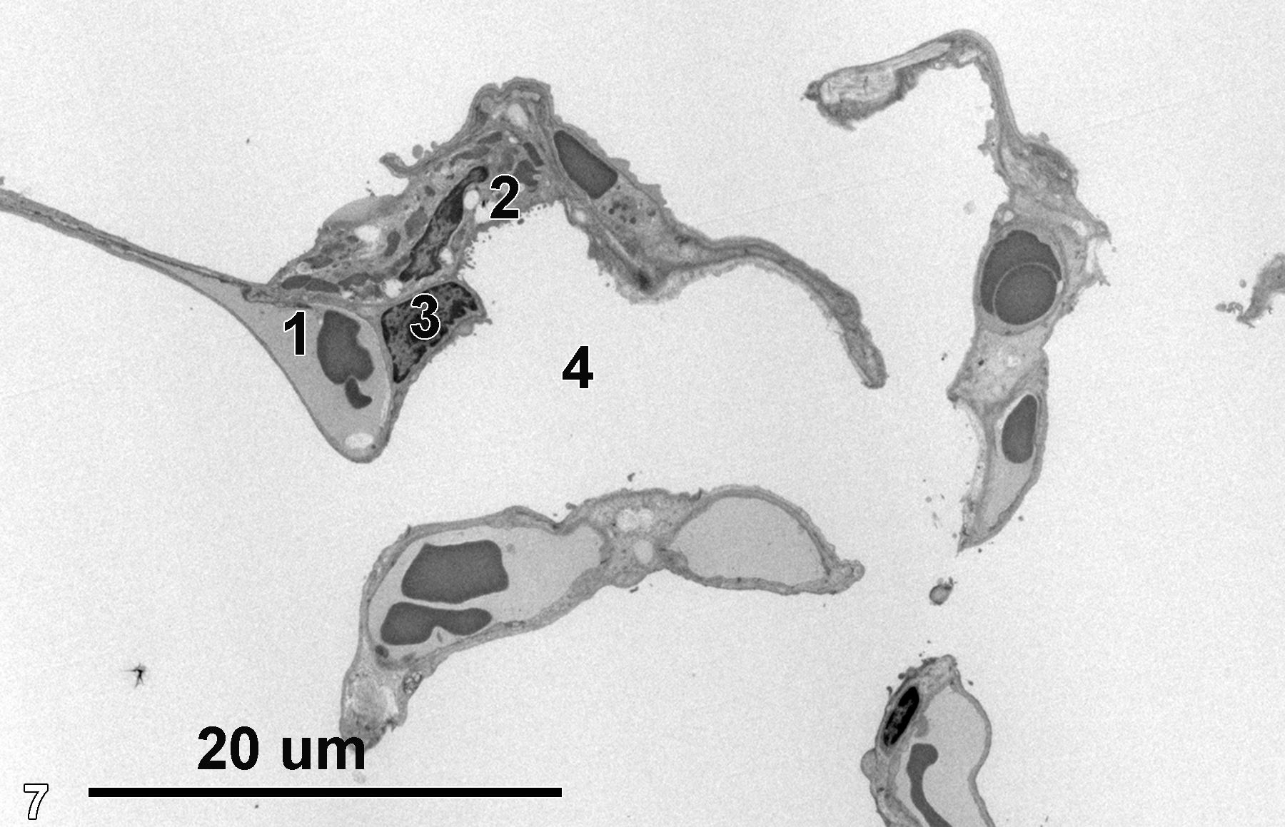
Figure 7. An alveolar septum containing a capillary with erythrocytes (1), type II cells with electron-lucent surfactant (2), and adjacent type I cells (3) lining the alveolar airway (4). 1900x.
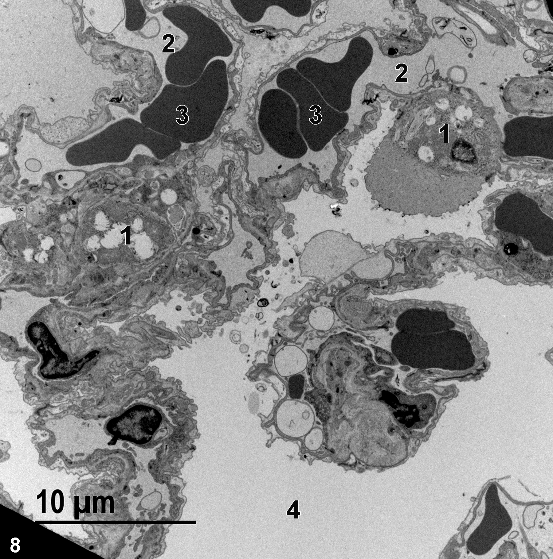
Figure 8. Another image of an airway (4) next to an alveolar septum composed of type II cells (1) and capillaries (2) containing erythrocytes (3). 2900x.
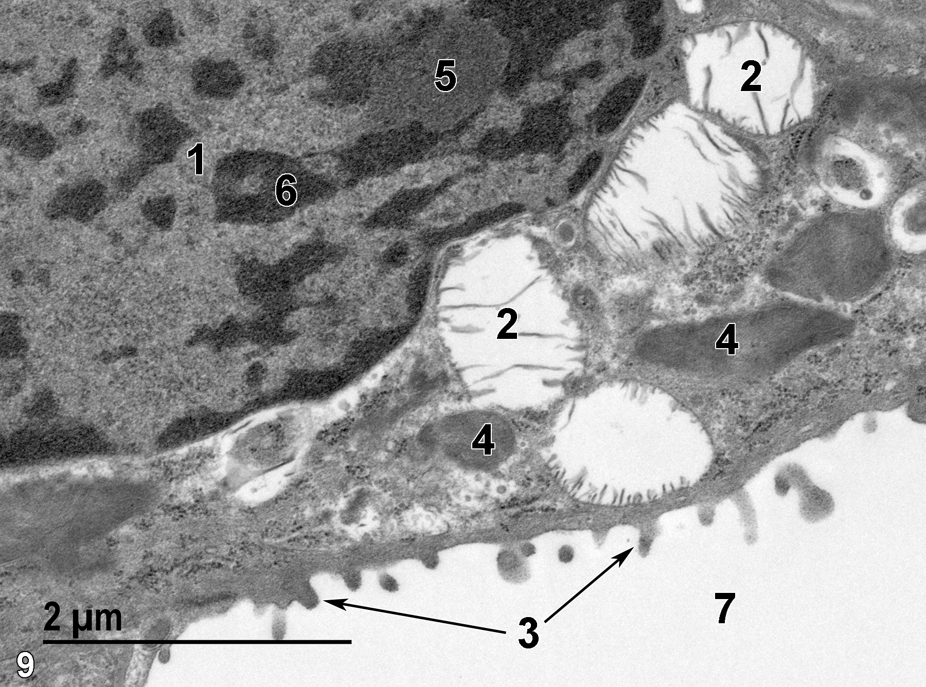
Figure 9. A high magnification view of a type II cell showing a nucleus (1) with a nucleolus (5) and scattered electron-dense heterochromatin (6). The surface of the type II cell in contact with the alveolar airway (7) has short microvilli (3, arrows). Mitochondria (4) and multilamellar bodies (2) containing surfactant are present. 23000x.
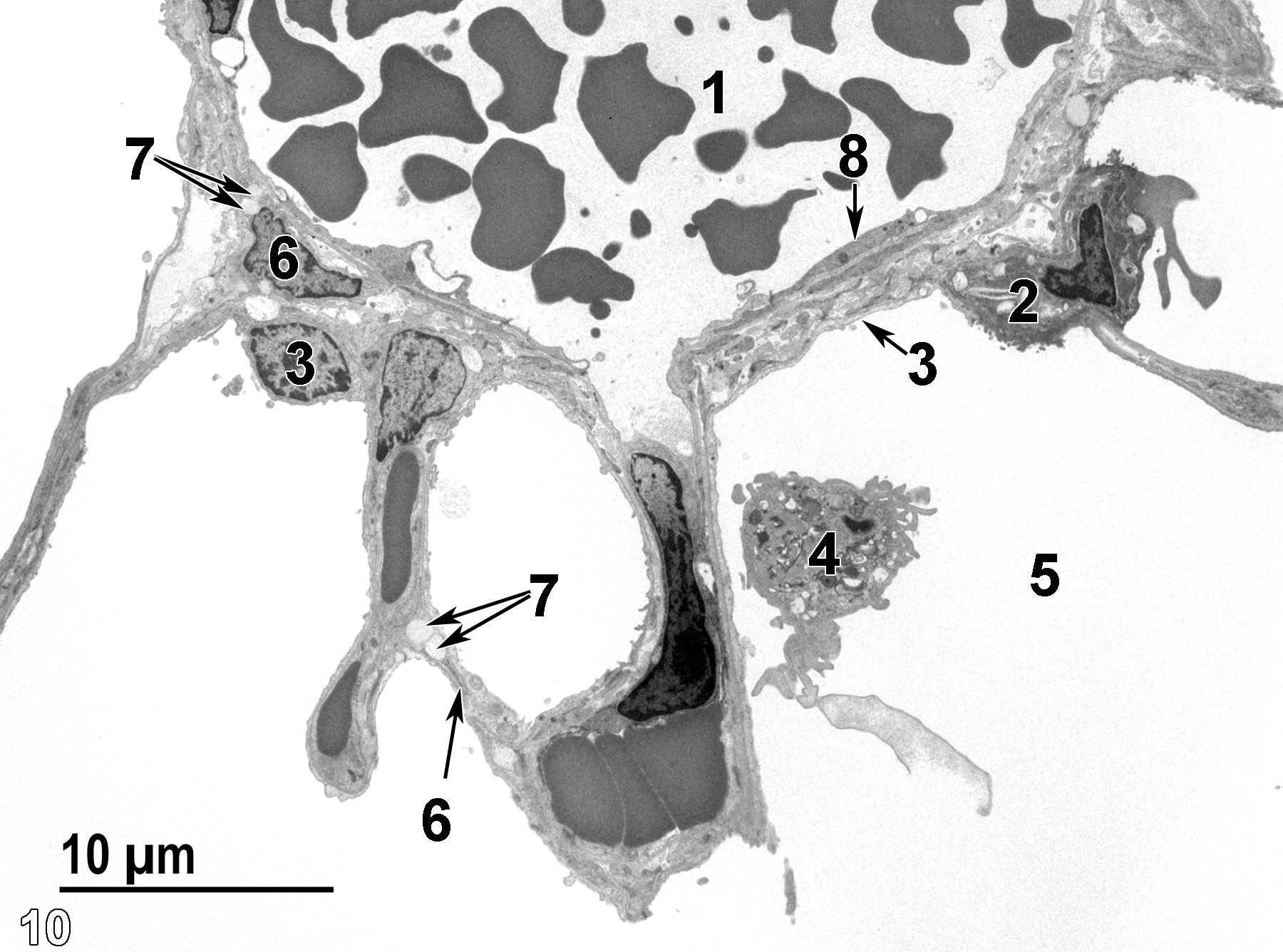
Figure 10. Another view of alveolar structure, with a blood vessel (1) with erythrocytes lined with a thin layer of endothelial cells (8, arrow). A type II cell (2) with a thin type I cell (3, arrow) are present. A pulmonary macrophage (4) is located in the airway (5). Occasional lipofibroblasts (6) are identified by the presence of lipid bodies (7, arrows). 2900x.
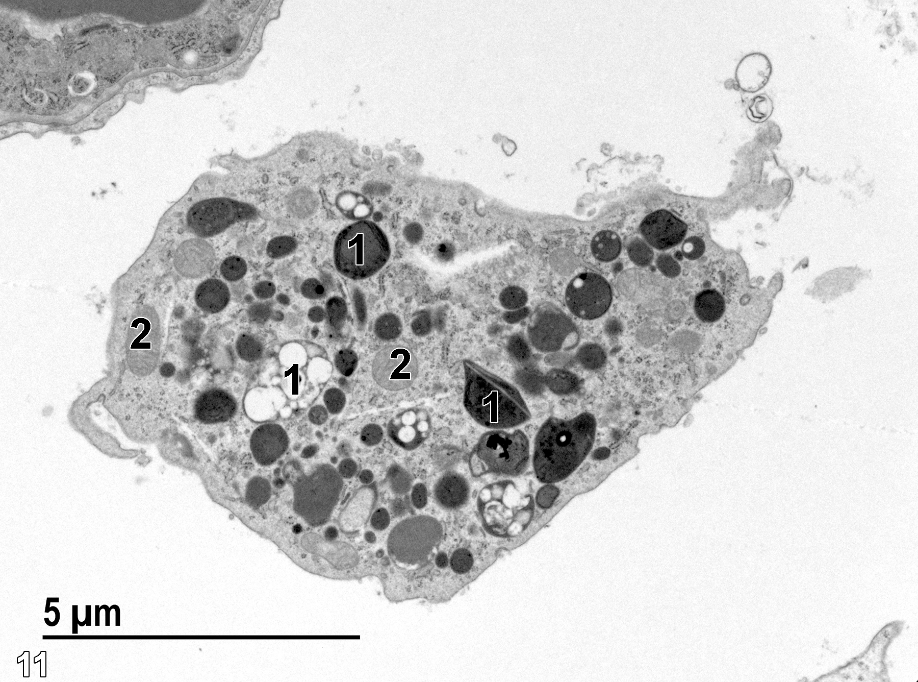
Figure 11. A higher magnification view of a pulmonary macrophage in the alveolar airway. The macrophage contains numerous electron-dense pleomorphic lysosomes (1) with varying contents. Less electron-dense mitochondria (2) are also present. 9300x.
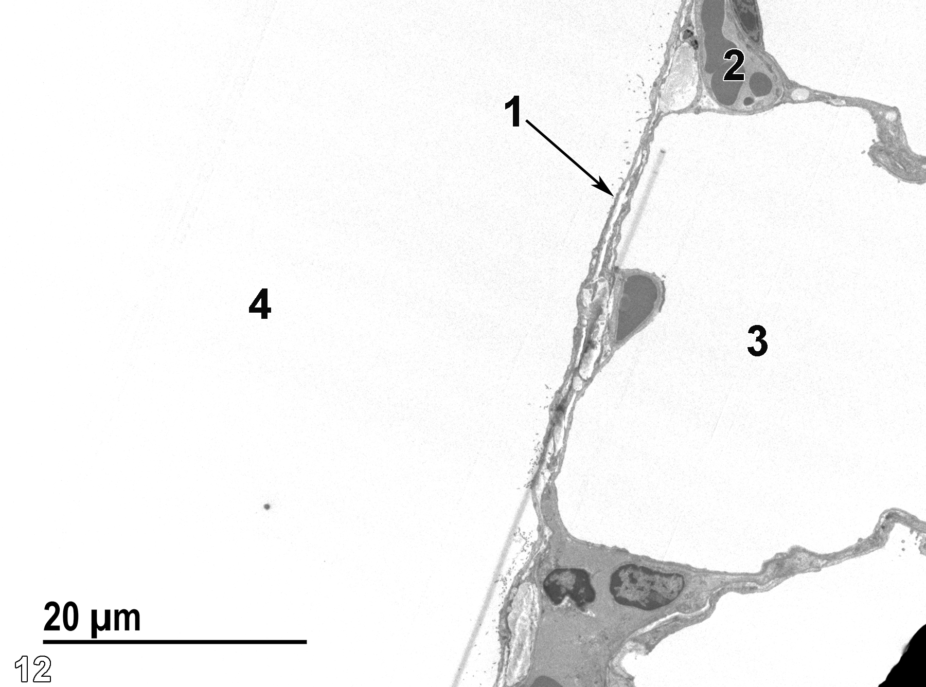
Figure 12. A low magnification view of the pleural surface. A thin layer of pleural mesothelial cells (1, arrow) is adjacent to the pleural cavity (4) and overlies an alveolus (3). A capillary (2) is present in the alveolar septum. The lightly stained linear structure extending from the pleural cavity through the mesothelial cell layer and into the alveolar space is an artifact. 1900x.
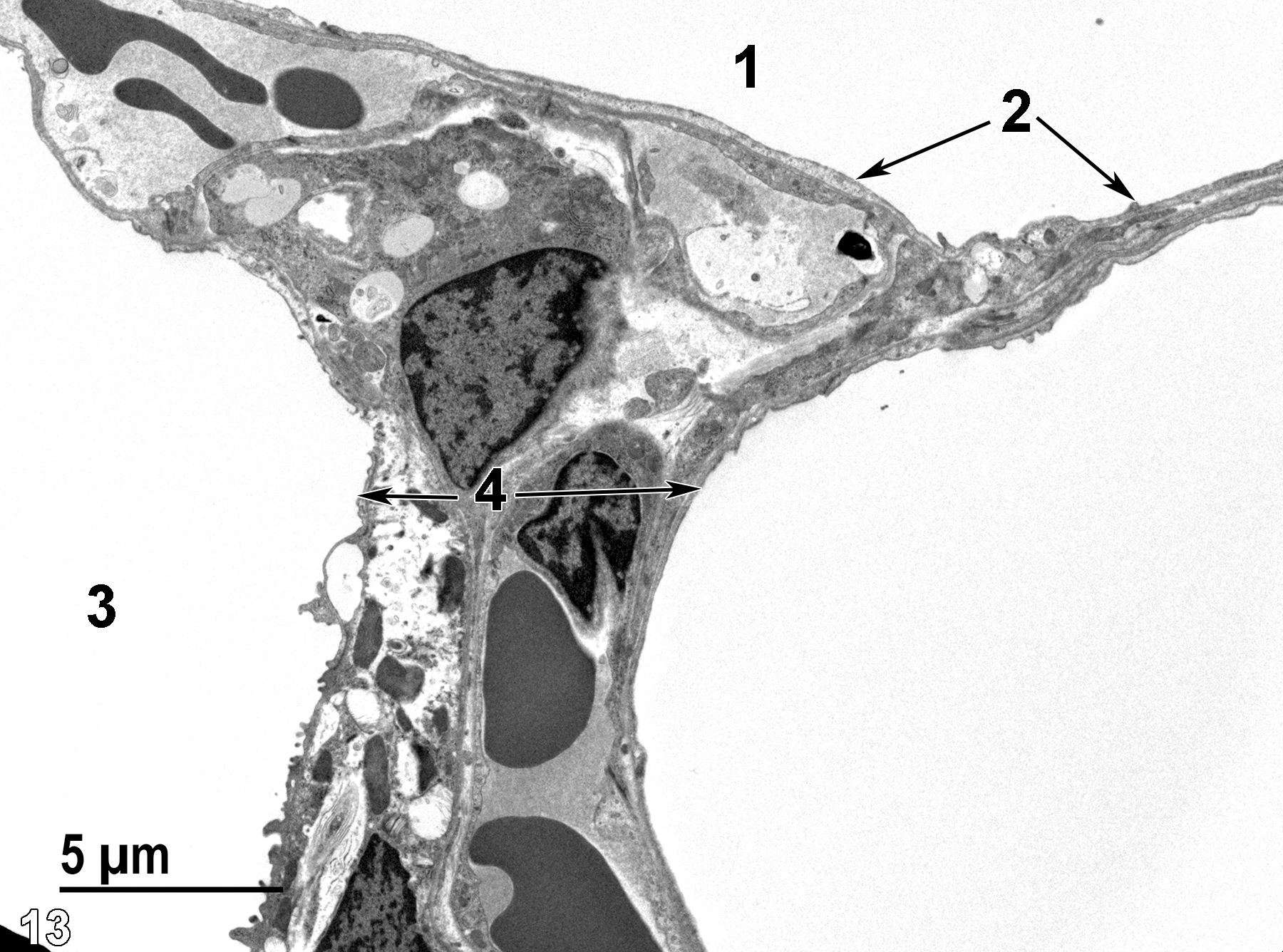
Figure 13. A higher magnification image of the pleural cavity (1), pleural mesothelial cells (2, arrows), the alveolar septum (4, arrows), and a pulmonary alveolus (3). 4800x.
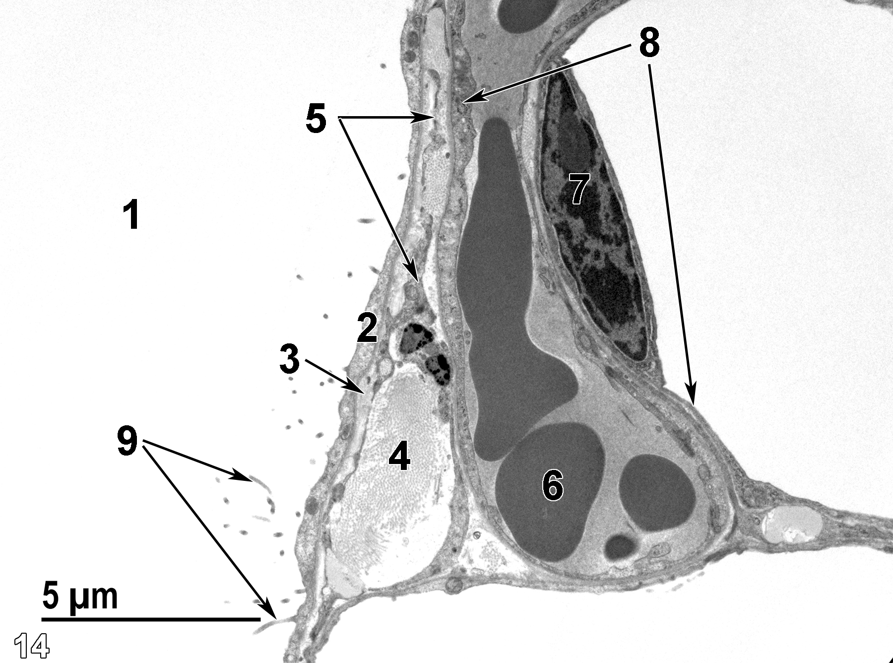
Figure 14. Another view of the pleural surface. The thin pleural mesothelial cell layer (2) has microvilli (9, arrows) extending into the pleural cavity(1). Directly below the mesothelial cells is an elastic membrane (3, arrow) that is underlain by collagen bundles (4). Cytoplasmic strands of fibroblasts (5, arrows) are within the layer of collagenous tissue. Endothelium (8, arrows) lines a capillary containing erythrocytes (6) that is adjacent to a larger vessel also lined by a thin endothelium (8) with an elongate nucleus (7). 6800x.
| Herbert RA, Janardhan KS, Pandiri AR, Cesta MF, Chen V, Miller RA. 2018. Chapter 23: Lung, pleura, and mediastinum. In Boorman’s Pathology of the Rat (Suttie AW, ed.). 2nd ed. London: Academic Press, 437−466. |
| McGowan SE, Torday JS. 1997. The pulmonary lipofibroblast (lipid interstitial cell) and its contributions to alveolar development. Ann Rev Physiol 59:43−62. |
| Peake JL, Pinkerton KE. 2015. Chapter 3: Gross and subgross anatomy of lungs, pleura, connective tissue septa, distal airways, and structural units. In Comparative Biology of Normal Lung (Parent RA, ed.). 2nd ed. San Diego, CA: Academic Press, 21−31. |
| Rhodin JAG. 1974. Histology: A Text and Atlas. New York: Oxford University Press. |
| Ross MH, Kaye GI, Pawlina W. 2003. Histology: A Text and Atlas. 4th ed. Philadelphia: Lippincott Williams & Wilkins. |
| Weiss L, ed. 1988. Cell and Tissue Biology: A Textbook of Histology. 6th ed. Baltimore: Urban & Schwarzenberg. |
All Images


