

Reproductive System, Male
Testis
Narrative
A thick capsule (tunica albuginea) of connective tissue containing numerous vascular elements surrounds the testis. The testicular parenchyma is divided into lobules by fibrous septa. The parenchyma is made up of convoluted seminiferous tubules with stratified germinal epithelium surrounded by a basal lamina, collagen, elastic fibers, and a single layer of squamous epithelial myoid cells that make up the lamina propria. The seminiferous tubules empty into the tubulus recti, which connect to the channels of the rete testis (Whitney and Suttie 2018). Interstitial spaces have connective tissue, lymph and blood vessels, fibroblasts, Leydig cells (interstitial endocrine cells that produce androgens), and macrophages.
At the basal aspect of seminiferous tubules are many type A spermatogonia, typically flattened on the basal lamina. They contain ovoid nuclei, few mitochondria, and have dispersed chromatin. The type A spermatogonia undergo mitosis, yielding 50% type A spermatogonia and 50% type B spermatogonia. In lesser numbers, Sertoli cells (sustentacular cells) are located among the type A spermatogonia on the basal lamina of the seminiferous tubules. Sertoli cells have cytoplasmic extensions filling spaces between adjacent spermatogenic cells and contain large amounts of rough endoplasmic reticulum and large nuclei with dispersed chromatin. Type B spermatogonia have round nuclei with marginated heterochromatin and more mitochondria than that found in type A spermatogonia. These cells undergo mitosis to produce primary spermatocytes. Primary spermatocytes have few mitochondria and many free ribosomes. Primary spermatocytes undergo meiosis to produce early (round) spermatids. Early spermatids are characterized by dispersed heterochromatin, prominent smooth endoplasmic reticulum, numerous free ribosomes, developing acrosome (consisting of the acrosomal vesicle and acrosomal granule), and small marginal mitochondria arranged along the cell membrane. The nuclei of early spermatids are round, and then the nuclei elongate during spermiogenesis. The spermatids differentiate into spermatozoa, with a head containing a nucleus with extremely condensed chromatin and an acrosome, and a neck (connecting piece) connecting the head to the tail. The neck contains paired centrioles and nine segmented rings of fibrous material. The tail consists of a middle piece, a principal piece, and an end piece. The middle piece has a 9 + 2 assemblage of microtubules (axoneme) surrounded by nine outer dense fibril masses. A mitochondrial sheath surrounds the middle piece. The principal piece is narrower than the middle piece and contains the axoneme, outer dense fibril masses, and an outer fibrous sheath. The end piece consists only of the axoneme of the flagellum.
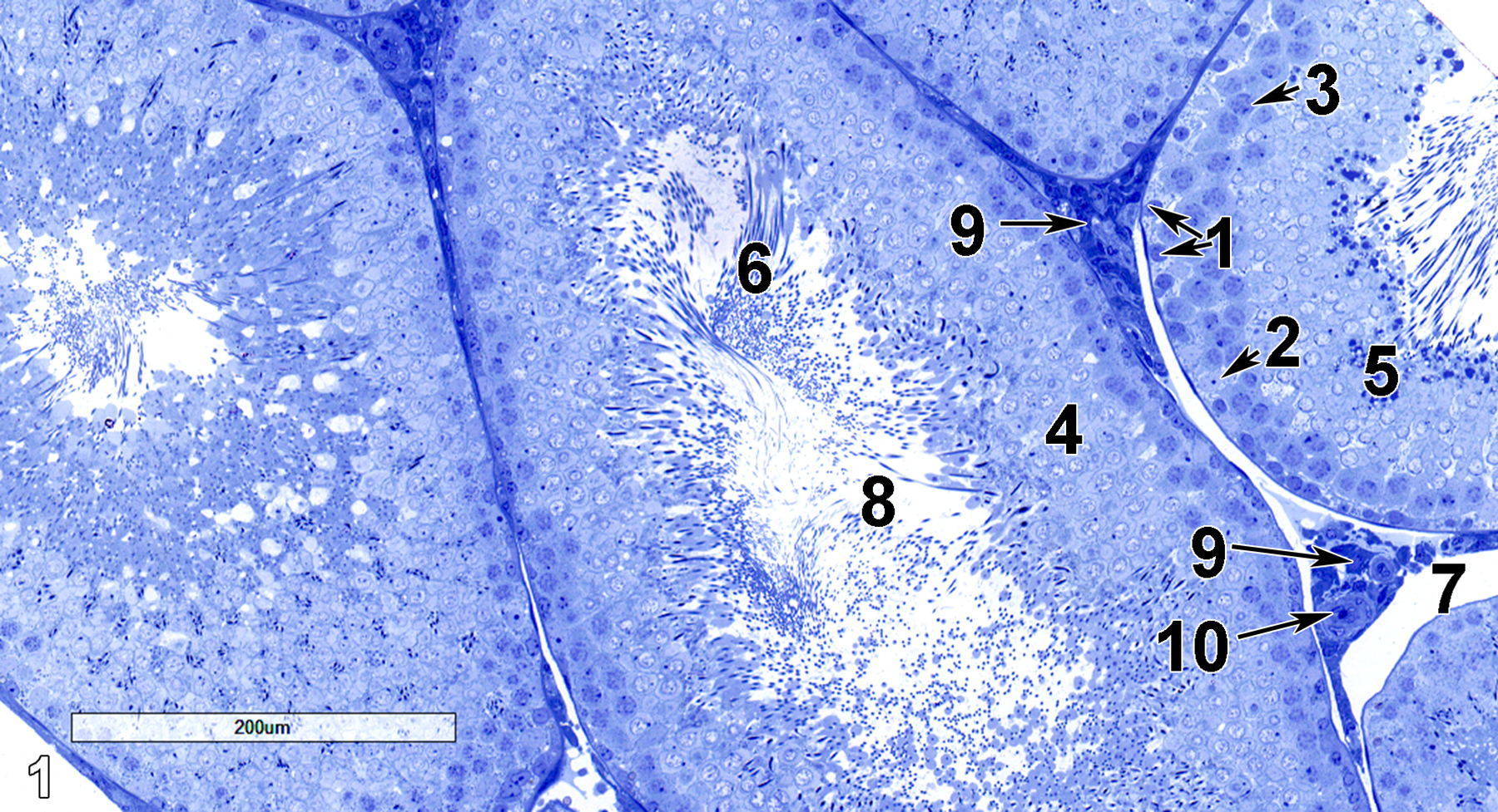
Figure 1. A semithin section (0.5 micrometer thick) of a toluidine blue O-stained portion of a testis, showing several seminiferous tubules. Spermatogonia (1) with round nuclei (arrows) lie on the basal lamina underlain by the lamina propria. In lesser numbers are Sertoli cells (2), with larger, less stained nuclei (arrow). Primary spermatocytes (3) form several layers above the basal cell layers and have round, relatively dark nuclei (arrow). Early spermatids (4) have less dense and smaller nuclei than the primary spermatocytes lying beneath them. Late spermatids (5) have accumulations of densely staining material. Spermatozoa (6) have flagellar shafts extending into the seminiferous tubule lumen (8). The peritubular lymphatic space (7) is located between the seminiferous tubules, along with connective tissue, Leydig cells (9, arrow), and occasional lymphocytes (10, arrow). 13x.
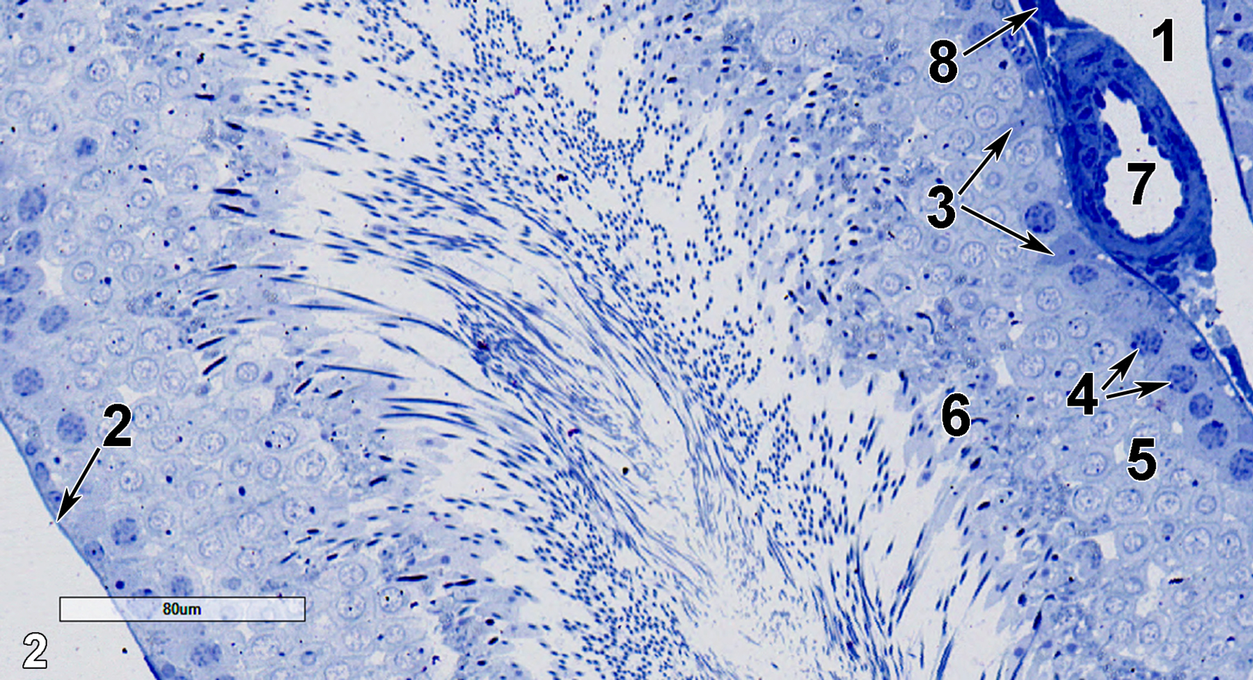
Figure 2. Another semithin section of a portion of a seminiferous tubule at slightly higher magnification. An interstitial lymphatic space (1) is next to an arteriole (7), which has an adjacent Leydig cell (8, arrow). Sertoli cells with relatively pale nucleoplasm (3, arrows) are adjacent to the lamina propria (2, arrow), as are the primary spermatocytes (4, arrows). Above the basal layer of cells are early spermatids (5), which lie beneath late spermatids (6) that contain dense staining material that eventually forms acrosomes. 25x.
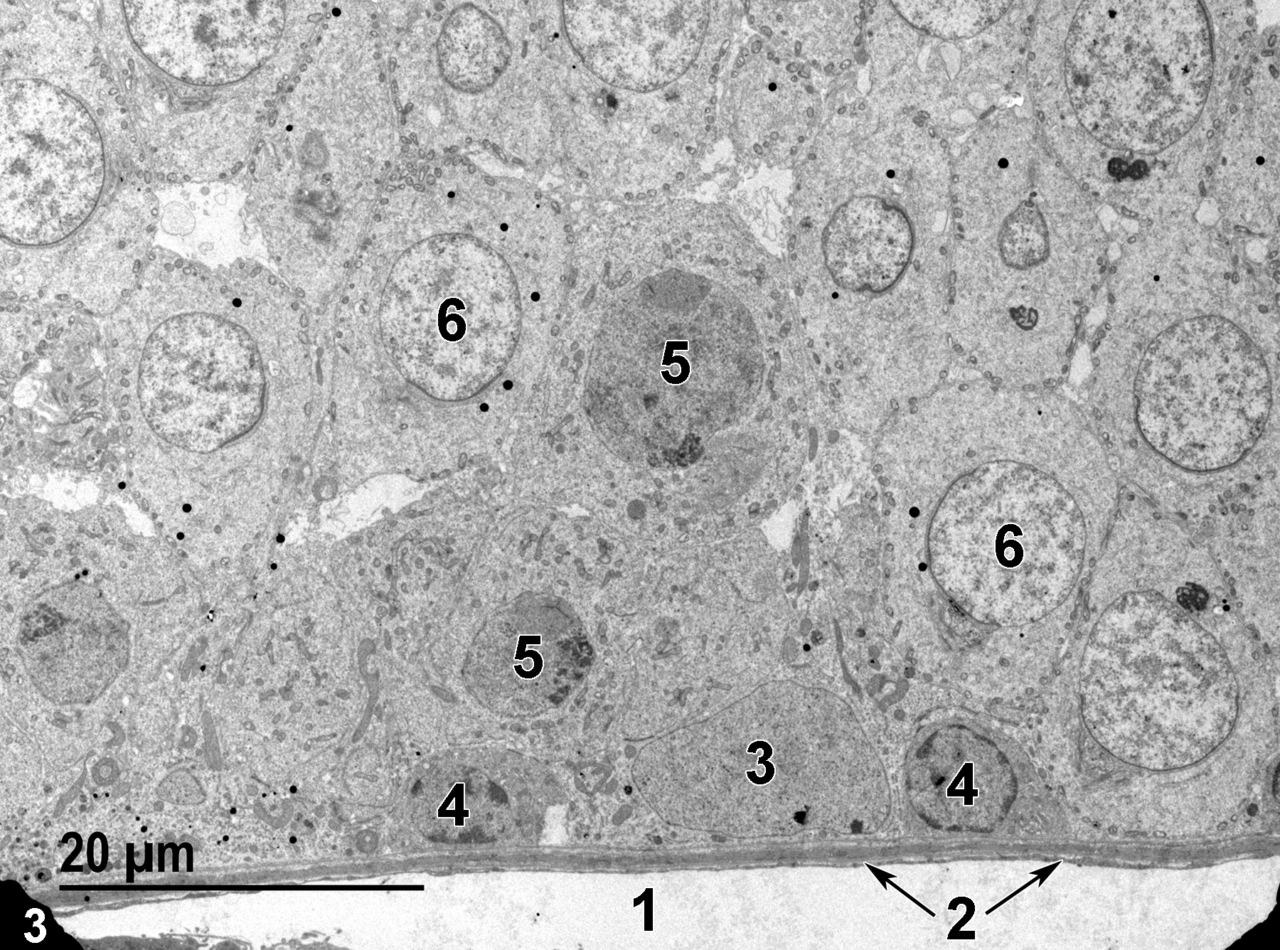
Figure 3. A low magnification electron micrograph of the basal region of a seminiferous tubule. The lamina propria is separated from the lumen of the peritubular lymphatic space (1) by a thin endothelial cell layer (2, arrows). A Sertoli cell with a large nucleus (3) and dispersed chromatin is sitting on the basal lamina of the seminiferous tubule. Type A spermatogonia with smaller, round nuclei (4) show marginated heterochromatin. Primary spermatocytes (5) have slightly more mitochondria than that of spermatogonia and are not located on the basal lamina. Early spermatids (6) are characterized by the numerous small mitochondria located along the plasma membrane and the relatively dispersed chromatin. 1900x.
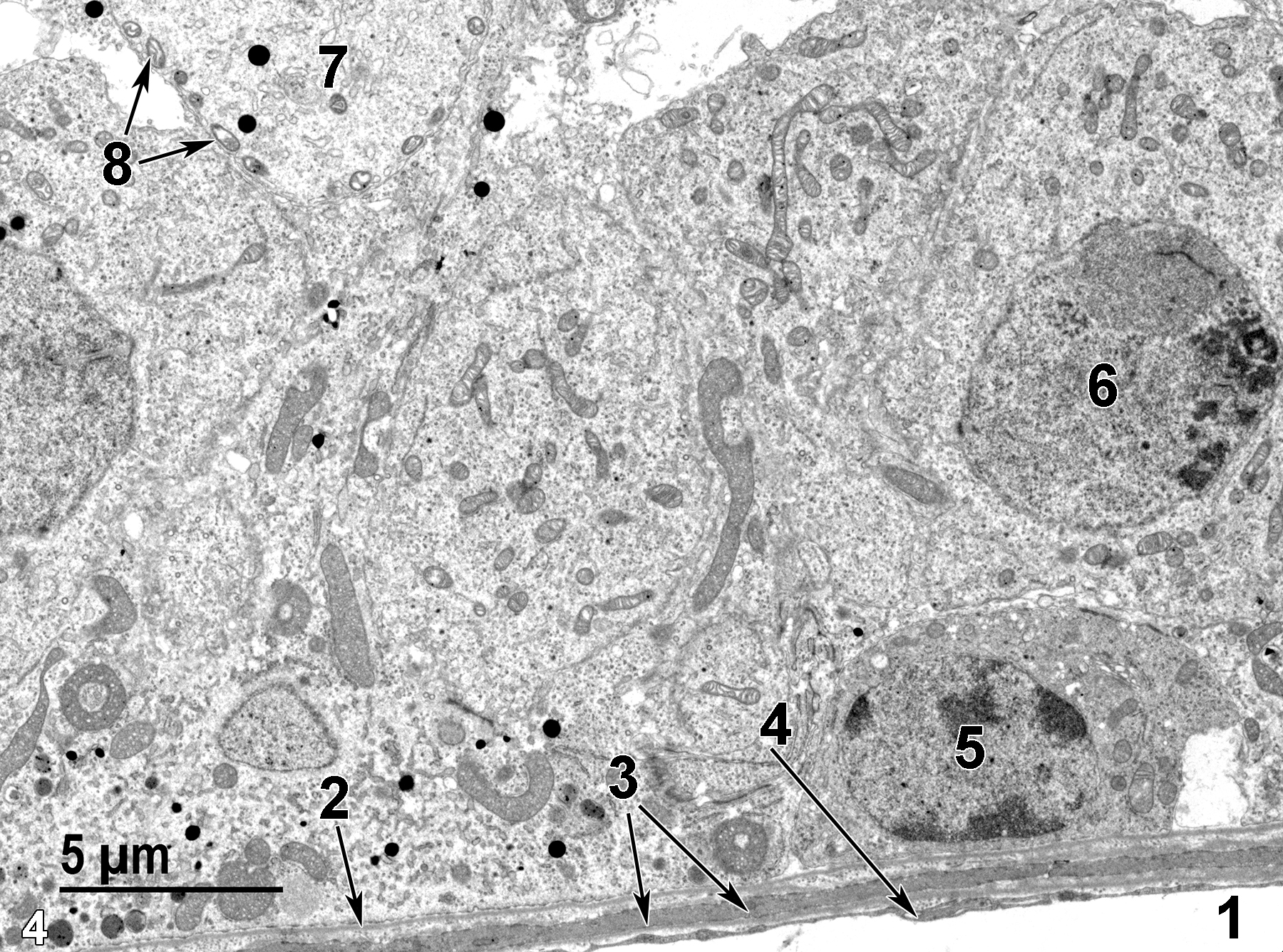
Figure 4. Another view of the basal region of a seminiferous tubule, showing the peritubular lymphatic space (1) lined with a thin endothelial cell layer (4, arrow). The base of the seminiferous tubule has a thin basal lamina (2, arrow) over a small band of connective tissue and a single layer of peritubular myoid cells (3, arrows). A single type A spermatogonium (5) is closely appressed to the basal lamina and contains an ovoid nucleus with marginated heterochromatin. Above the spermatogonium is a primary spermatocyte cell with a labeled nucleus (6). In the upper left of the image is a portion of an early spermatid (7) with its characteristic small mitochondria (8, arrows) arranged along the plasma membrane. 4800x.
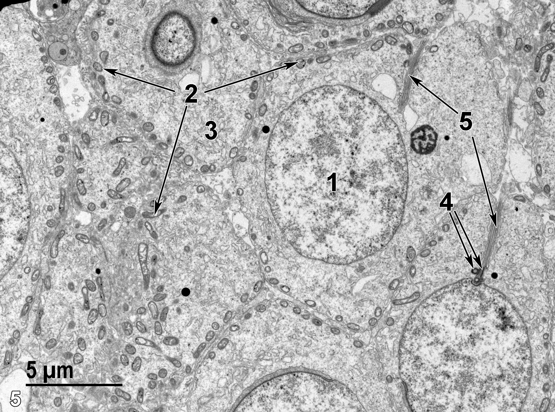
Figure 5. More early spermatids with large round nuclei with dispersed chromatin (1), numerous small mitochondria (2, arrows) located along the cell membrane, pools of smooth endoplasmic reticulum (3), paired centrioles (4, arrows), and axonemes (5, arrows). 4800x.
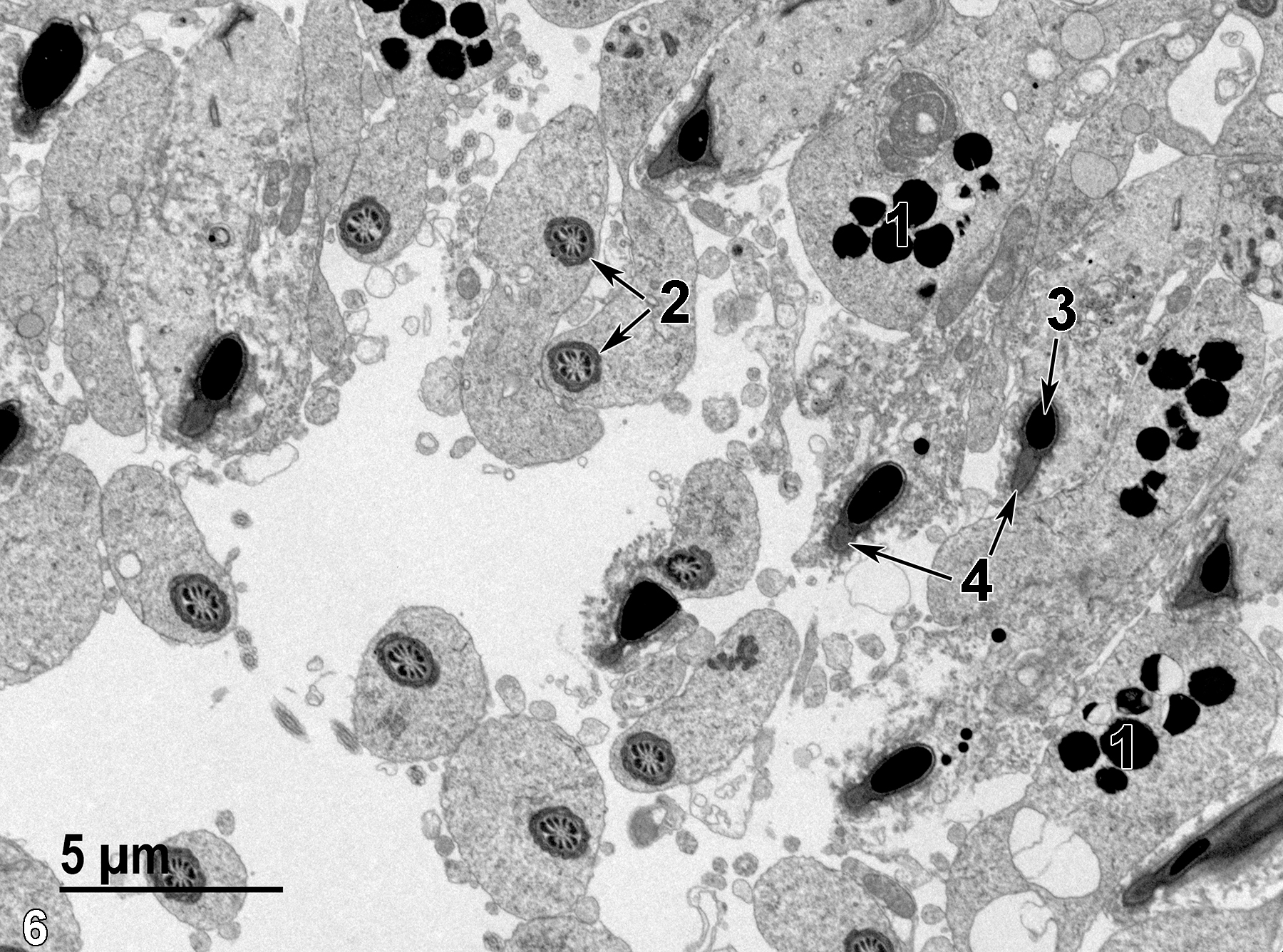
Figure 6. A view of the luminal surface of a seminiferous tubule with deep electron-dense acrosomal granules (1) in spermatids. Several middle pieces of spermatozoa (2, arrows) are present, along with portions of spermatozoon nuclei (3, arrow) and acrosomes (4, arrows). 4800x.
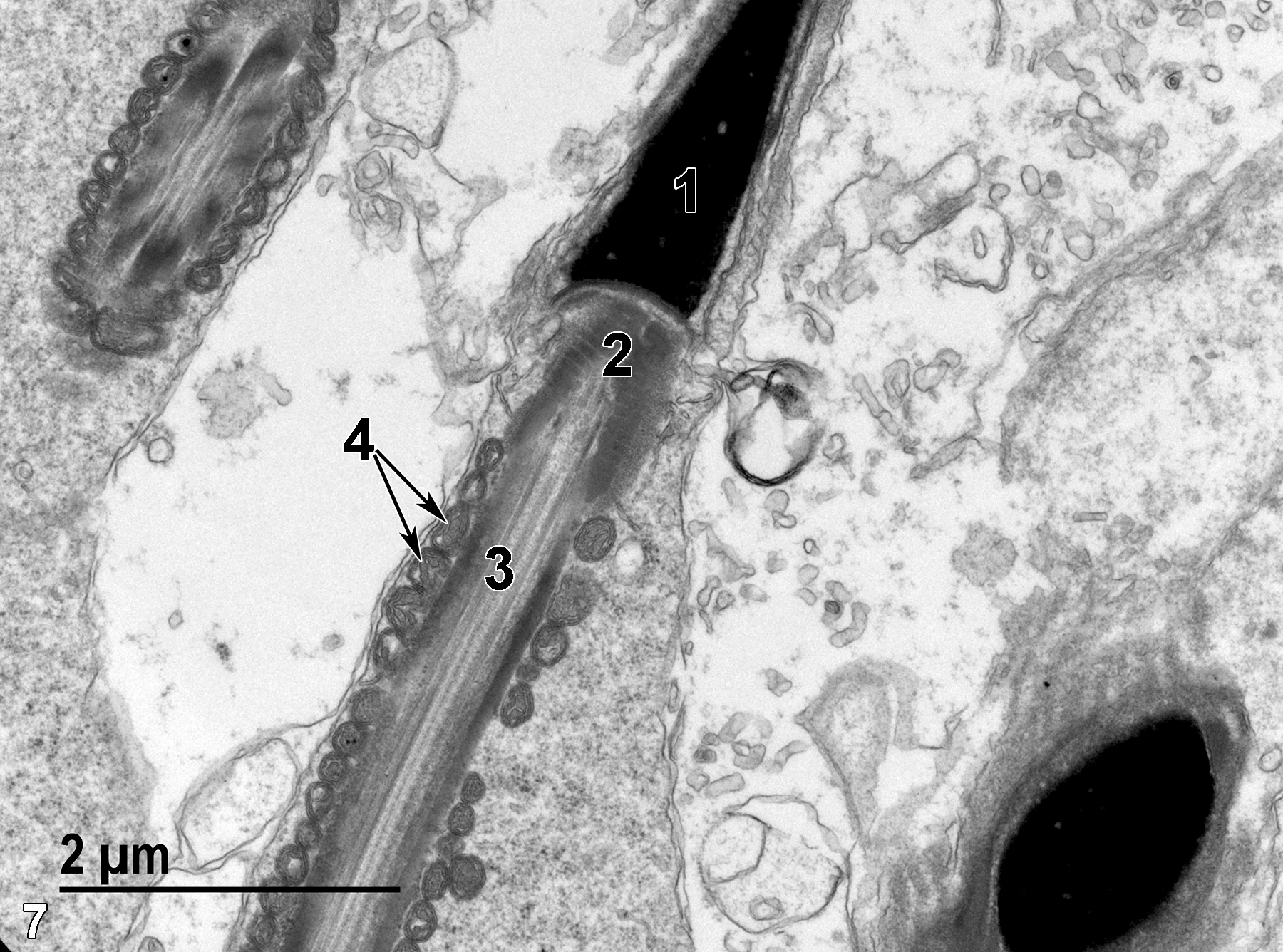
Figure 7. A longitudinal view of the apical region of a spermatozoon. The extremely electron-dense nucleus (1) is underlain by the neck (2), followed by the middle piece with a central axoneme (3) surrounded by numerous round mitochondria (4, arrows). 18500x.
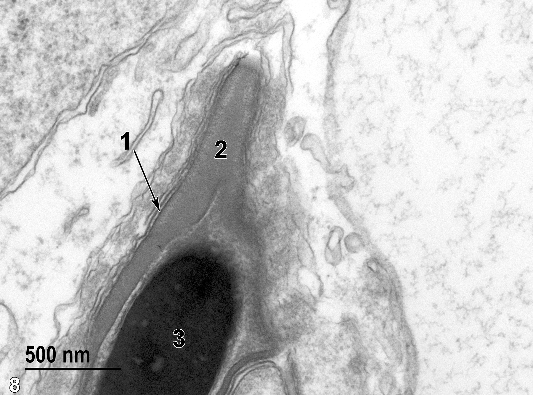
Figure 8. A longitudinal view of the spermatozoon head with the cell membrane (1, arrow) surrounding the acrosomal cap (2), which surrounds the apical portion of the extremely electron-dense nucleus (3). 49000x.
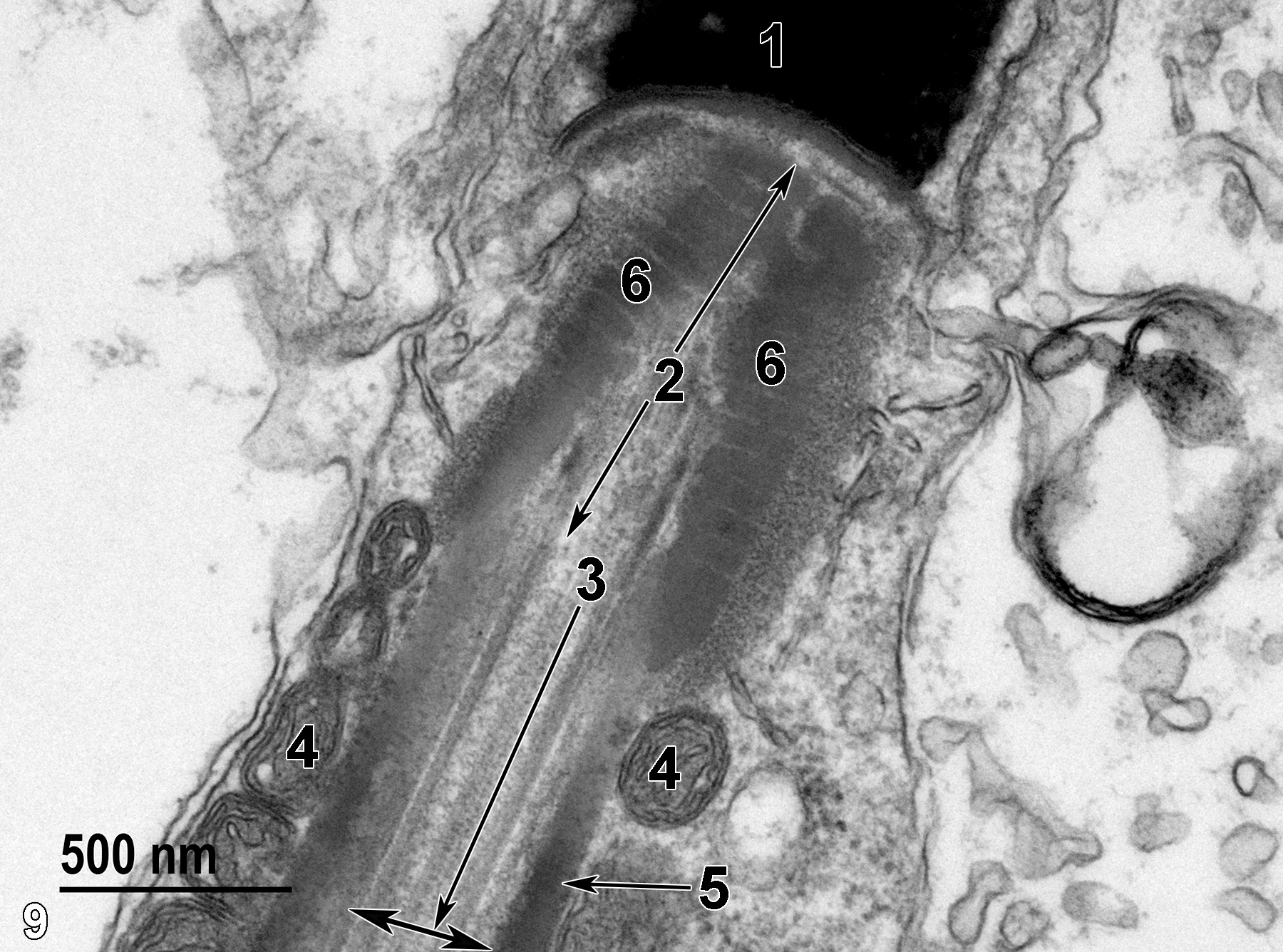
Figure 9. The region directly beneath Figure 8. The base of the nucleus (1) is adjacent to the neck region (2, arrows) with outer segmented rings of fibrous material (6) surrounding the axoneme that extends down the center of the spermatozoon. The beginning (3) of the middle piece (arrow) consists of the axoneme (two-headed arrow) surrounded by a relatively thin fibrous sheath (5, arrow), which is, in turn, surrounded by mitochondria (4). 49000x.
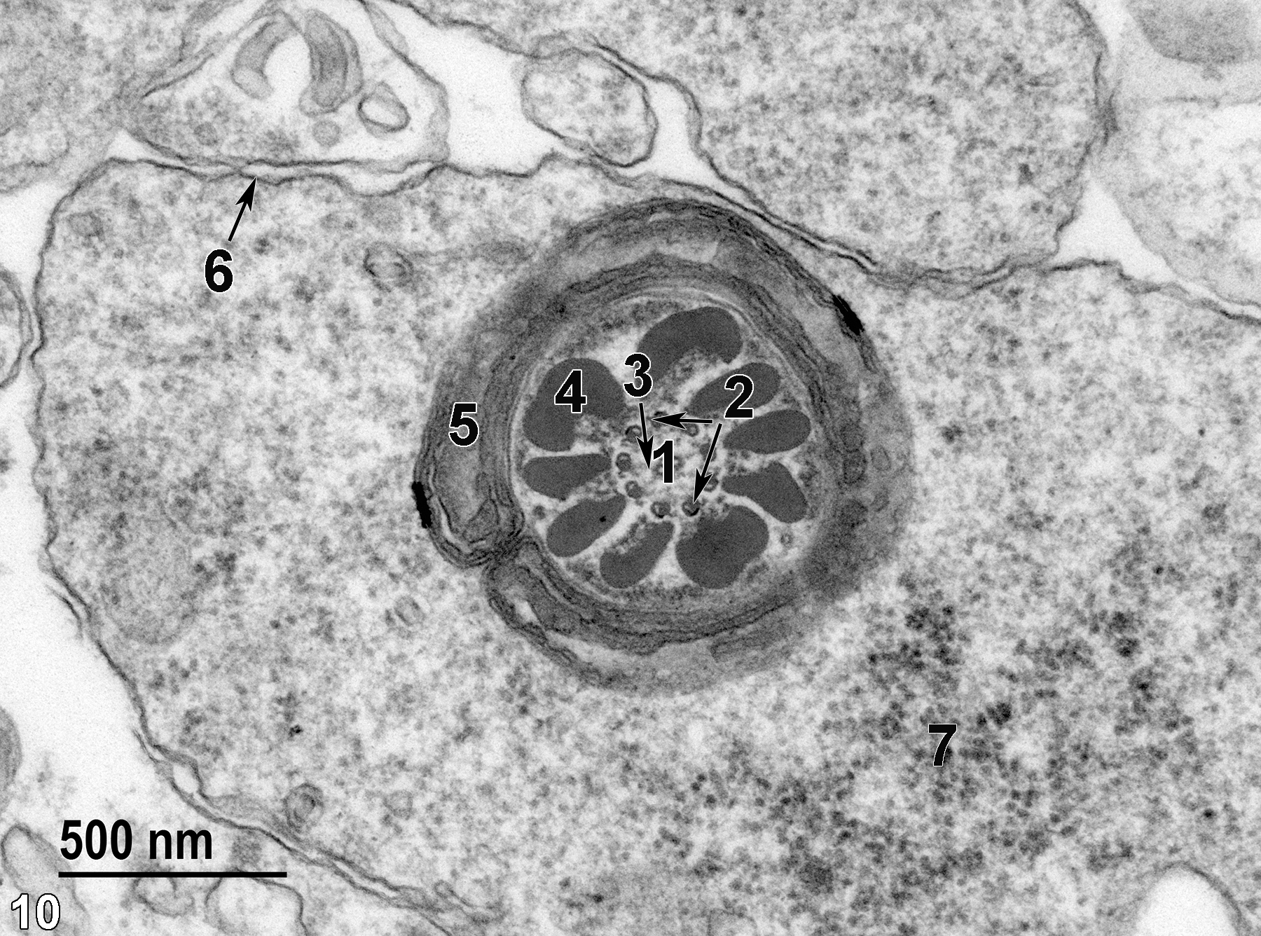
Figure 10. A transverse view of the middle piece of a spermatozoon. The axoneme consists of two central microtubules (1) surrounded by nine doublet microtubules (2, arrows), with radial spokes (3) extending from the central tubules to the peripheral tubules (arrow). Nine outer dense fibrils (4) are surrounded by a sheath of mitochondria (5). The spermatozoon cytoplasm bound by the plasma membrane (6, arrow) contains accumulations of ribosomes (7). 49000x.
| Dellmann HD, Eurell J, eds. 1998. Textbook of Veterinary Histology. 5th ed. Philadelphia: Lippincott Williams & Wilkins. |
| Rhodin JAG. 1974. Histology: A Text and Atlas. New York: Oxford University Press. |
| Ross MH, Kaye GI, Pawlina W. 2003. Histology: A Text and Atlas. 4th ed. Philadelphia: Lippincott Williams & Wilkins. |
| Weiss L, ed. 1988. Cell and Tissue Biology: A Textbook of Histology. 6th ed. Baltimore: Urban & Schwarzenberg. |
All Images


