

Alimentary System
Stomach
Narrative
The stomach has two portions that are divided by the limiting ridge. The forestomach (nonglandular stomach) has similar tissue layers to those of the esophagus: a keratinized squamous epithelium underlain with a lamina propria of connective tissue with a few lymphocytes, neutrophils, mast cells, and plasma cells. The muscularis mucosae is beneath the lamina propria and overlies the submucosa, which contains lymphatics and nerves. The tunica muscularis consists of three smooth muscle cell layers, and the final layer is the tunica adventitia.
Forestomach
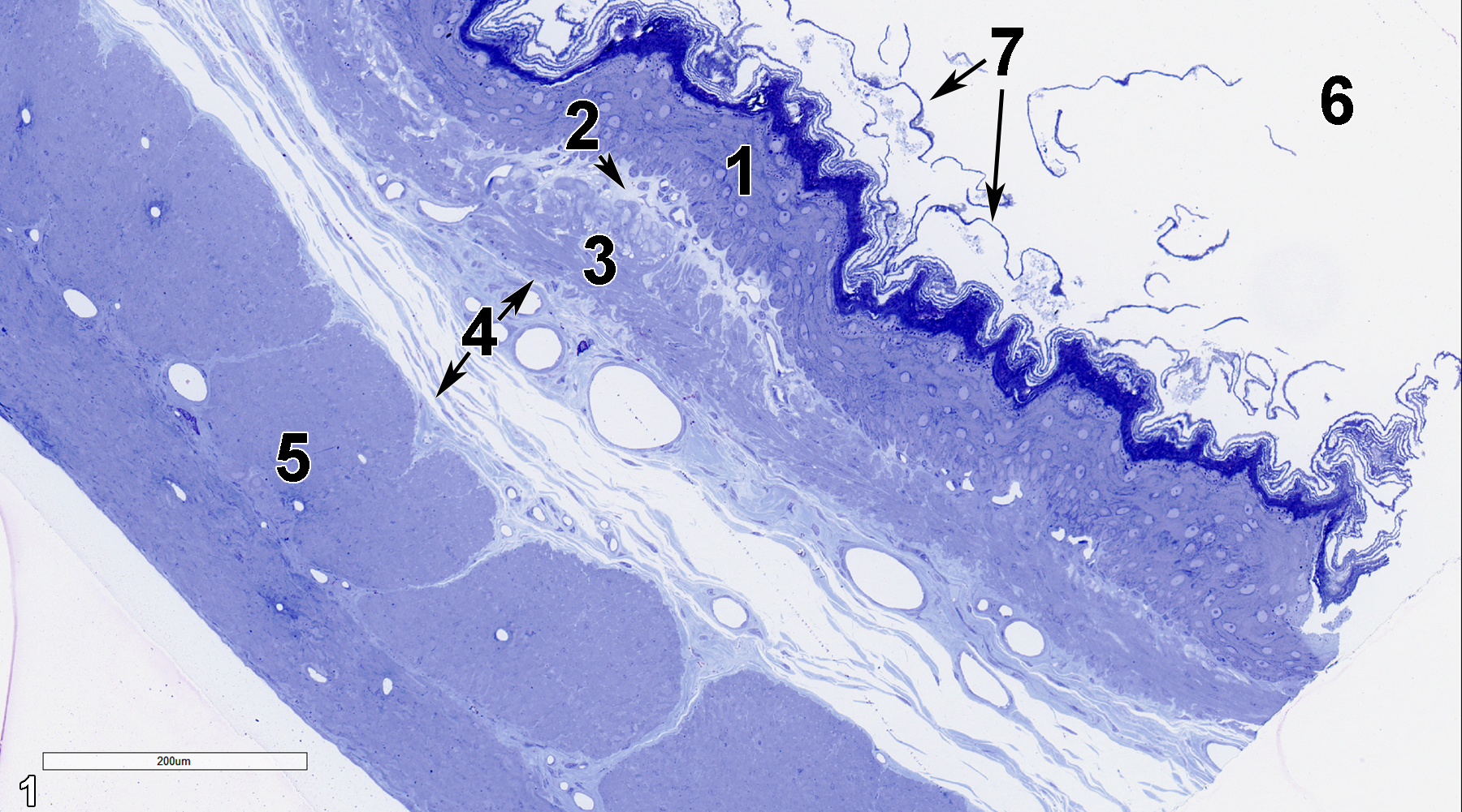
Figure 1. A semithin section (0.5 micrometer thick) of a toluidine blue O-stained portion of the nonglandular forestomach. The epithelial layer (1) is composed of stratified squamous cells. Directly beneath it is the lamina propria (2, arrow), which is mostly composed of a collagenous matrix with fibroblasts and vessels. Below that layer is the muscularis mucosae (3), which is primarily composed of smooth muscle cells and collagen fibrils. The submucosa (4, arrows) contains fibroblasts, collagen fibrils, and prominent vessels as major components. The tunica muscularis (5) consists of smooth muscle cells, with one layer oriented transversely (circular) and the other longitudinally. The lumen of the forestomach (6) shows keratinized epithelial cells sloughing into it (7, arrows). 15x.
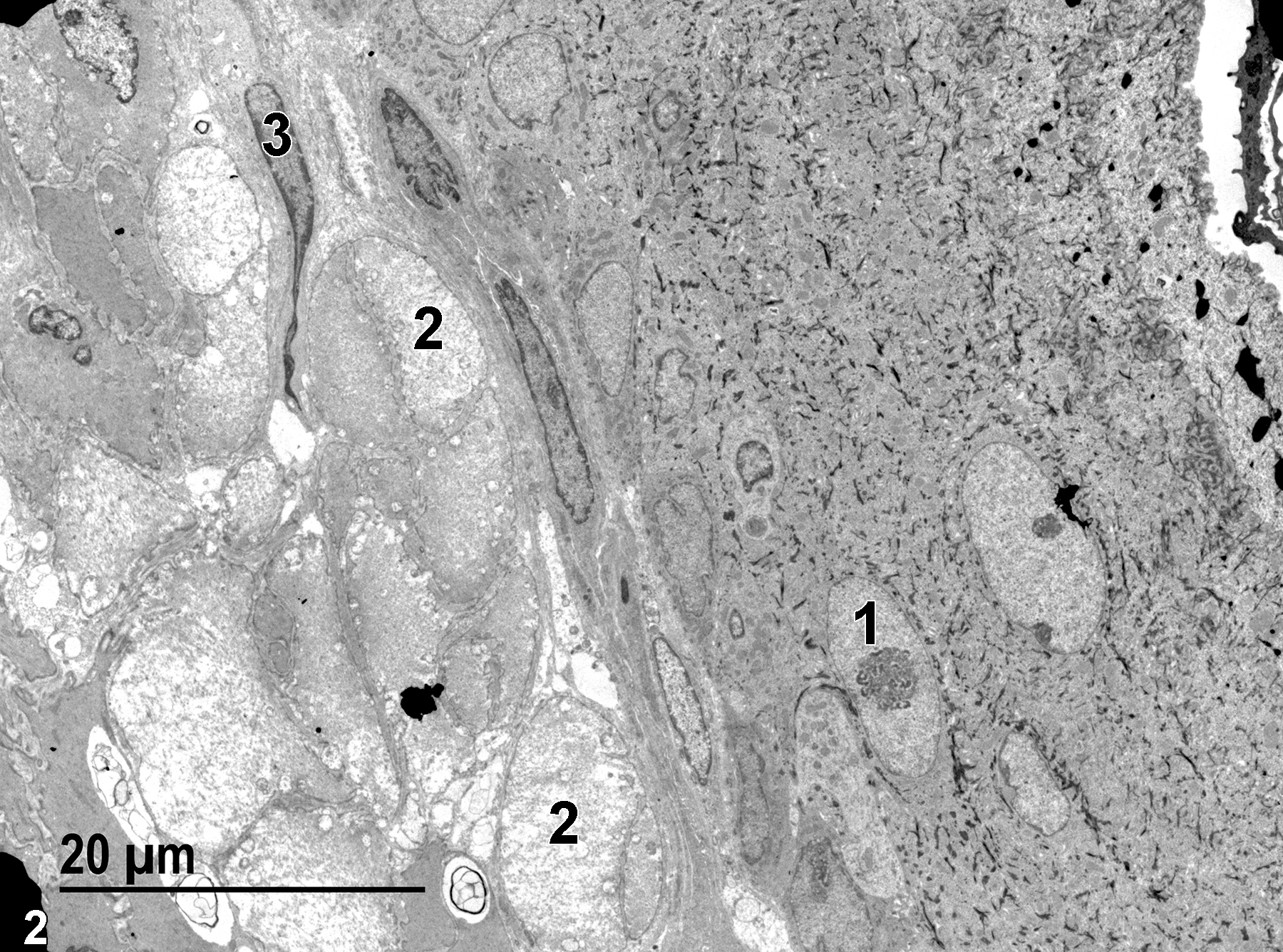
Figure 2. An ultrastructural view of the epithelium and lamina propria of the nonglandular forestomach. A nucleus (1) of one of the stratified squamous epithelial cells is shown, as is a nucleus of a fibroblast (3) in the lamina propria, along with bundles of collagen (2). 1900x.
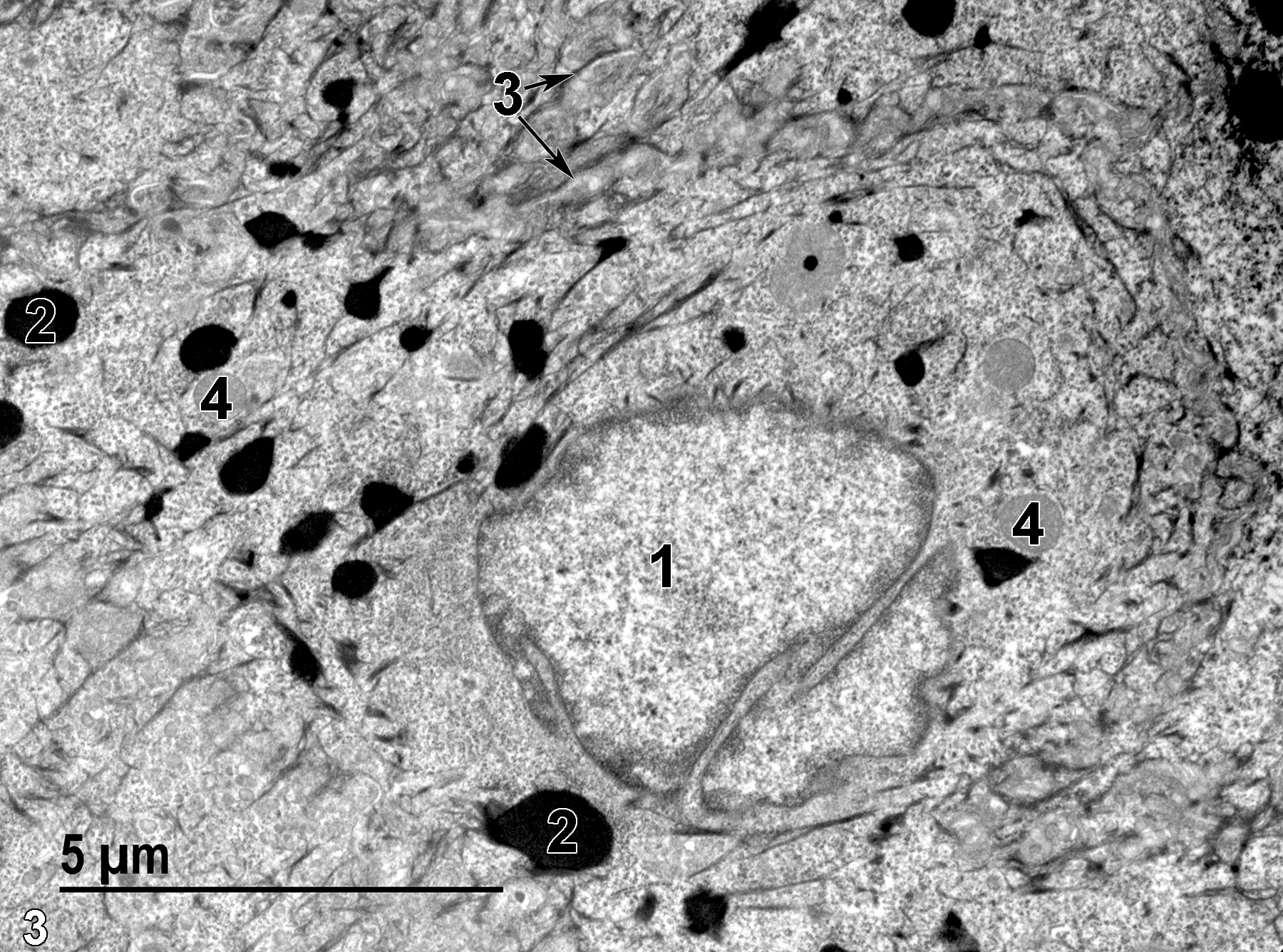
Figure 3. A higher magnification view of an epithelial cell nucleus (1), electron-dense melanosomes (2), tonofilament bundles (3, arrows), and mitochondria (4). 9300x.
Glandular Stomach
The glandular stomach of the rat has surface ridges (rugae), but no villi. The lumen of the glandular stomach is lined with a mucous layer that is 95% water with glycoproteins. This layer is 10-20 times thicker than the epithelium beneath. The epithelium of the fundic (gastric) glands consists of columnar cells that form gastric pits with glands at the base. Located between the gastric pit and the glandular basal region is the isthmus region, which contains cells that replicate, with some migrating upward to become epithelial cells, whereas others migrate downward to form chief (zymogen) cells, parietal (oxyntic) cells, and mucous cells. Parietal cells are found mostly in the upper and middle part of the gastric glands, whereas chief cells are found primarily in the basal half of the gastric glands. The submucosa, tunica muscularis, and adventitia are similar to the forestomach (Uehara et al. 2018).
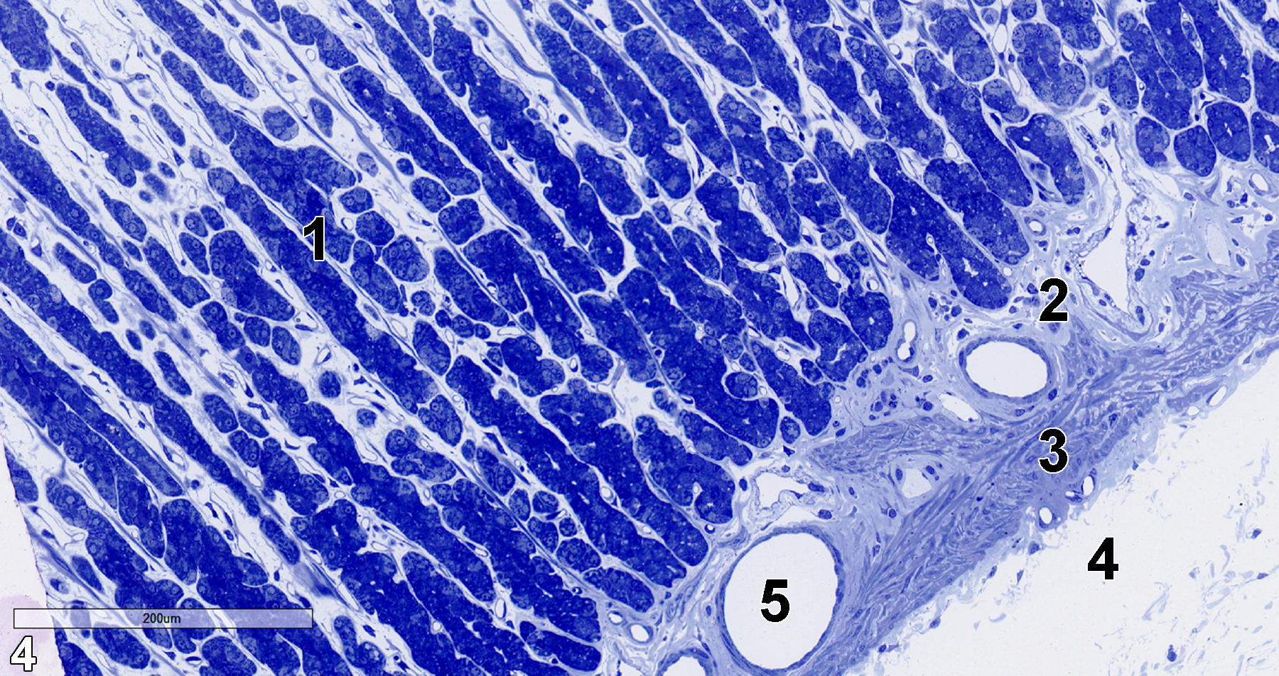
Figure 4. A semithin section (0.5 micrometer thick) of a toluidine blue O-stained portion of the glandular stomach showing the gastric pits (1) lined with columnar epithelial cells. Beneath them is the lamina propria (2), which overlies the muscularis mucosae (3) that, in turn, overlies the submucosa (4). A lymphatic vessel (5) is located within the lamina propria. 15x.
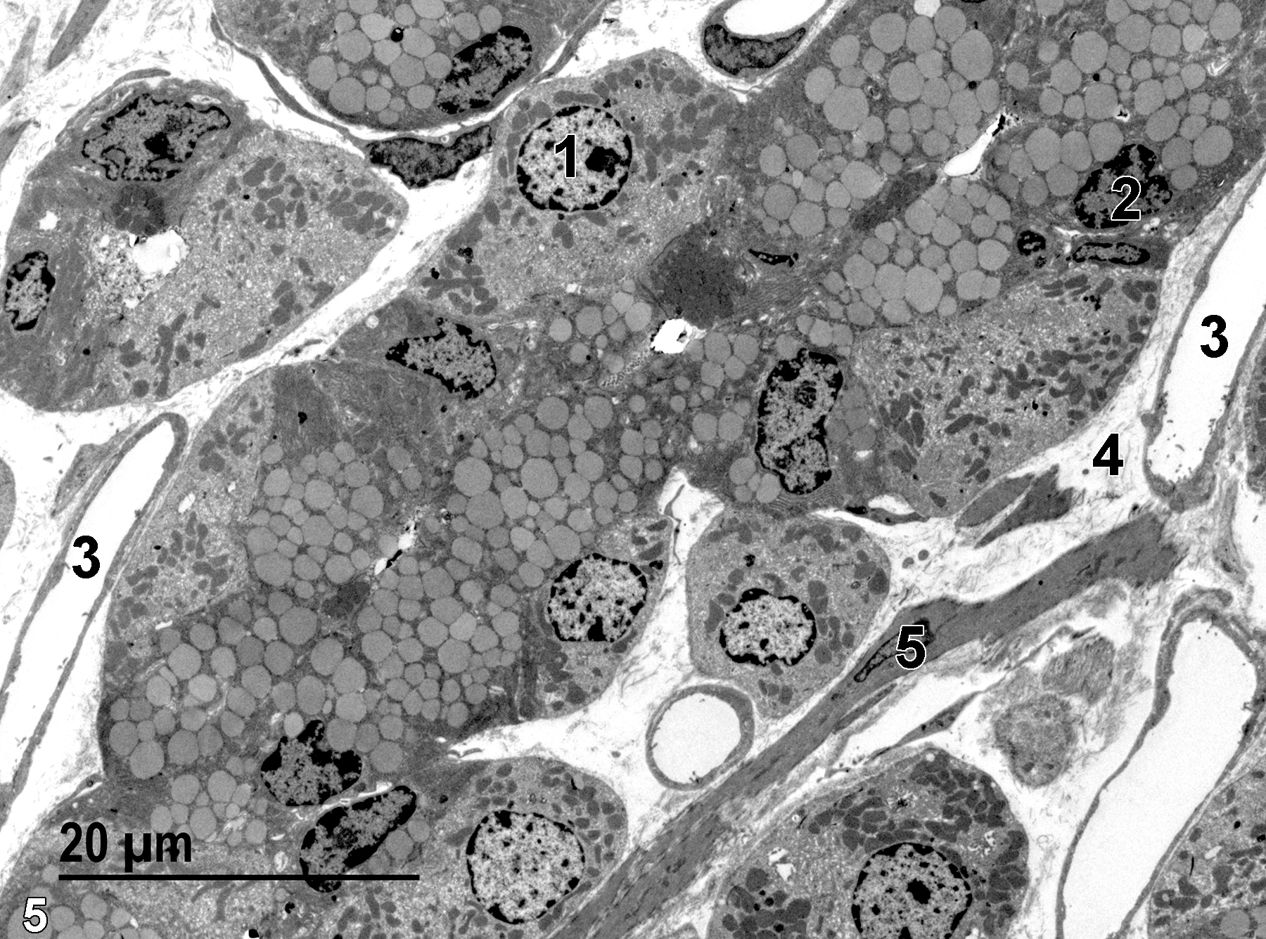
Figure 5. A low magnification view of the glandular stomach near the base of a gastric pit. A nucleus (1) of a parietal cell can be seen, as well as a nucleus of a chief (zymogen) cell (2). Capillaries (3) are located between the epithelial cells of the gastric pits, along with collagen fibrils (4) and one smooth muscle cell (5). 1900x.
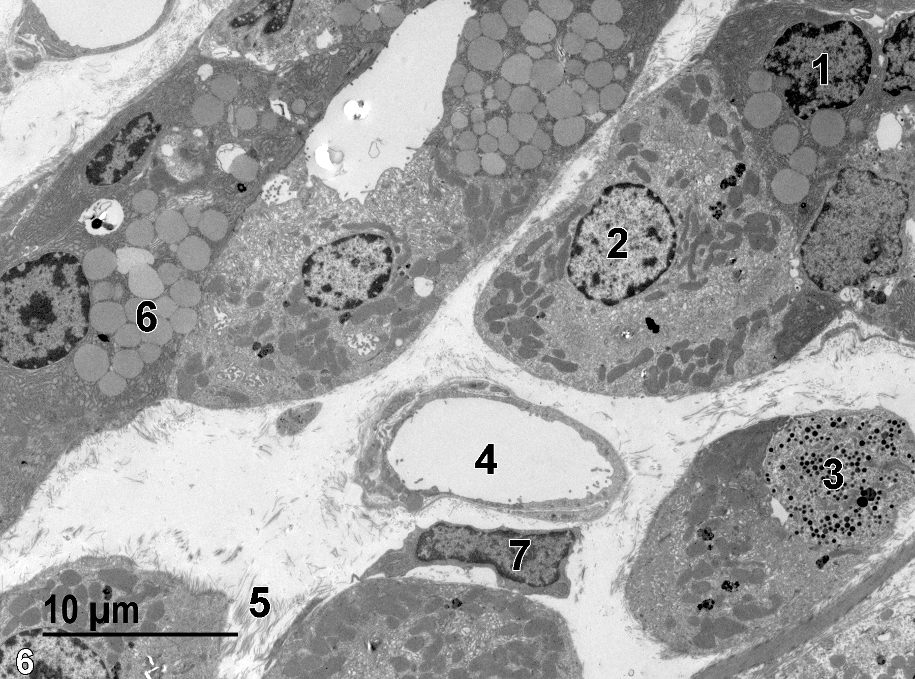
Figure 6. A view of another area in a gastric pit, with a chief cell nucleus (1) and a parietal cell nucleus (2). Chief cells contain numerous zymogen granules (6), whereas the parietal cells contain a few electron-dense lysosomes, numerous mitochondria, and vesicular profiles (intracellular canaliculi). One enteroendocrine cell (3) with small secretory granules with electron-dense content is present. A blood vessel (4) surrounded by a loose collagenous matrix (5) has a fibroblast (7) directly beneath it. 2900x.
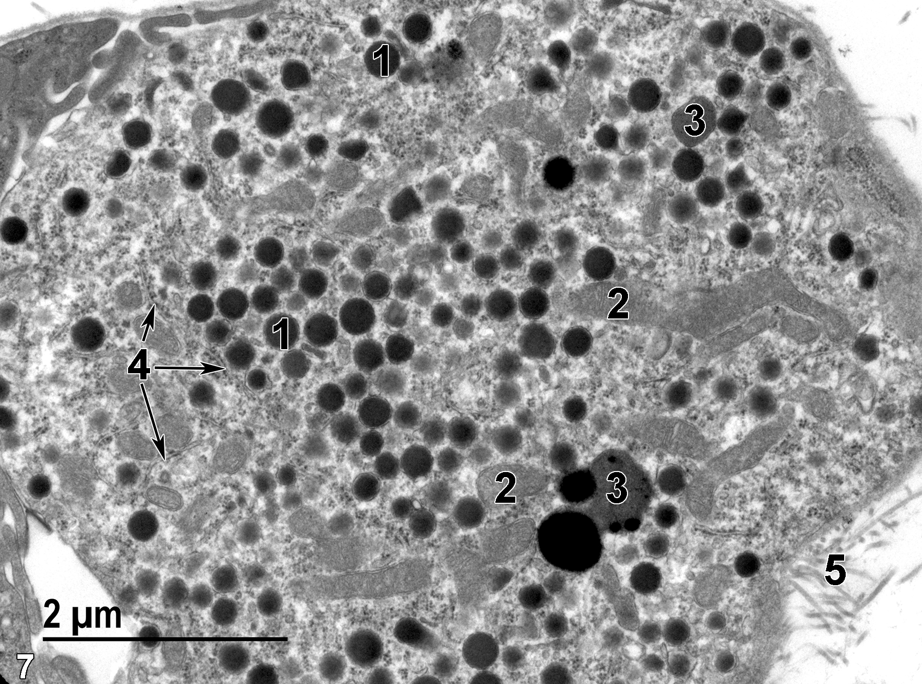
Figure 7. A higher magnification view of an enteroendocrine cell containing electron-dense secretory granules (1), mitochondria (2), lysosomes (3), and rough endoplasmic reticulum (4, arrows). Collagen fibrils (5) can be seen in the space outside the glandular epithelium. 18500x.
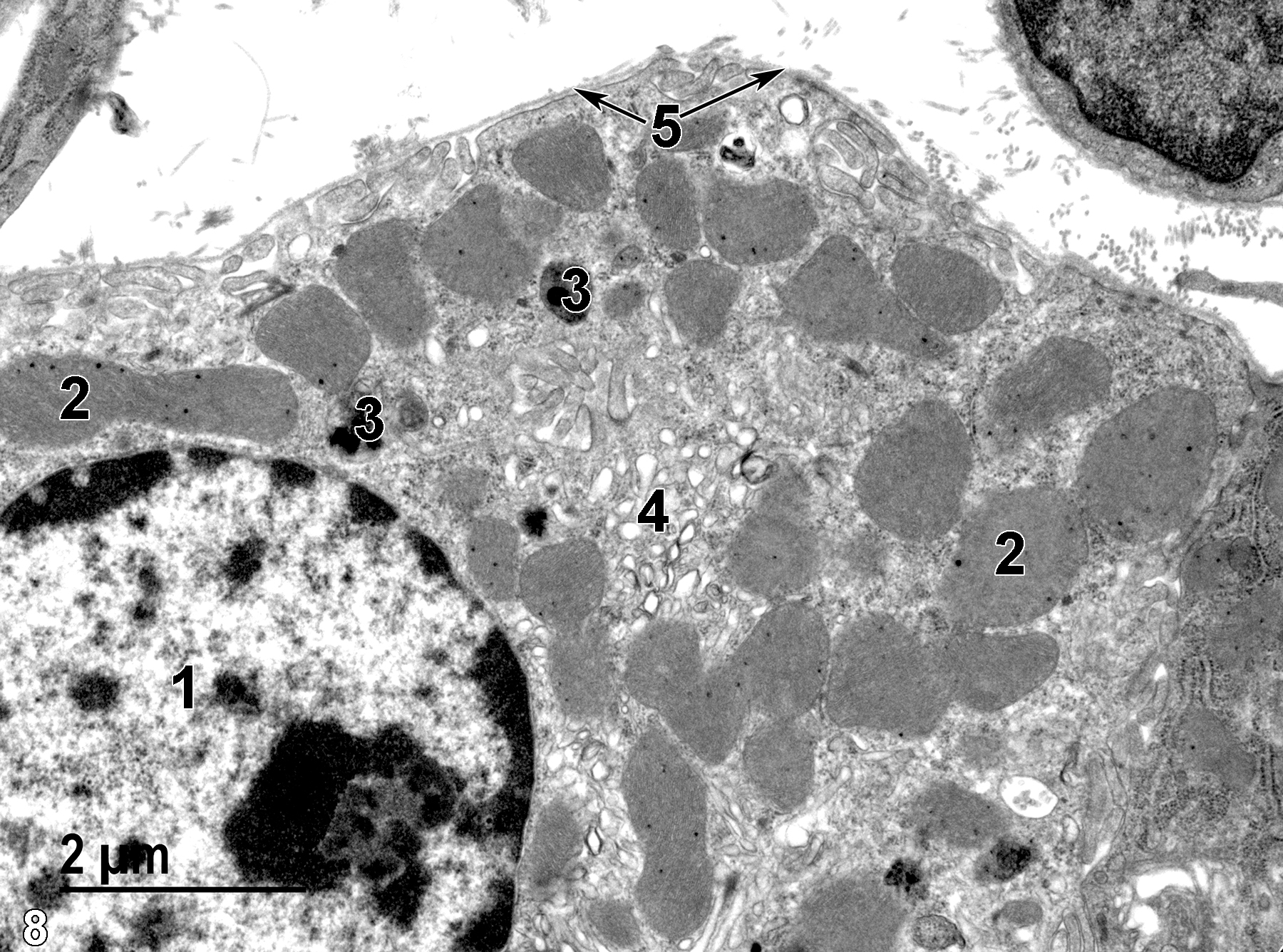
Figure 8. A view of a parietal cell. The nucleus (1) contains a single nucleolus with marginated heterochromatin. Numerous pleomorphic mitochondria (2) are present, along with small lysosomes (3) and vesicular profiles (4) of the intracellular canaliculi characteristic of this cell type. The cell has a basal lamina at its surface (5, arrows). 13000x.
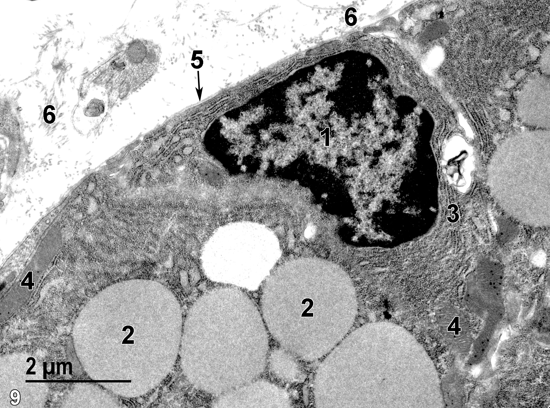
Figure 9. The details of a chief cell. The nucleus (1) is surrounded by rough endoplasmic reticulum (3) elements. Large zymogen granules (2) are present, along with a few mitochondria (4). The surface of the cell is covered with a thin basal lamina (5, arrow). Bundles of collagen fibrils (6) can be seen in the gastric pit lumen. 13000x.
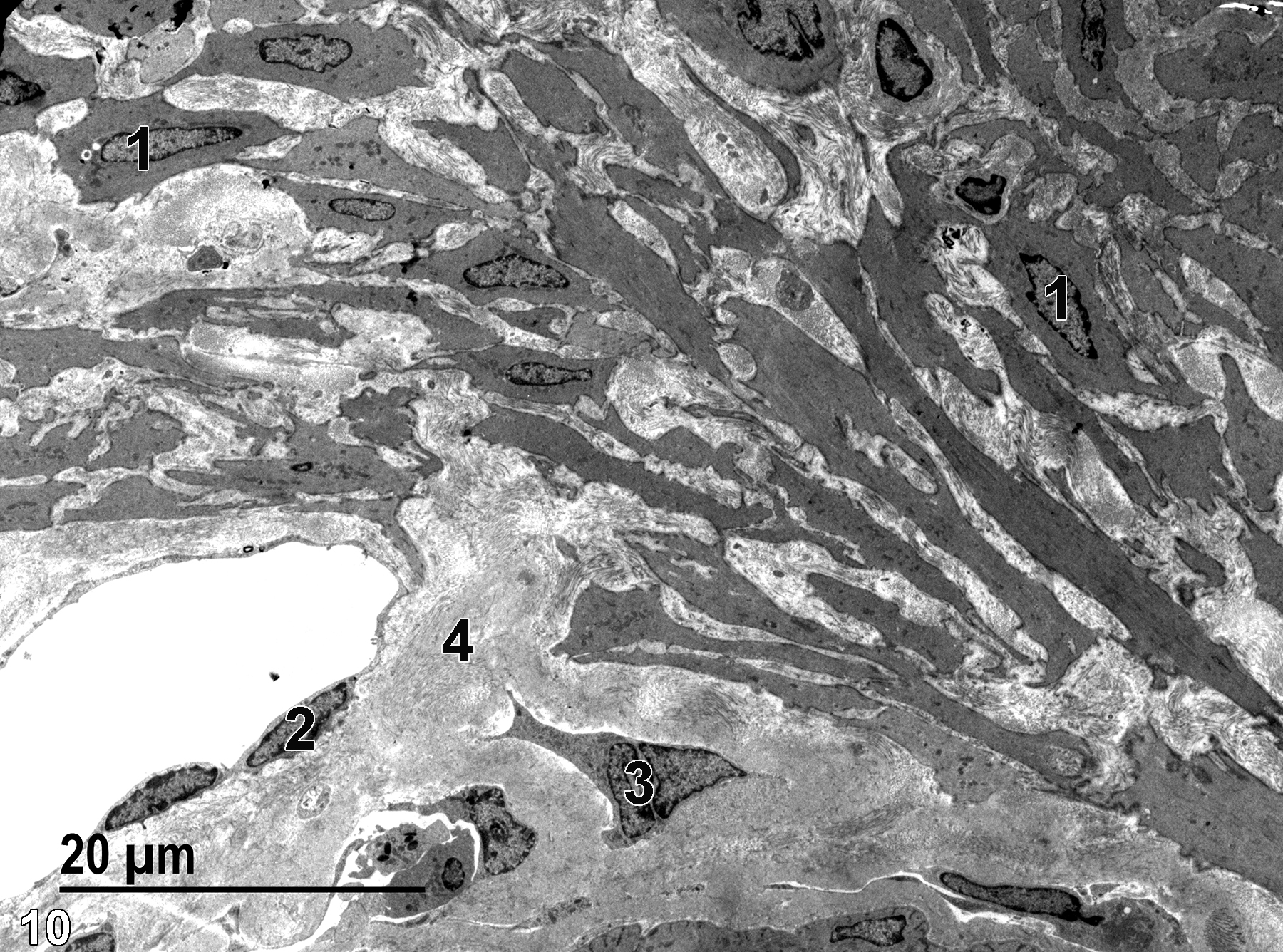
Figure 10. A low magnification view of the muscularis mucosae, with numerous elongate smooth muscle cells with elongate nuclei (1). A capillary (2) is present, lined with a thin endothelial cell layer with elongate nuclei. A small portion of the submucosa is present, composed of fibroblasts (3) and large amounts of collagen fibrils (4). 1900x.
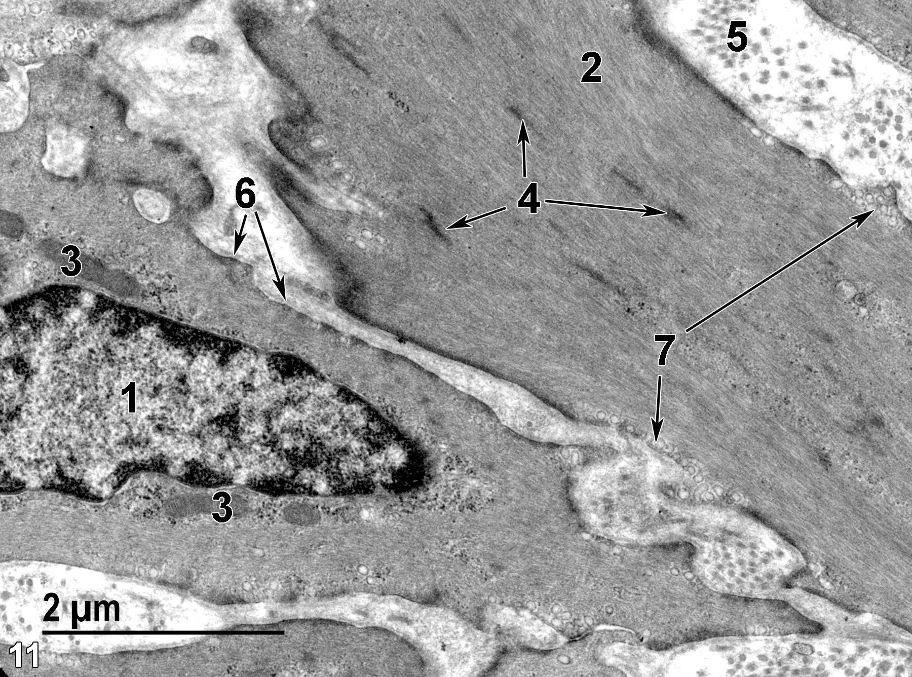
Figure 11. A higher magnification view of the smooth muscle cells of the muscularis mucosae. Each cell contains a single nucleus (1), a small number of mitochondria (3), large quantities of cytoplasmic protein fibrils (2) composed of actin and myosin, and more electron-dense patches consistent with alpha-actinin (4, arrows). Each cell is surrounded by a basal lamina (6, arrows). The surface of the smooth muscle cells exhibits numerous micro-pinocytotic vesicles (7, arrows). The extracellular spaces contain collagen fibrils (5). 18500x.
| Boorman GA, Eustis SL, Elwell MR, Montgomery CA, Jr., MacKenzie WF, eds. 1990. Pathology of the Fischer Rat: Reference and Atlas. New York: Academic Press. |
| Cross PC, Mercer KL. 1993. Cell and Tissue Ultrastructure: A Functional Perspective. New York: W.H. Freeman and Company. |
| Dellmann HD, Eurell J, eds. 1998. Textbook of Veterinary Histology. 5th ed. Philadelphia: Lippincott Williams & Wilkins. |
| Rhodin JAG. 1974. Histology: A Text and Atlas. New York: Oxford University Press. |
| Ross MH, Kaye GI, Pawlina W. 2003. Histology: A Text and Atlas. 4th ed. Philadelphia: Lippincott Williams & Wilkins. |
| Uehara T, Elmore SA, K.A. Szabo KA. 2017. Chapter 6: Esophagus and stomach. In Boorman’s Pathology of the Rat (Suttie AW, ed). 2nd ed. London: Academic Press, 35-50. |
| Weiss L, ed. 1988. Cell and Tissue Biology: A Textbook of Histology. 6th ed. Baltimore: Urban & Schwarzenberg. |
All Images


