

Endocrine System
Parathyroid Gland
Narrative
The parathyroid glands are surrounded by thin capsules of connective tissue that contain numerous fat cells, blood vessels, lymphatic vessels, and nerves. The capsule is continuous, with septa that penetrate and subdivide the parenchyma into incomplete lobules. The parenchyma consists of densely packed epithelial cells organized into a loose network of cords or plates that are surrounded by a thin basal lamina. Two types of cells typically predominate within the parenchyma - chief (principal) cells and oxyphil cells. However, oxyphil cells are not present in the rat (Mense and Rosol 2018). Chief cells are 4-8 micrometers in diameter with a small central nucleus, with rare or absent secretory granules, minimal rough endoplasmic reticulum and Golgi bodies, and variable amounts of glycogen, lipid, and lysosomes. Some chief cells have prominent rough endoplasmic reticulum and secretory granules near the plasma membrane. Oxyphil cells (observed in other species) are less common than chief cells. They are typically found in small clusters, are 6-10 micrometers in diameter and have smaller nuclei than those found in chief cells. The cytoplasm of oxyphil cells is packed with mitochondria but has only small amounts of endoplasmic reticulum and Golgi bodies. Secretory granules are typically absent.
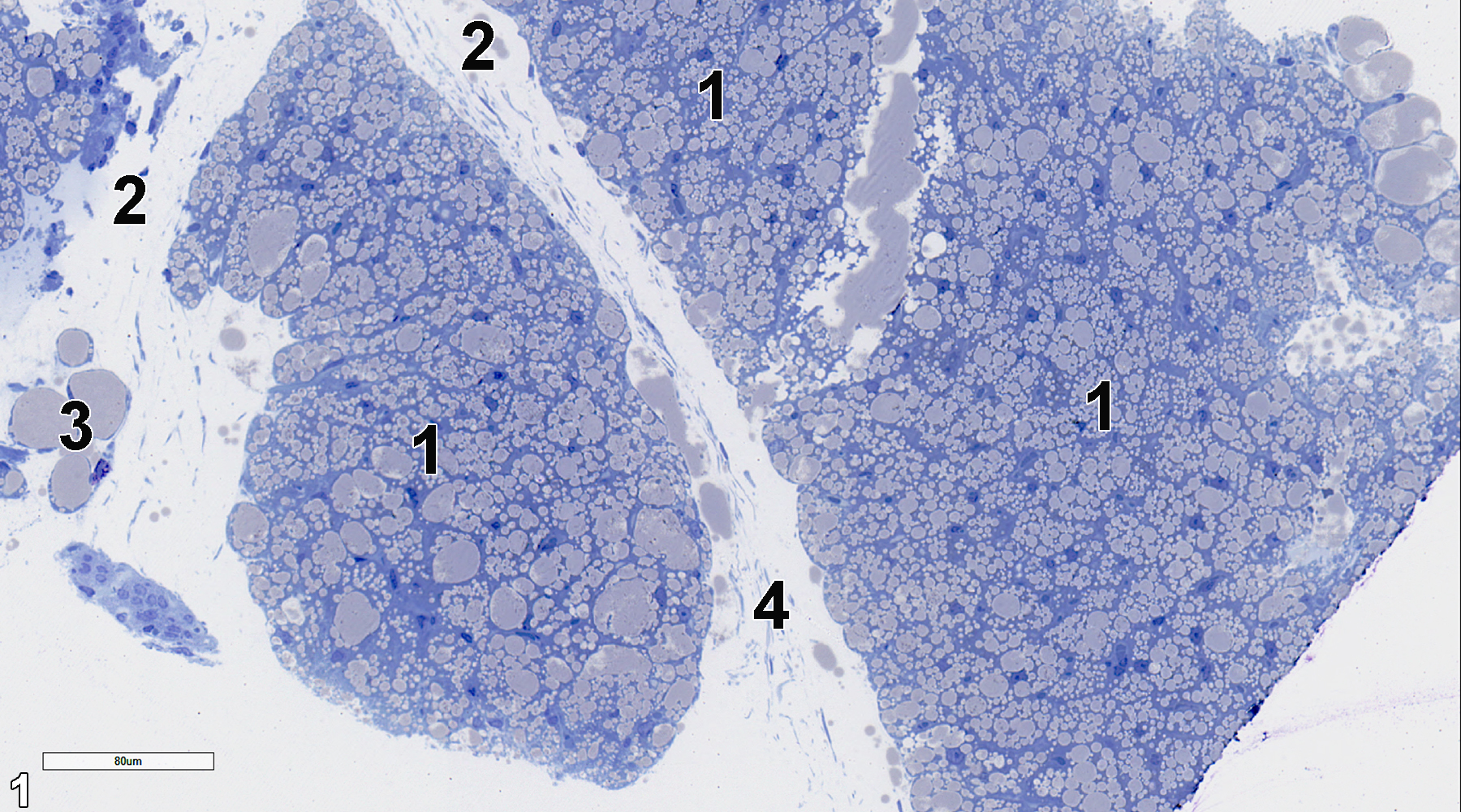
Figure 1. A semithin section (0.5 micrometer thick) of a toluidine blue O-stained section showing lobules of the parathyroid parenchyma, which consists of chief cells (1). The lobules are separated by septa (2), which consists of connective tissue with fat cells (3) and collagen (4). 25x.
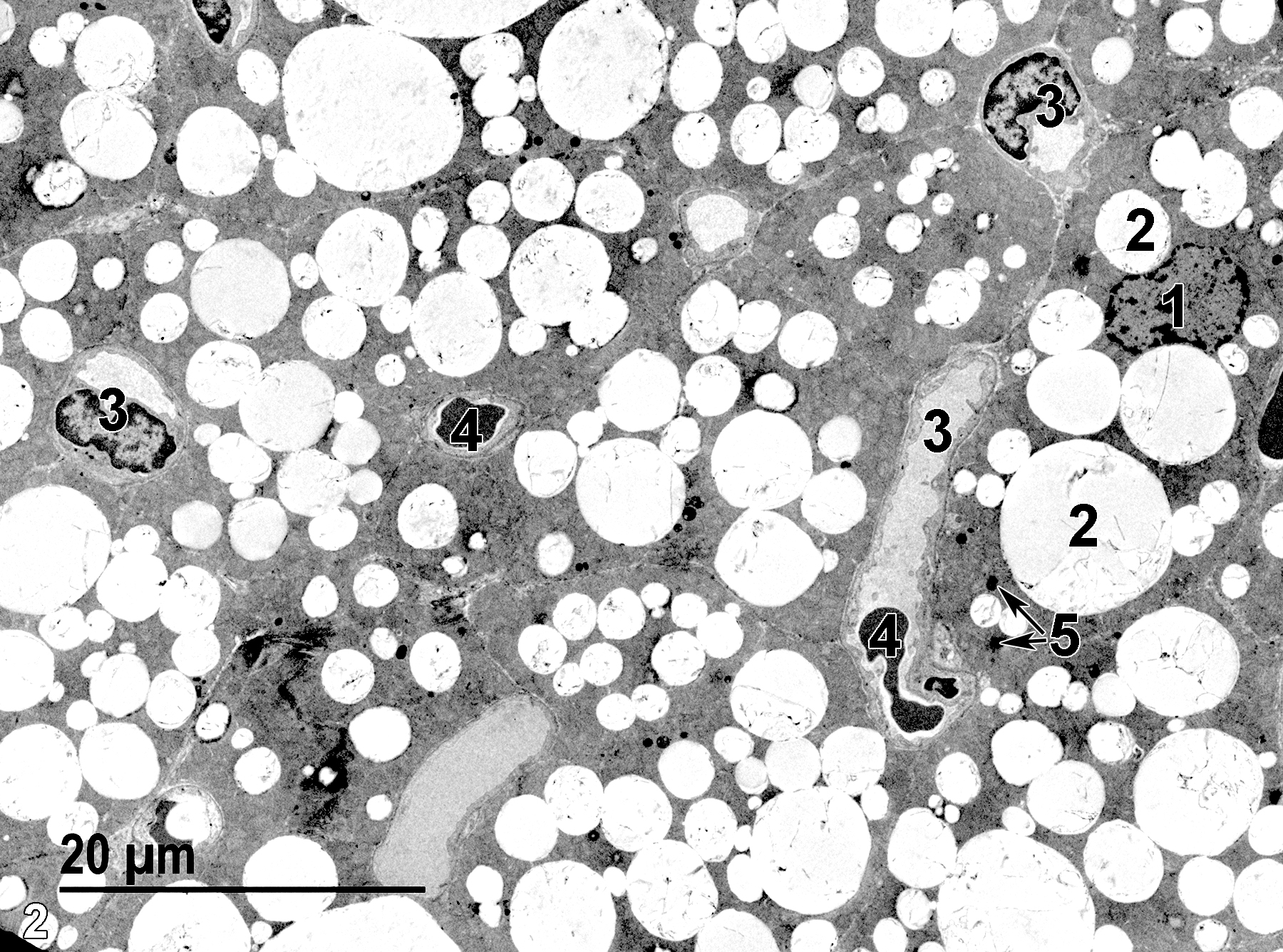
Figure 2. A low magnification ultrastructural view of the parathyroid parenchyma showing a nucleus of a chief cell (1), a large number of variably sized lipid bodies (2) within chief cells, and a number of septal capillaries (3), some containing erythrocytes (4). Low numbers of lysosomes (5) are present in the chief cells. 1900x.
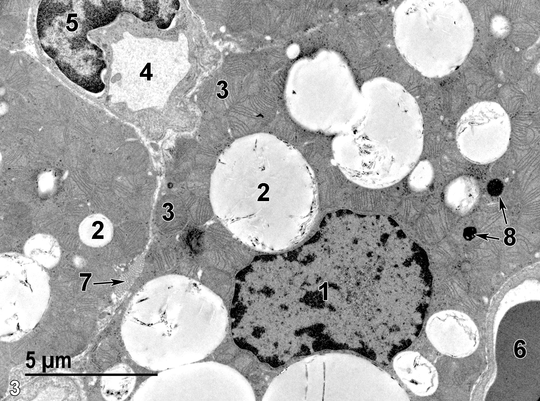
Figure 3. A higher magnification of chief cells showing a single nucleus (1) with marginated heterochromatin, lipid bodies of variable size (2), and numerous mitochondria (3). Endothelial cells, with a single visible nucleus (5) line a capillary lumen (4). Another capillary contains an erythrocyte (6). The capillaries are in the septa, along with collagen bundles (7, arrow). Two lysosomes are present in the cytoplasm of one of the chief cells (8, arrows). 6800x.
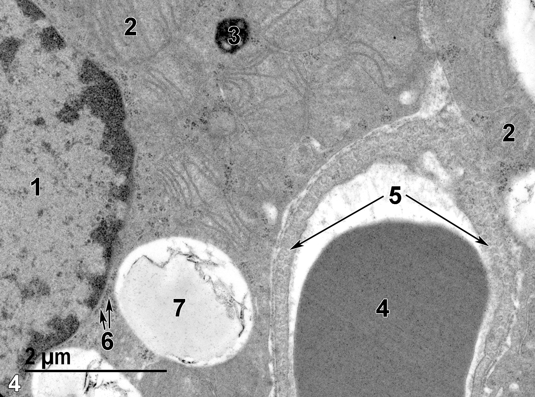
Figure 4. An even higher magnification of chief cells with an adjacent capillary. The left side of the image shows a chief cell nucleus (1). Mitochondria are numerous and show flattened cristae (2). A single lysosome (3) is present. The capillary contains an erythrocyte (4) and the endothelial cells that surround the capillary lumen have characteristic micro-pinocytotic vesicles (5, long arrows). Small accumulations of glycogen are present (6, short arrows), along with a prominent lipid body (7). 18500x.
| Dellmann HD, Eurell J, eds. 1998. Textbook of Veterinary Histology. 5th ed. Philadelphia: Lippincott Williams & Wilkins. |
| Mense MJ, Rosol TJ. 2018. Chapter 34: Parathyroid gland. In Boorman’s Pathology of the Rat (Suttie AW, ed.). 2nd ed. London: Academic Press; 687–693. |
| Rhodin JAG. 1974. Histology: A Text and Atlas. New York: Oxford University Press. |
| Ross MH, Kaye GI, Pawlina W. 2003. Histology: A Text and Atlas. 4th ed. Philadelphia: Lippincott Williams & Wilkins. |
| Weiss L, ed. 1988. Cell and Tissue Biology: A Textbook of Histology. 6th ed. Baltimore: Urban & Schwarzenberg. |
All Images


