

Endocrine System
Thyroid Gland
Narrative
The thyroid is an endocrine gland that secretes thyroxine and triiodothyronine into follicles. The perifollicular capillaries then carry the hormones to their sites of action. C cells (parafollicular cells) occur in small numbers among the follicular cells and are responsible for producing calcitonin. The thyroid gland has a thin capsule of connective tissue. The parenchymal tissue is further subdivided into lobules by septa of connective tissue. The lobules contain follicles that consist of a central cavity surrounded by a cuboidal epithelium that produces the colloid filling the central cavity of the follicle. Each follicle is surrounded by a basal lamina. The epithelial cells of the follicle contain large amounts of rough endoplasmic reticulum and free ribosomes that are involved in the production of colloid.
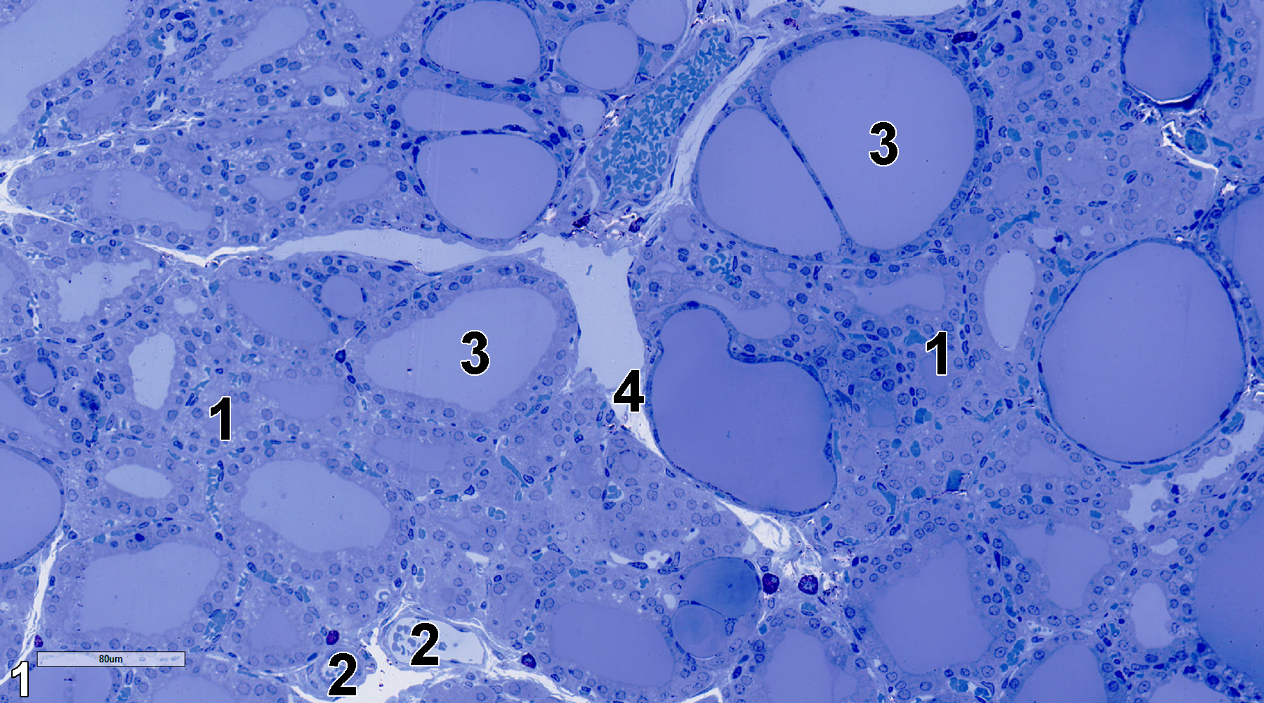
Figure 1. A semithin section (0.5 micrometer thick) of a toluidine blue O-stained area of the thyroid gland. Lobules (1) separated by septa composed of connective tissue (4) have follicles with cuboidal epithelial cells surrounding luminal spaces (3) containing colloid. Capillaries (2) containing erythrocytes. 25x.
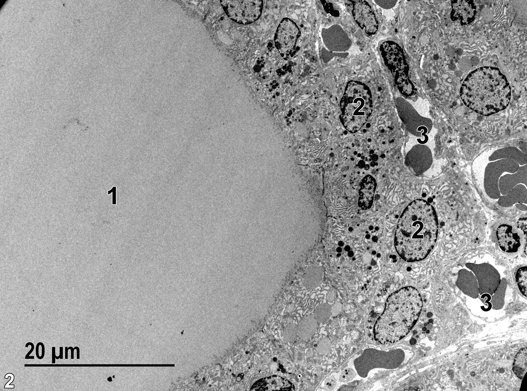
Figure 2. An electron micrograph showing a thyroid follicle (1) with colloid surrounded by a follicular epithelium composed of cuboidal cells with basal nuclei (2). Capillaries (3) containing erythrocytes are located at the base of the epithelial cells. 1900x.
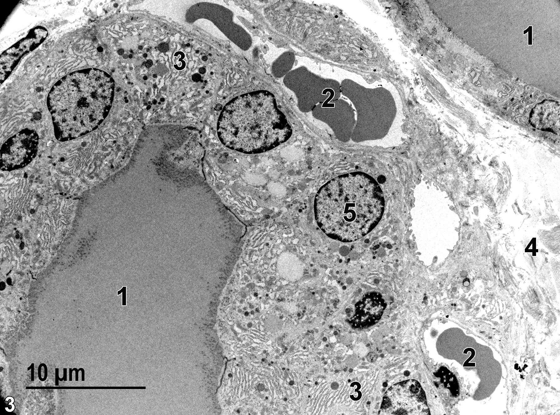
Figure 3. Another view of a follicle containing colloid (1). The follicle has capillaries (2) with erythrocytes located at the base of the epithelial cells. The epithelial cells have spherical nuclei (5) and large patches of rough endoplasmic reticulum (3). A septum of connective tissue (4), made up primarily of collagen, separates adjacent thyroid lobules. 2900x.
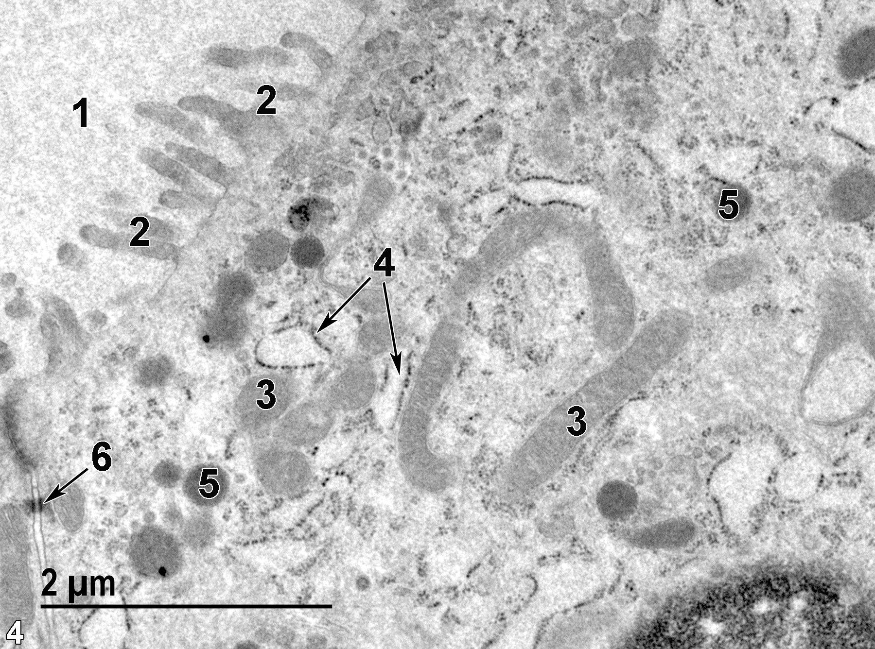
Figure 4. A high magnification view of a follicular epithelial cell. The follicular lumen (1) contains somewhat flocculent colloid. Microvilli (2) protrude into the follicular lumen. Inside the epithelial cell, there are elongated mitochondria (3), as well as rough endoplasmic reticulum profiles (4 arrows) and lysosomes (5). A desmosome (6, arrow) is located at the base of a junctional complex, binding the apical regions of adjacent epithelial cells together. 23000x.
| Dellmann HD, Eurell J, eds. 1998. Textbook of Veterinary Histology. 5th ed. Philadelphia: Lippincott Williams & Wilkins. |
| Rhodin JAG. 1974. Histology: A Text and Atlas. New York: Oxford University Press. |
| Weiss L, ed. 1988. Cell and Tissue Biology: A Textbook of Histology. 6th ed. Baltimore: Urban & Schwarzenberg. |
All Images


