

Immune System
Spleen
Narrative
The spleen is covered with a capsule composed of smooth muscle cells, fibroblasts, reticular fibrils, collagen, and elastic fibers. Trabeculae are extensions of the capsular components into the splenic parenchyma. Reticular tissue that makes up the stroma of the parenchymal tissue is a network of reticular cells that are similar to fibroblasts but have bundles of cytoplasmic filaments and reticular fibrils. The stroma extends throughout the parenchymal tissue. The parenchymal tissue is made up of white pulp, consisting of a compact lymphoid tissue arranged around an arterial system (peri-arteriolar lymphatic sheaths [PALS]) and lymphoid follicles. Reticular fibers separate the marginal zone from the PALS. The marginal zone (composed of more diffuse lymphatic tissue) surrounds the white pulp and merges into the red pulp. The red pulp makes up most of the splenic parenchyma and is composed of a reticular sinus network of venules, arterioles, capillaries, venous sinuses, and splenic cords.
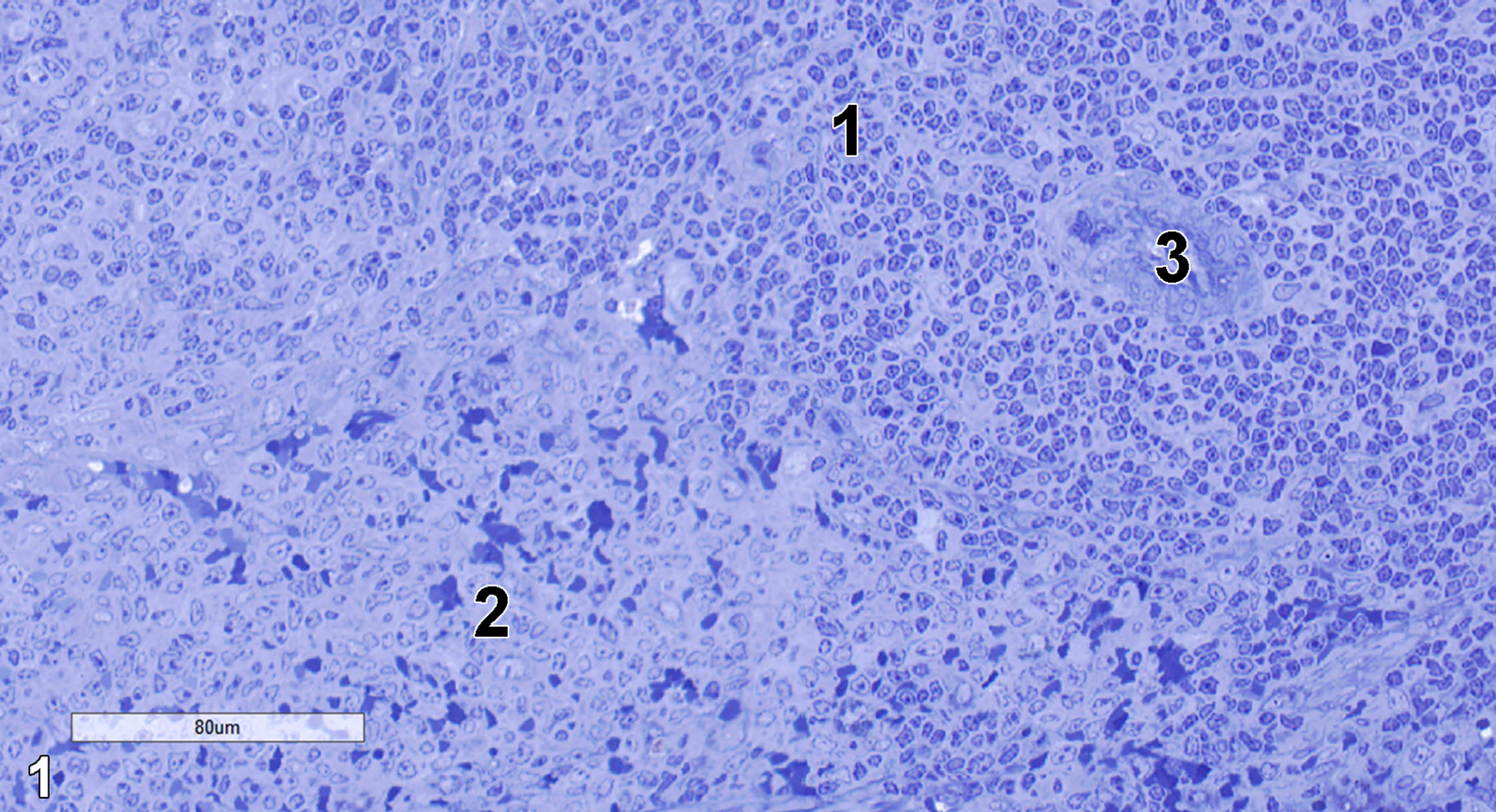
Figure 1. A semithin section (0.5 micron thick) of a toluidine blue O-stained portion of the spleen showing white pulp (1) that consists of a dense lymphocyte population and red pulp (2) with a more diffuse lymphocytic population and numerous erythrocytes. A central arteriole (3) of the white pulp is present. 25x.
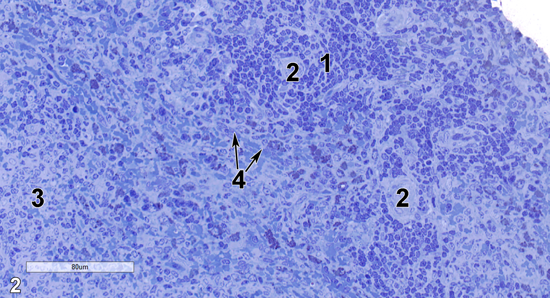
Figure 2. Another semithin section showing white pulp (1), central arterioles (2) of the white pulp, a portion of the marginal zone (3) where the white pulp begins merging with the red pulp, and small stretches of the reticular meshwork (4, arrows). 25x.
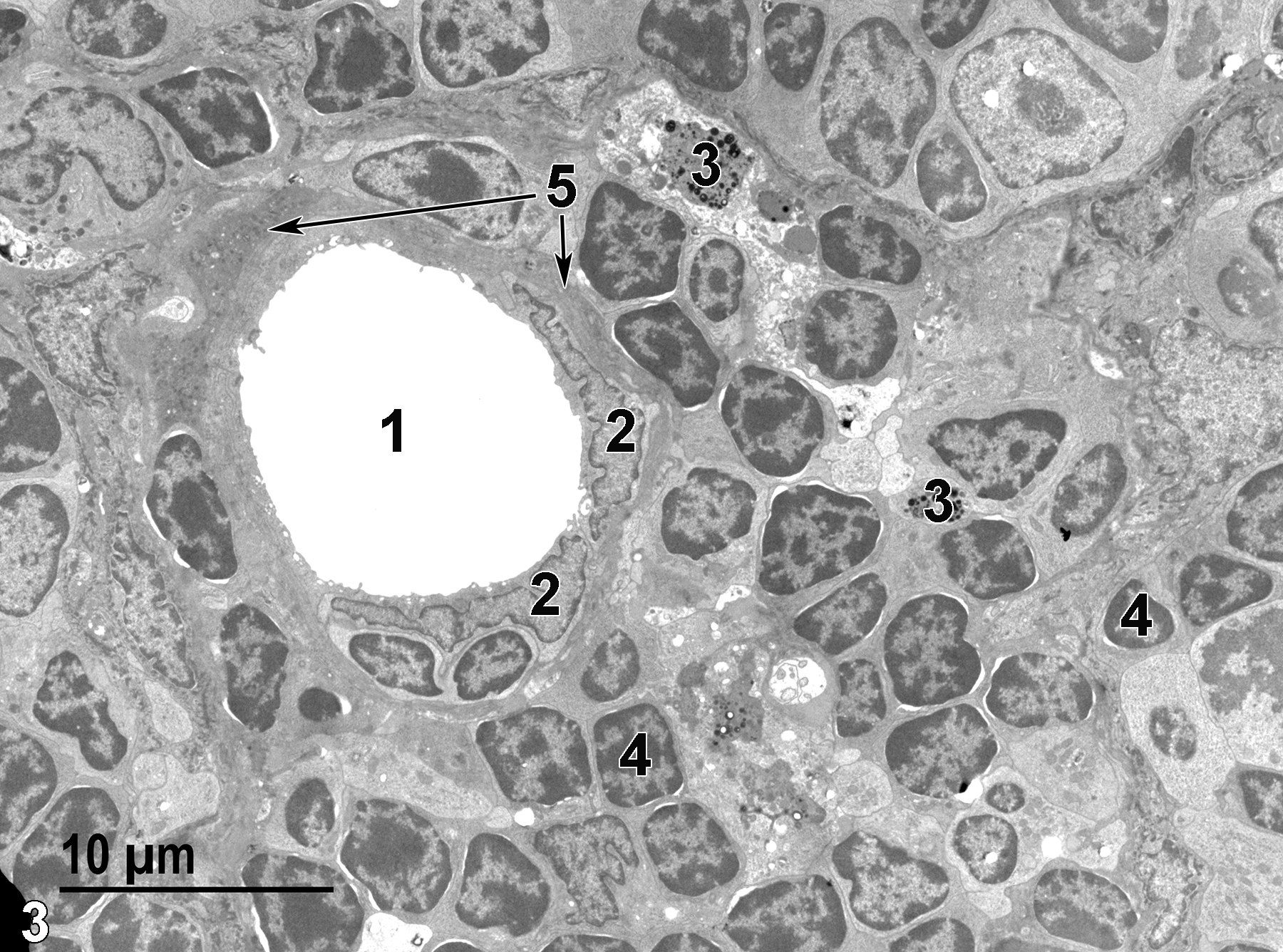
Figure 3. A low magnification ultrastructural image of the white pulp. A central arteriole lumen (1) is surrounded by endothelial cells with flattened nuclei (2), with a thin band of smooth muscle cells (5, arrows) surrounding the endothelial cells. Macrophages (3) containing lysosomes are present among the lymphocytes (4). 2900x.
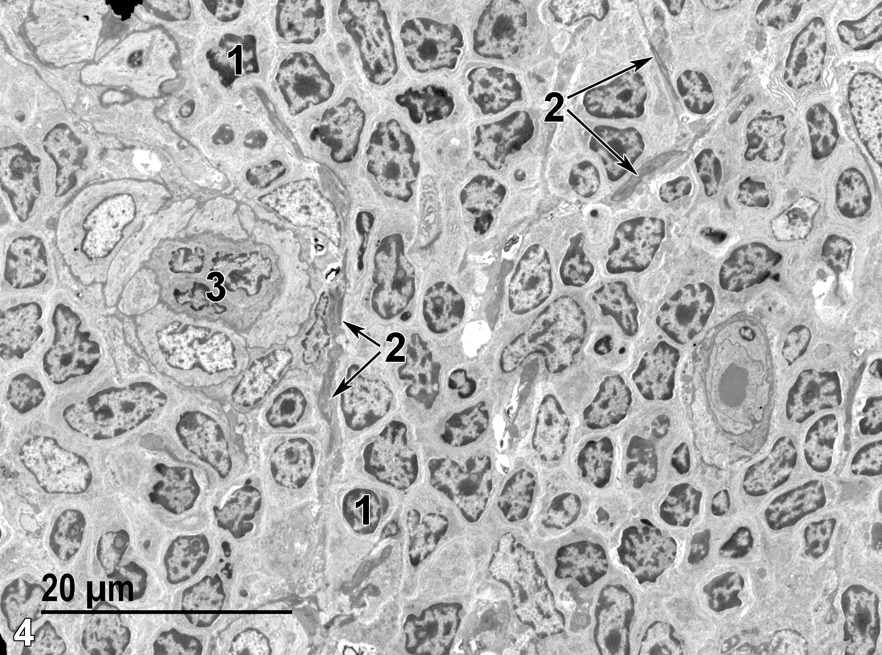
Figure 4. A low magnification view of the splenic white pulp. There are numerous densely packed lymphocytes (1), electron-dense portions of the reticular meshwork (2, arrows), and a central arteriole (3). 1900x.
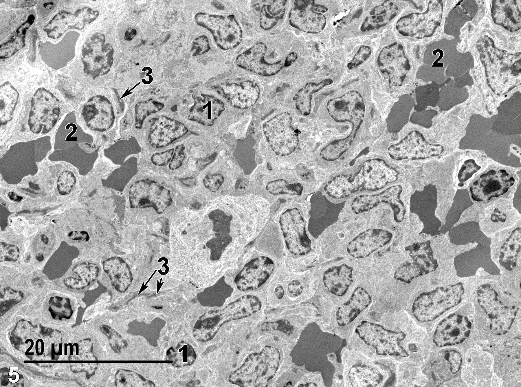
Figure 5. A portion of the red pulp. Lymphocytes (1) are present, along with a number of erythrocytes (2). Small fragments of the reticular meshwork (3, arrows) are present. 1900x.
| Dellmann HD, Eurell J, eds. 1998. Textbook of Veterinary Histology. 5th ed. Philadelphia: Lippincott Williams & Wilkins. |
| Rebelatto MC. 2018. Chapter 24: Spleen, lymph nodes, and thymus. In Boorman’s Pathology of the Rat (Suttie AW, ed.). 2nd ed. London: Academic Press, 469-491. |
| Rhodin JAG. 1974. Histology: A Text and Atlas. New York: Oxford University Press. |
| Ross MH, Kaye GI, Pawlina W. 2003. Histology: A Text and Atlas. 4th ed. Philadelphia: Lippincott Williams & Wilkins. |
| Weiss L, ed. 1988. Cell and Tissue Biology: A Textbook of Histology. 6th ed. Baltimore: Urban & Schwarzenberg. |
All Images


