

Respiratory System
Trachea
Narrative
The trachea is composed of a mucosal layer with a pseudostratified epithelium and a fibrous lamina propria. The mucosal layer is populated primarily with ciliated columnar cells with long microvilli and mucous cells with short microvilli and mucous granules. Rarer brush cells are present, which are in contact with afferent nerve endings and have blunt microvilli. There are also rare small granule cells with granules that exhibit dense granule cores. The granule cells have thin cytoplasmic processes that sometimes extend to the trachea lumen. Basal cells are located in a row near the basal lamina of the mucosal layer. Below the mucosal layer is the lamina propria, which is composed of loose connective tissue with lymphocytes, fibroblasts, eosinophils, mast cells, and plasma cells. The submucosa below the lamina propria has slightly denser connective tissue than that found in the lamina propria and contains vessels and lymphatics, as well as submucosal acinar glands with associated serous demilunes. Under the submucosa is a layer of cartilage, which is underlain by adventitious tissue made up primarily of collagen that binds the trachea to adjacent tissues, such as the esophagus.
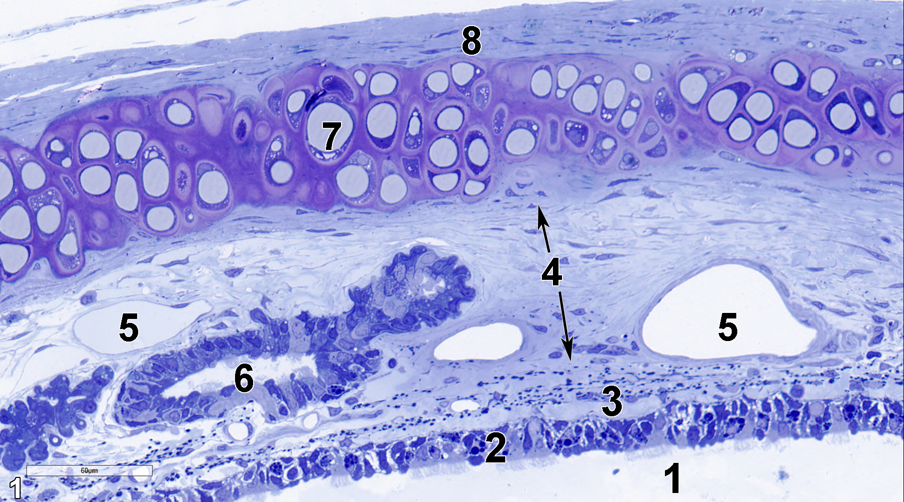
Figure 1. A semithin section (0.5 micrometer thick) of a toluidine blue O-stained portion of the trachea. The tracheal lumen (1) is lined with a pseudostratified epithelium (2) that consists of ciliated columnar cells, mucous cells, and basal cells. The lamina propria (3) is composed primarily of collagen and fibroblasts. Below the lamina propria is the submucosal layer (4, arrows), which is also composed primarily of collagen and fibroblasts, along with vessels (5), and submucosal glands (6). The next layer is cartilage (7), with the fibrous adventitia (8) below. 40x.
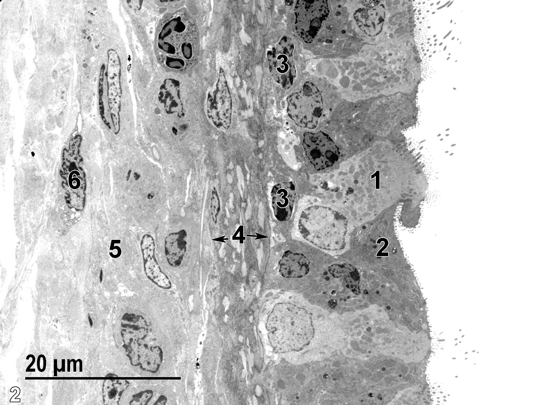
Figure 2. A low magnification electron micrograph of the tracheal mucosa and submucosa, with the tracheal lumen present on the right of the image. The pseudostratified epithelium shown is made up of ciliated cells (1), non-ciliated mucous cells (2), and basal cells (3). At the base of the mucosal layer is the lamina propria (4, arrows), which is composed primarily of bundles of collagen fibrils and fibroblasts. The submucosal layer is also composed of a collagenous matrix (5) with its associated fibroblasts (6). 1900x.
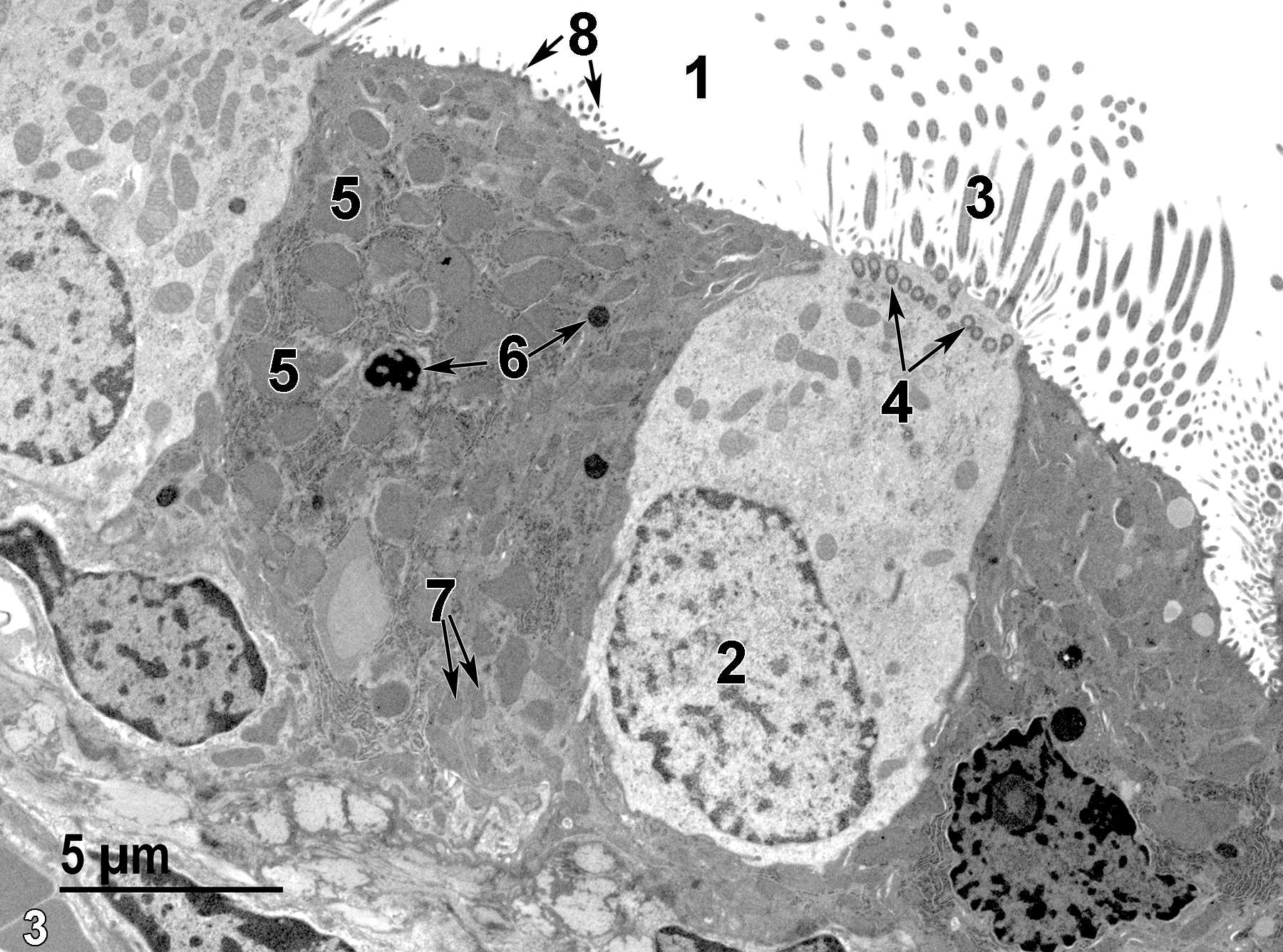
Figure 3. A higher magnification view of the epithelial surface. The tracheal lumen (1) is lined with the two major types of epithelial cells. A ciliated epithelial cell nucleus (2) is shown, as well as cilia (3) and apical basal bodies (4, arrows). The mucous cell shown contains numerous pleomorphic mucous granules (5), some lysosomes (6, arrows), and mitochondria (7, arrows). Short microvilli (8, arrows) are visible at the surface of the mucous cell. 4800x.
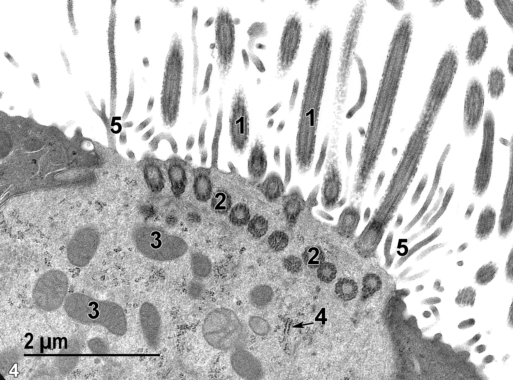
Figure 4. A high magnification view of the apical region of a ciliated epithelial cell that shows cilia (1), basal bodies (2) of the cilia, mitochondria (3), a segment of rough endoplasmic reticulum (4, arrow), and long and sometimes branched microvilli (5). Note that they are much longer than the microvilli on the surface of the two adjacent mucous cells with darker cytoplasm. 18500x.
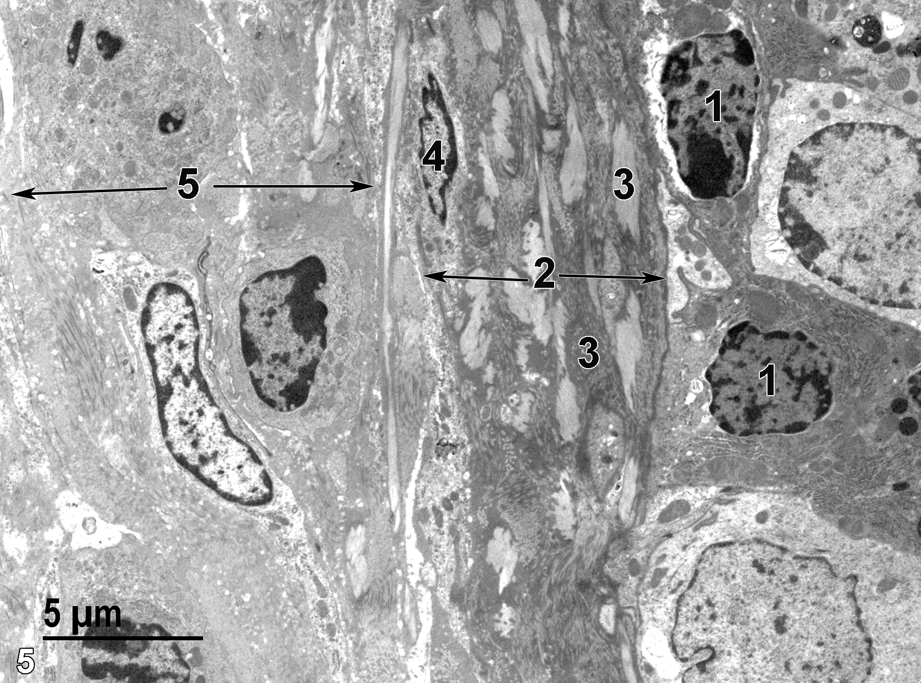
Figure 5. A view of the lamina propria (2, arrows) and submucosa (5, arrows) below the mucosal layer. Nuclei of two basal cells (1) in the epithelial cell layer are shown. Below them are bundles of collagen fibrils (3) and a fibroblast (4) in the lamina propria. The submucosal layer is also composed of a collagenous matrix with embedded fibroblasts. 4800x.
| Rhodin JAG. 1974. Histology: A Text and Atlas. New York: Oxford University Press. |
| Ross MH, Kaye GI, Pawlina W. 2003. Histology: A Text and Atlas. 4th ed. Philadelphia: Lippincott Williams & Wilkins. |
| Weiss L, ed. 1988. Cell and Tissue Biology: A Textbook of Histology. 6th ed. Baltimore: Urban & Schwarzenberg. |
All Images


