

Reproductive System, Female
Oviduct
Narrative
The two oviducts start with the infundibulum, which has a wide opening toward the peritoneal cavity with fimbriae. This is connected to the ampulla, which narrows toward the uterus to form the isthmus attached to the uterus. The surface of the oviduct is lined with a columnar epithelium that contains ciliated cells (approximately 50% of the cell population) and secretory cells that produce mucoprotein nutritive materials. Below the epithelial layer is a muscularis layer that consists of smooth muscle cells and connective tissue. The serosa of the oviduct is covered with a layer of squamous epithelial cells and connective tissue with fibroblasts.
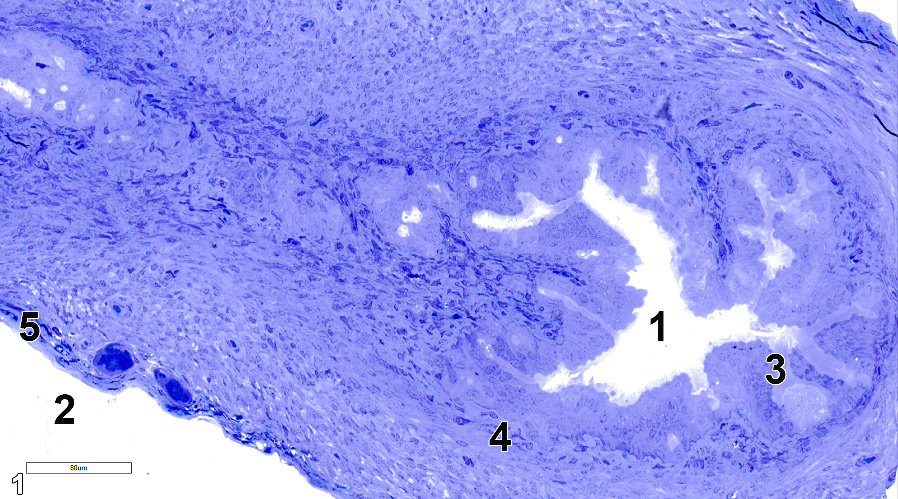
Figure 1. A semithin section (0.5 micrometer thick) of a toluidine blue O-stained section of the ampulla of the oviduct. The oviduct lumen (1) is lined with a columnar epithelium (3) that consists of ciliated cells and secretory cells with microvilli. Below the epithelial layer is the muscularis (4) that consists of smooth muscle cells and connective tissue. The serosa (5) has a thin squamous epithelium and associated connective tissue. The peritoneal cavity (2) is at the lower left corner of the image. 25x.
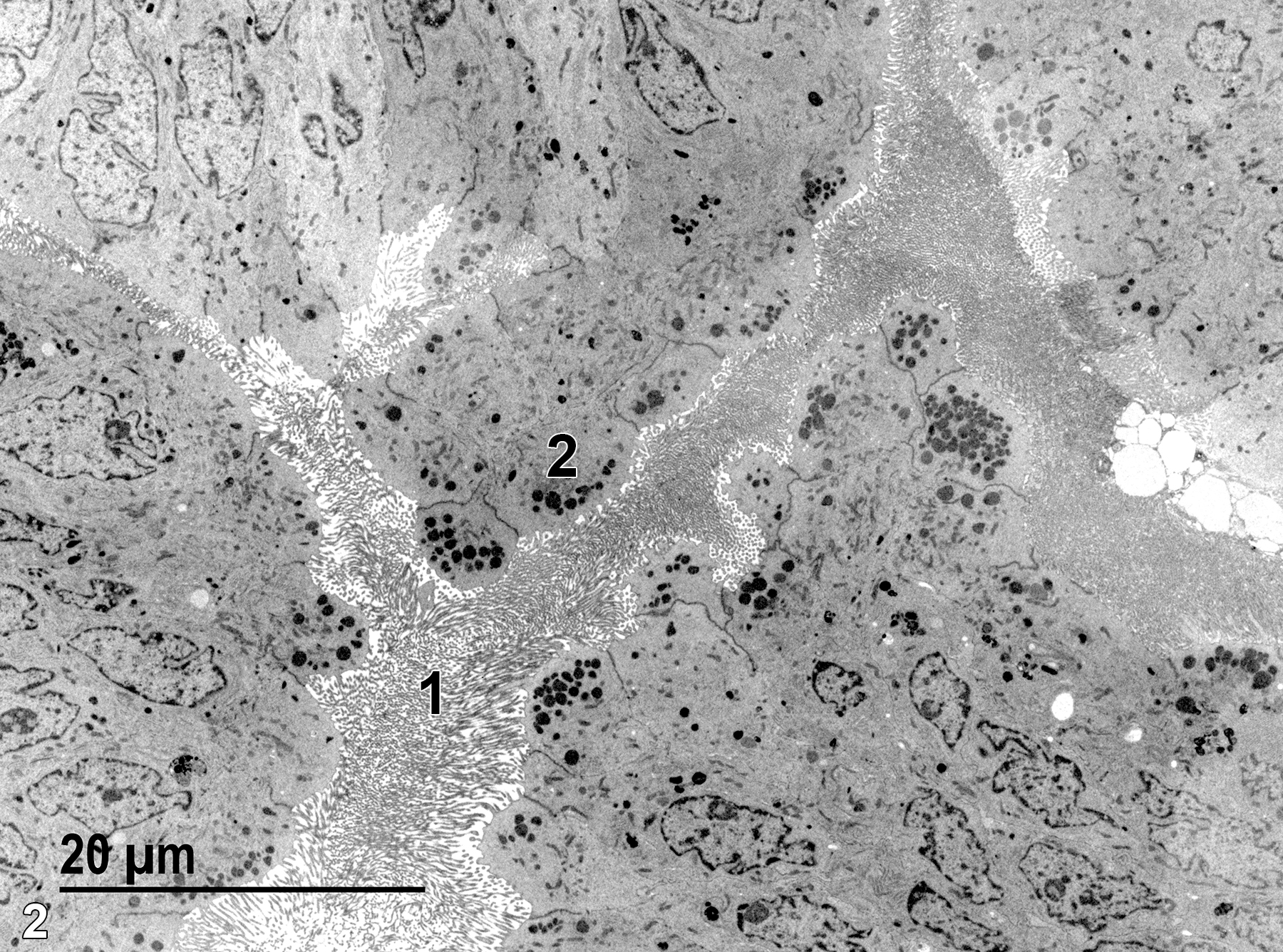
Figure 2. A low magnification electron micrograph showing the ampulla lumen (1) filled with microvilli and an epithelial cell (2) with secretory granules. 1900x.
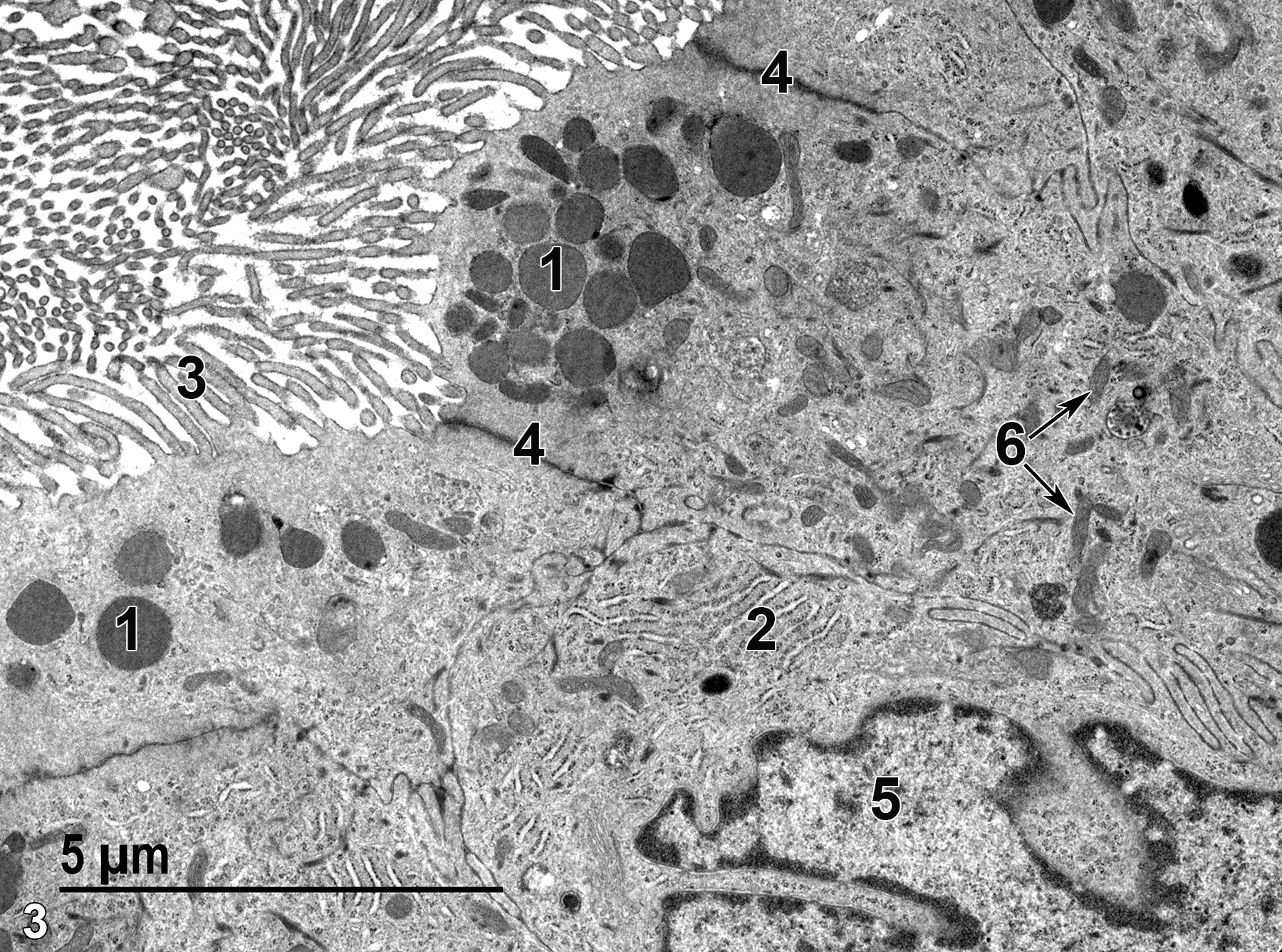
Figure 3. A higher magnification view of epithelial cells that show secretory granules (1), accumulations of rough endoplasmic reticulum (2), microvilli (3) lining the ampulla lumen, junctional complexes (4) between adjacent epithelial cells, a single nucleus (5), and mitochondria (6, arrows). 9300x.
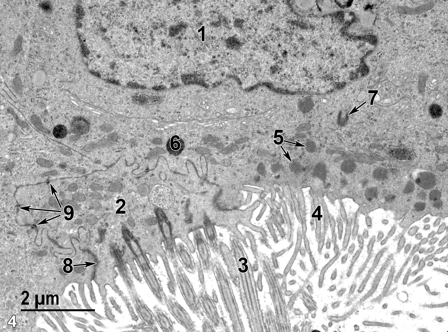
Figure 4. A view of the ampulla epithelial surface, lined with both a ciliated cell (2) with cilia (3) at the surface, next to two secretory cells that only have microvilli (4) at their surfaces. Within a secretory cell is a secretory granule (6), mitochondria (5, arrows), a single centriole (7, arrow) and a nucleus (1). A junctional complex (8, arrow) binds the apical borders of two adjacent cells. Desmosomes (9) also hold the borders of adjacent cells together (arrows). 11000x.
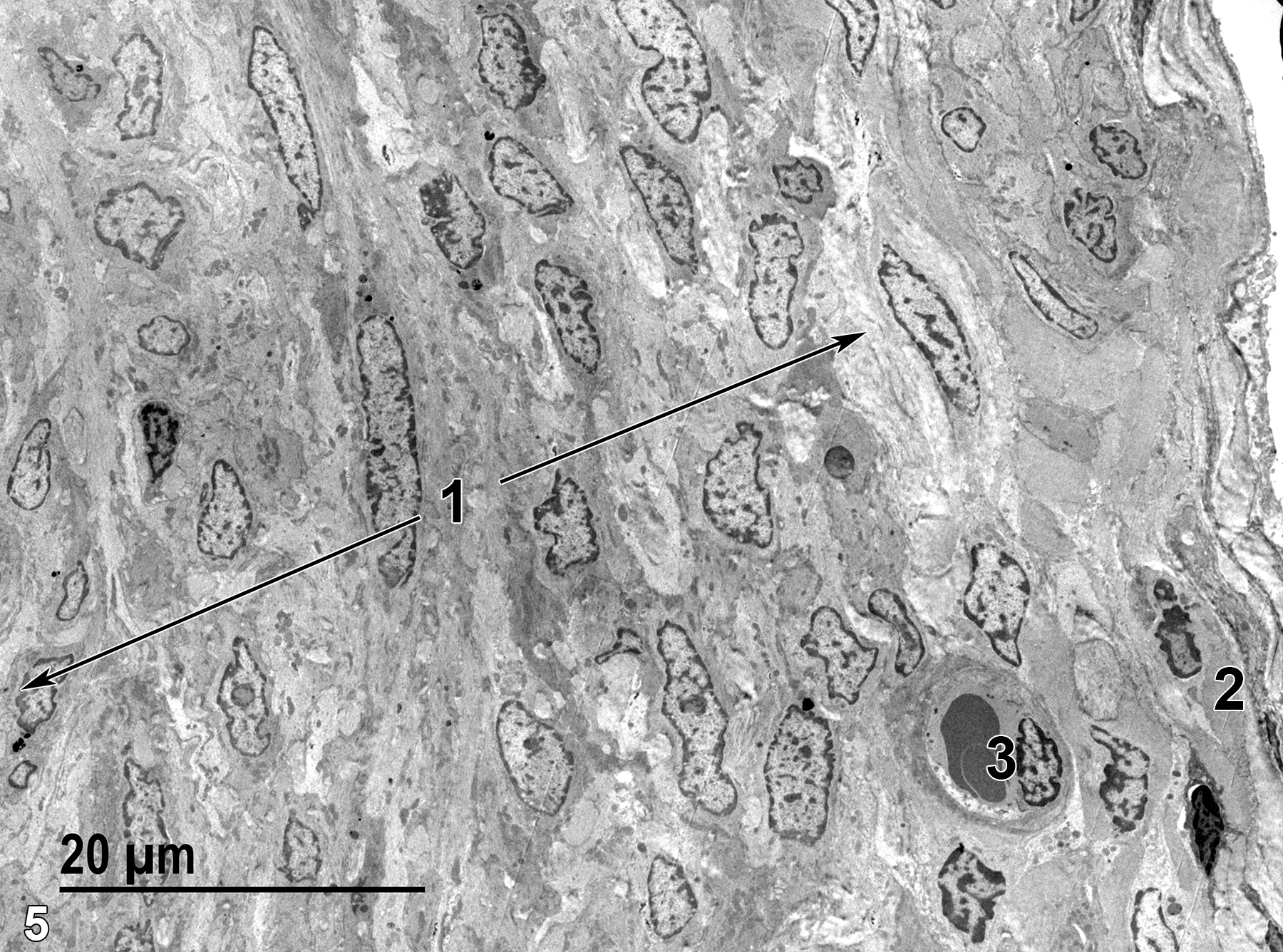
Figure 5. The muscularis layer, which consists of smooth muscle cells and a collagenous matrix (1, arrows) with a thin band of serosa (2) at the far right of the image. One capillary (3) with a single endothelial nucleus and two erythrocytes is present. 1900x.
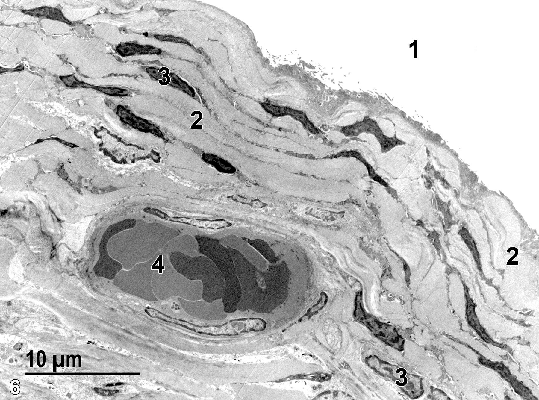
Figure 6. Another view of the serosal surface of the ampulla, showing the peritoneal cavity (1), the serosa, which consists of a thin squamous epithelium, and adjacent connective tissue. Fibroblast nuclei (3) are evident along with bundles of collagen (2). A small venule (4) that contains erythrocytes is present. 2900x.
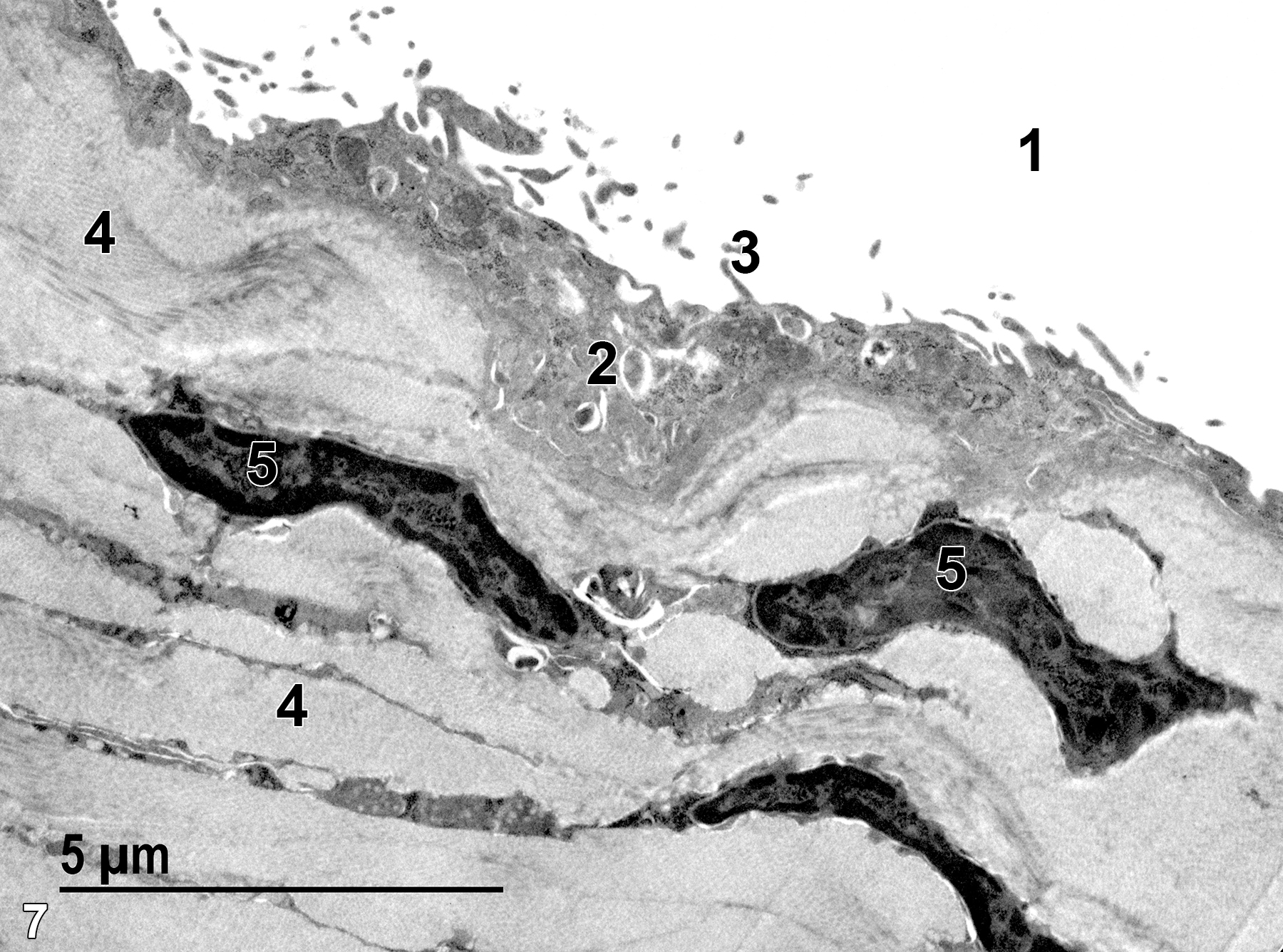
Figure 7. A higher magnification image of Figure 6. The peritoneal cavity (1) lies just outside of the thin squamous epithelium (2), which has short microvilli (3) on the surface of the epithelial cells. Bundles of collagen fibrils (4) with embedded fibroblasts (5) make up the serosal matrix. 9300x.
| Boorman GA, Eustis SL, Elwell MR, Montgomery CA, Jr., MacKenzie WF, eds. 1990. Pathology of the Fischer Rat: Reference and Atlas. New York: Academic Press. |
| Dellmann HD, Eurell J, eds. 1998. Textbook of Veterinary Histology. 5th ed. Philadelphia: Lippincott Williams & Wilkins. |
| Rhodin JAG. 1974. Histology: A Text and Atlas. New York: Oxford University Press. |
| Weiss L, ed. 1988. Cell and Tissue Biology: A Textbook of Histology. 6th ed. Baltimore: Urban & Schwarzenberg. |
All Images


