

Musculoskeletal System
Muscle
Narrative
Skeletal and cardiac muscles (striated muscles) are characterized by the presence of actin and myosin filaments organized into sarcomeres, whereas smooth muscle cells lack this organization. Individual striated muscles are surrounded by a dense connective tissue (epimysium). The muscles contain groups of myocytes (fascicles). Each fascicle is surrounded by connective tissue (perimysium). Each myocyte within the fascicles is surrounded by a relatively thin layer of connective tissue called the endomysium, and a basal lamina is located just outside the plasma membrane (sarcolemma).
Smooth muscle cells have both actin and myosin filaments, but these are not organized into sarcomeres. Each smooth muscle cell is surrounded by a basal lamina, except where desmosomes are formed between adjacent cells.
Cardiac muscle has individual myocytes connected by intercalated disks made up of junctional complexes that are specialized attachment sites between adjacent cells. Most of the intercalated disk consists of fascia adherens, which are similar to the zonula adherens of epithelial cells. The actin filaments of the terminal sarcomeres are attached to the sarcolemma here. Macula adherens (desmosomes) are also present, often at the periphery of the fascia adherens. Finally, gap junctions are found at the lateral surface of the intercalated disks, running primarily down the longitudinal axis of the myocyte. Muscle fibers of various lengths result because of cylindrical myocytes attached end to end. Branched muscle fibers can result from one cell joining two or more cells by the intercalated disks. Each myocyte contains a central nucleus (unlike the multiple marginal nuclei characteristic of skeletal muscle).
At the Z lines, T tubules are found, forming a dyad, rather than the triad characteristic of skeletal muscle. Branching sarcoplasmic reticulum elements are located on one side of the T tubules. The structure of the sarcomeres is the same as for skeletal muscle.
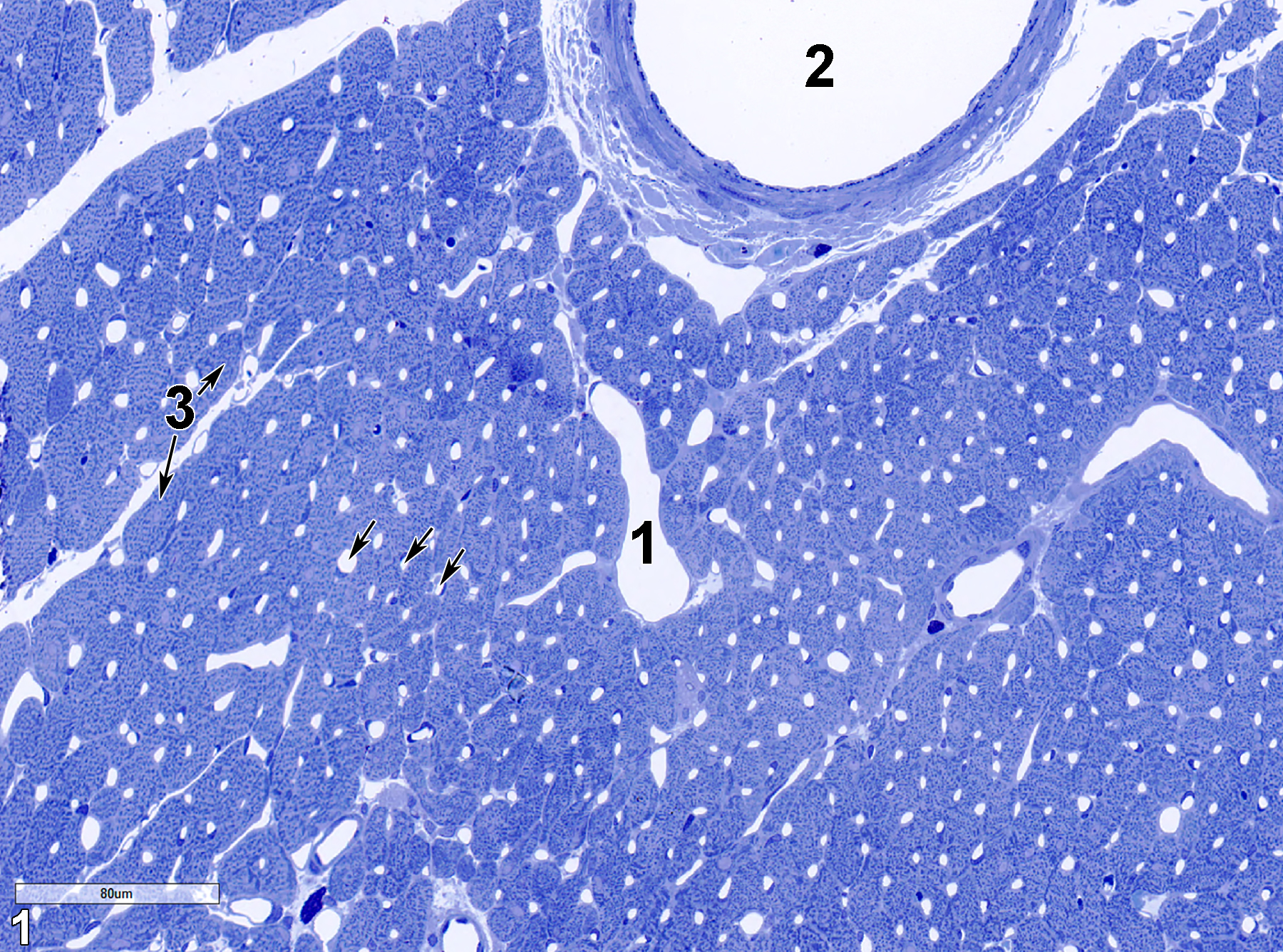
Figure 1. A toluidine blue O-stained section (0.5 micrometer thick) of cardiac muscle. The lumen of a vein (1) with a single endothelium cell layer lining contrasts with the lumen of an artery (2), again lined with a single endothelial cell layer, but then subtended with smooth muscle cells and connective tissue. Numerous small capillaries lined with a single endothelial cell layer are visible (arrows). Cardiac myocytes (3, double arrows) are seen in cross section. 25x.
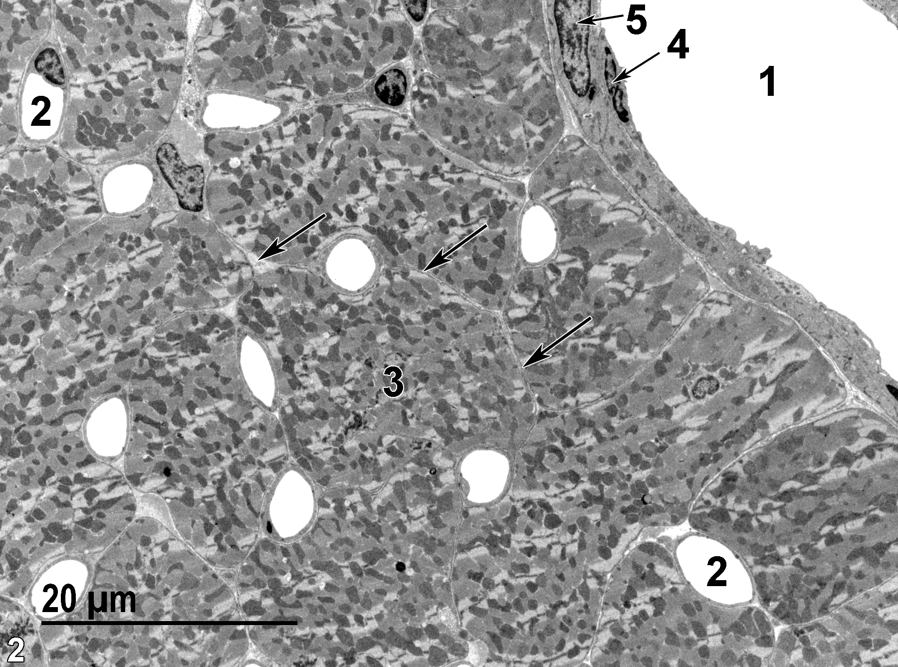
Figure 2. An ultrastructural image of cardiac muscle with a vein in the upper right (1) and a number of capillaries (2) seen in cross section. Myocyte cell (3) boundaries are defined by the endomysial space between them (arrows). Note the elongated central nucleus (5) of a smooth muscle cell and the elongated nucleus of an endothelial cell (4). 1900x.
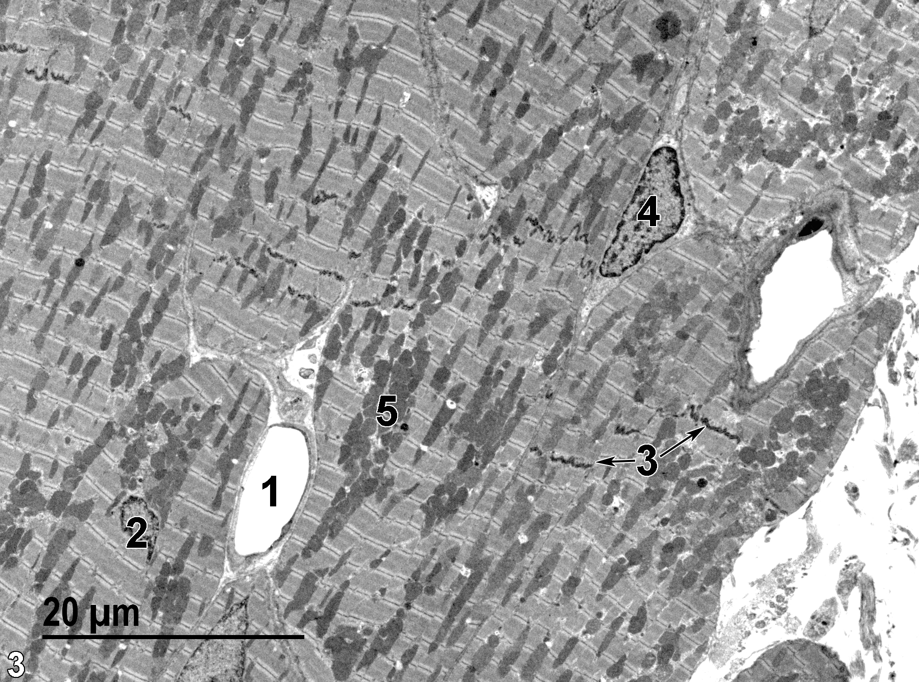
Figure 3. A low magnification view of a longitudinal section of cardiac muscle showing a capillary lumen (1), a centrally located myocyte nucleus (2), intercalated disks (3, double arrows), the nucleus of a connective tissue cell (4), and mitochondria (5). 1900x.
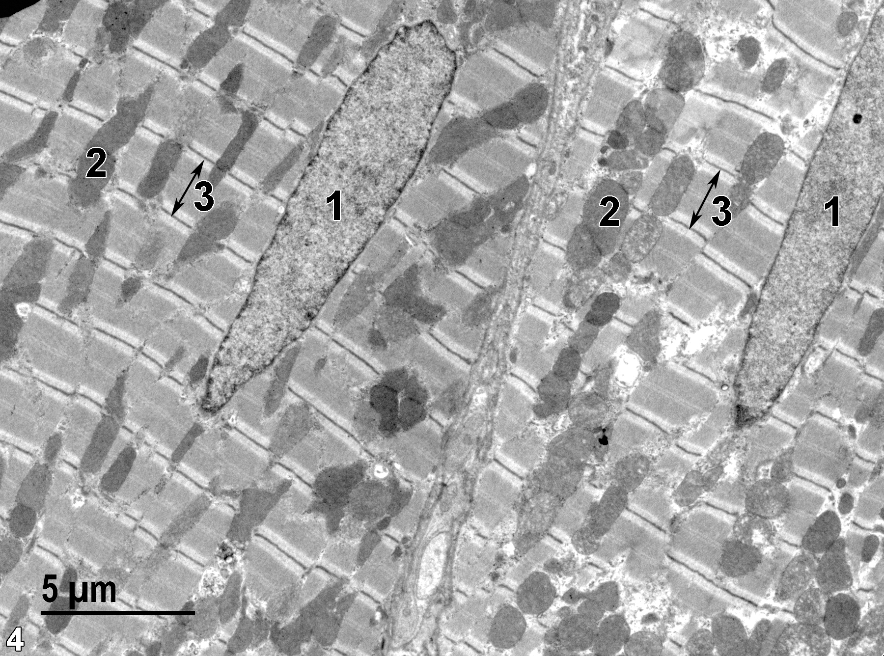
Figure 4. A longitudinal section of cardiac muscle showing two myocytes with primarily centrally located elongated nuclei (1), numerous mitochondria (2), and well-defined sarcomeres (3) delimited by Z lines at either end (double-headed arrows). 4800x.
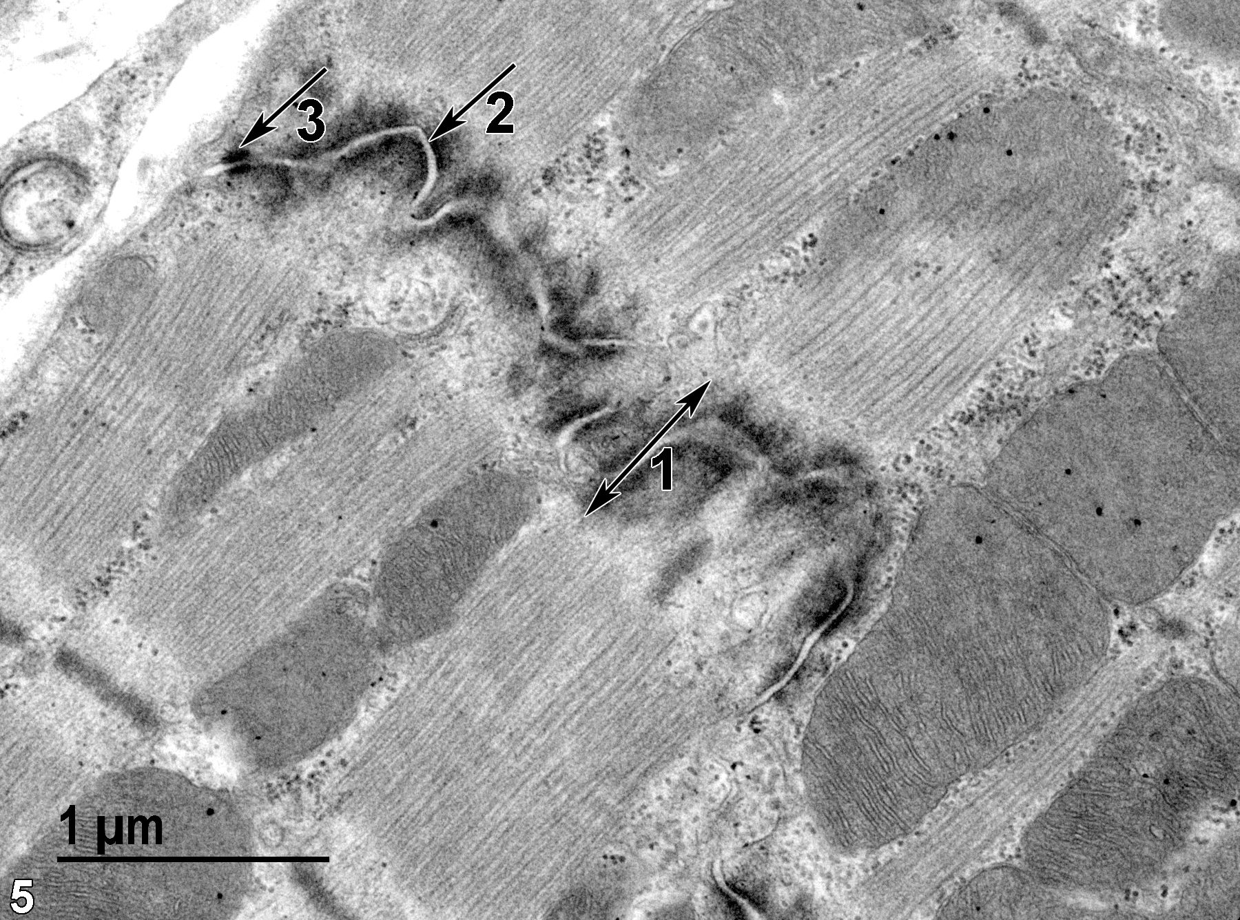
Figure 5. An intercalated disk (1, double-headed arrow) is primarily made up of fascia adherens or intermediate junctions (2, arrow), with macula adherens or desmosomes found primarily at the edges of the intercalated disk (3, arrow). 30000x.
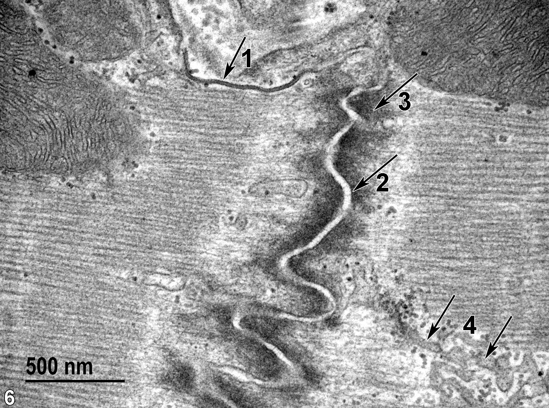
Figure 6. A higher magnification of an intercalated disk clearly showing a nexus or gap junction (1) running down the edge of the longitudinal axis of the sarcomere (arrow). As in the previous image, most of the intercalated disk is composed of fascia adherens (2, arrow) with associated webs of fine cytoplasmic filaments (3, arrow). Some of the sarcoplasmic reticulum can be seen (4, arrows). 49000x.
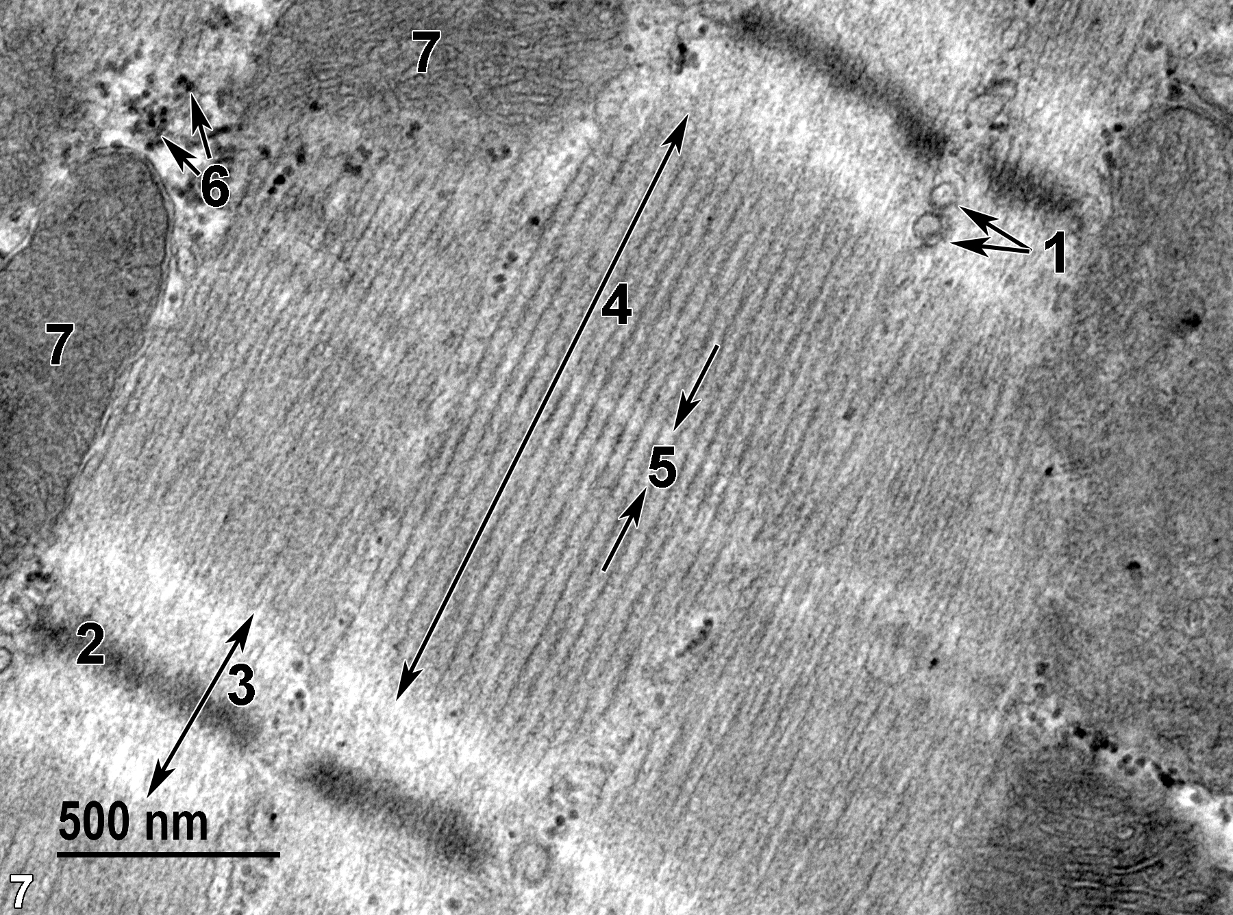
Figure 7. Two T tubules (1, double arrows) characteristic of cardiac muscle that are located near the Z line. A Z line (2) is found within the I band (3, double-headed arrow), and the M band (5, double arrow) located within the A band (4, long double-headed arrow). Glycogen (6, double arrows) is located near two mitochondria (7). 49000x.
Skeletal muscle has individual myocytes surrounded by the endomysium. The myocytes are organized into fascicles surrounded by the perimysium. Myofibrils organized into sarcomeres are composed of thick (myosin II) and thin (actin) myofilaments. These filaments overlap in the A band, whereas only thin filaments are found in the I band. Well-developed T tubules are found at the A-I junction. These consist of a triad with terminal cisternae. There are three T tubules (triads) per sarcomere. Each muscle fiber is a single myocyte containing multiple peripheral nuclei. Satellite cells that serve as progenitors to myocytes are found outside the myocyte sarcolemma but beneath the myocyte basal lamina.
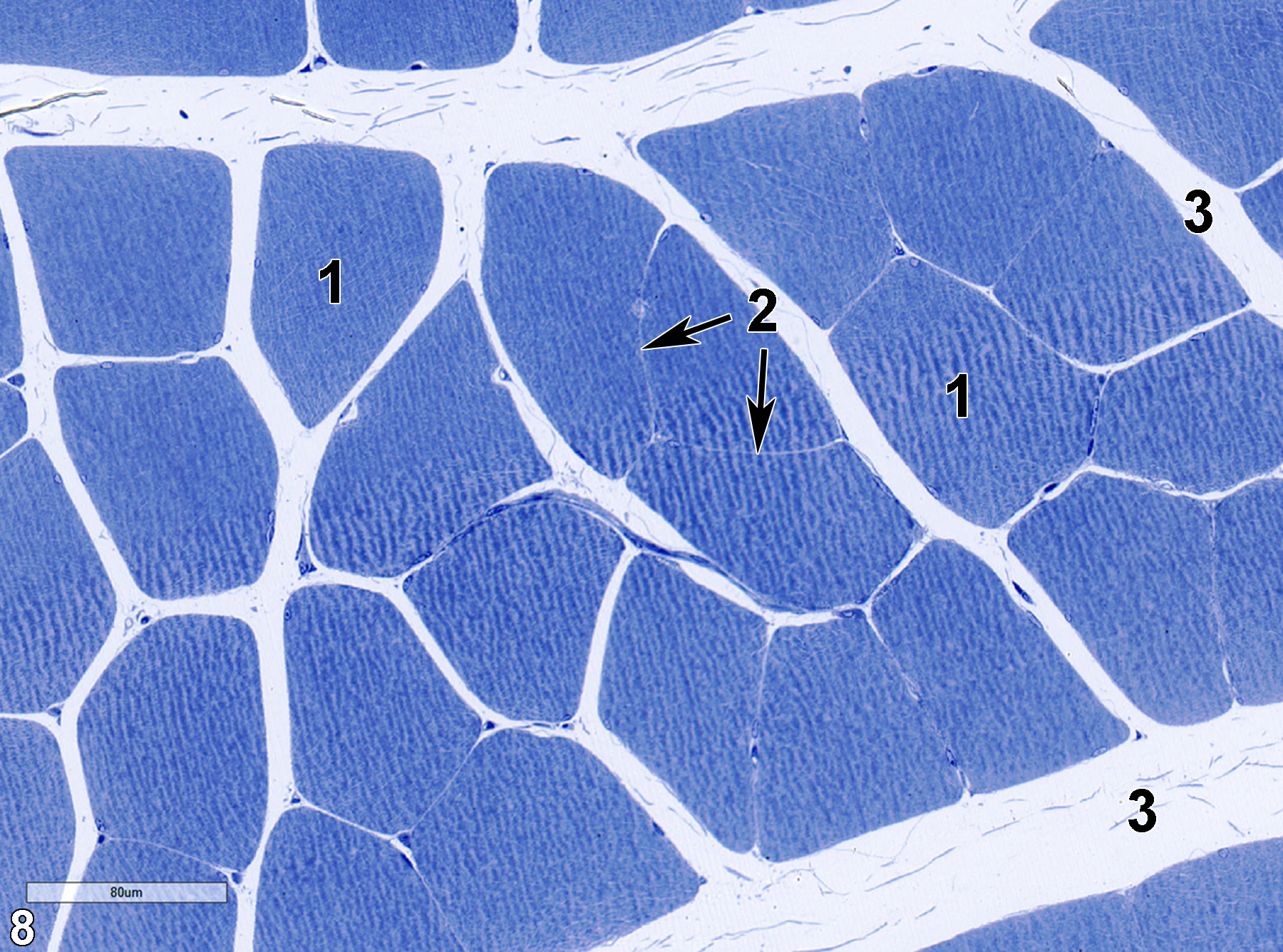
Figure 8. A toluidine blue O-stained semithin cross section of skeletal muscle showing myocytes (1) surrounded by an endomysium (2, double arrows) that is not resolvable at this magnification. The myocytes are grouped into fascicles surrounded by a perimysium (3). 25x.
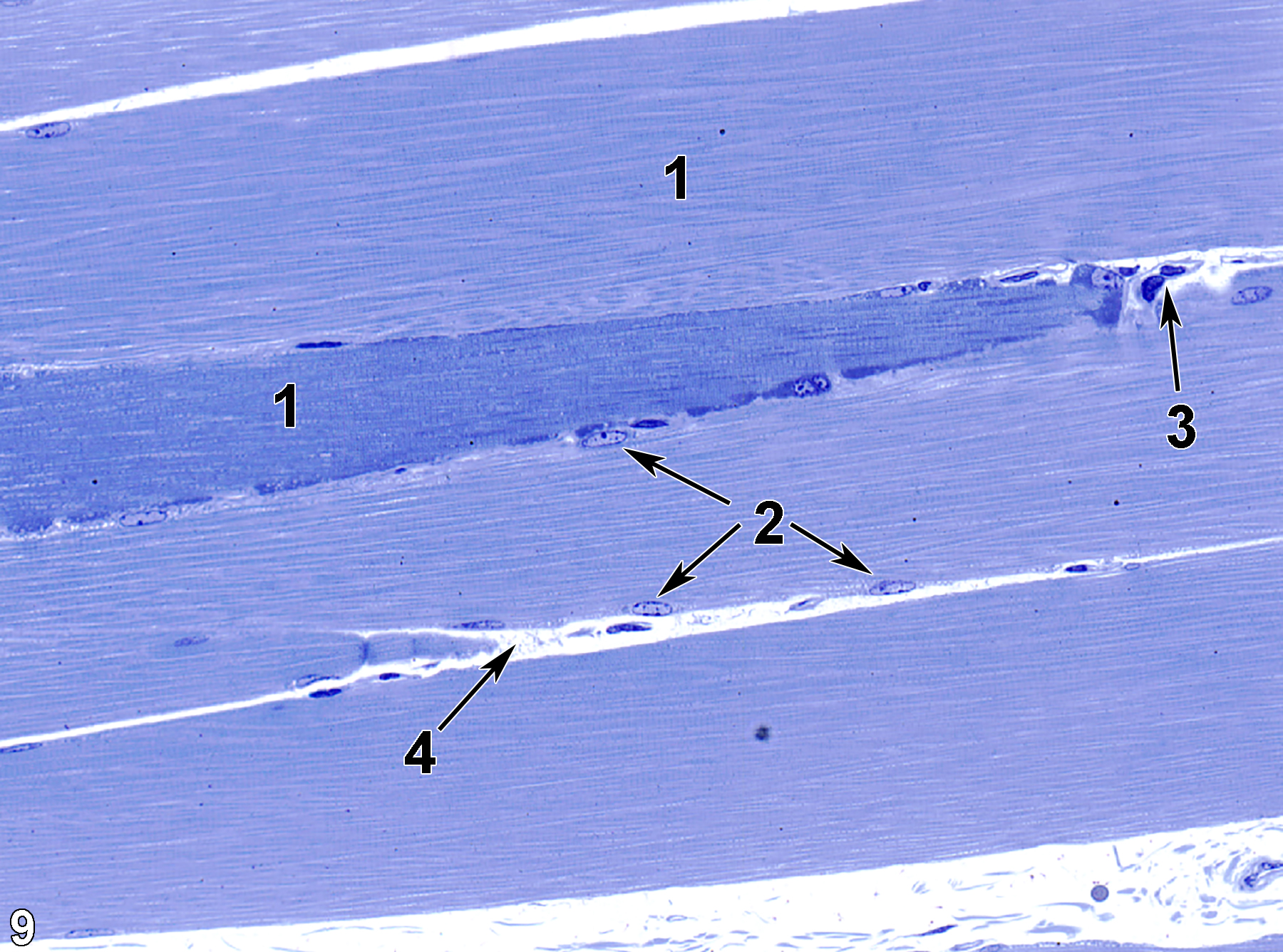
Figure 9. A toluidine blue O-stained semithin longitudinal section of skeletal muscle fiber (1), with multiple peripheral elongate nuclei (2, triple arrows). A capillary (3, arrow) is labeled, as well as the endomysium (4, arrow) between two adjacent myocytes. 25x.
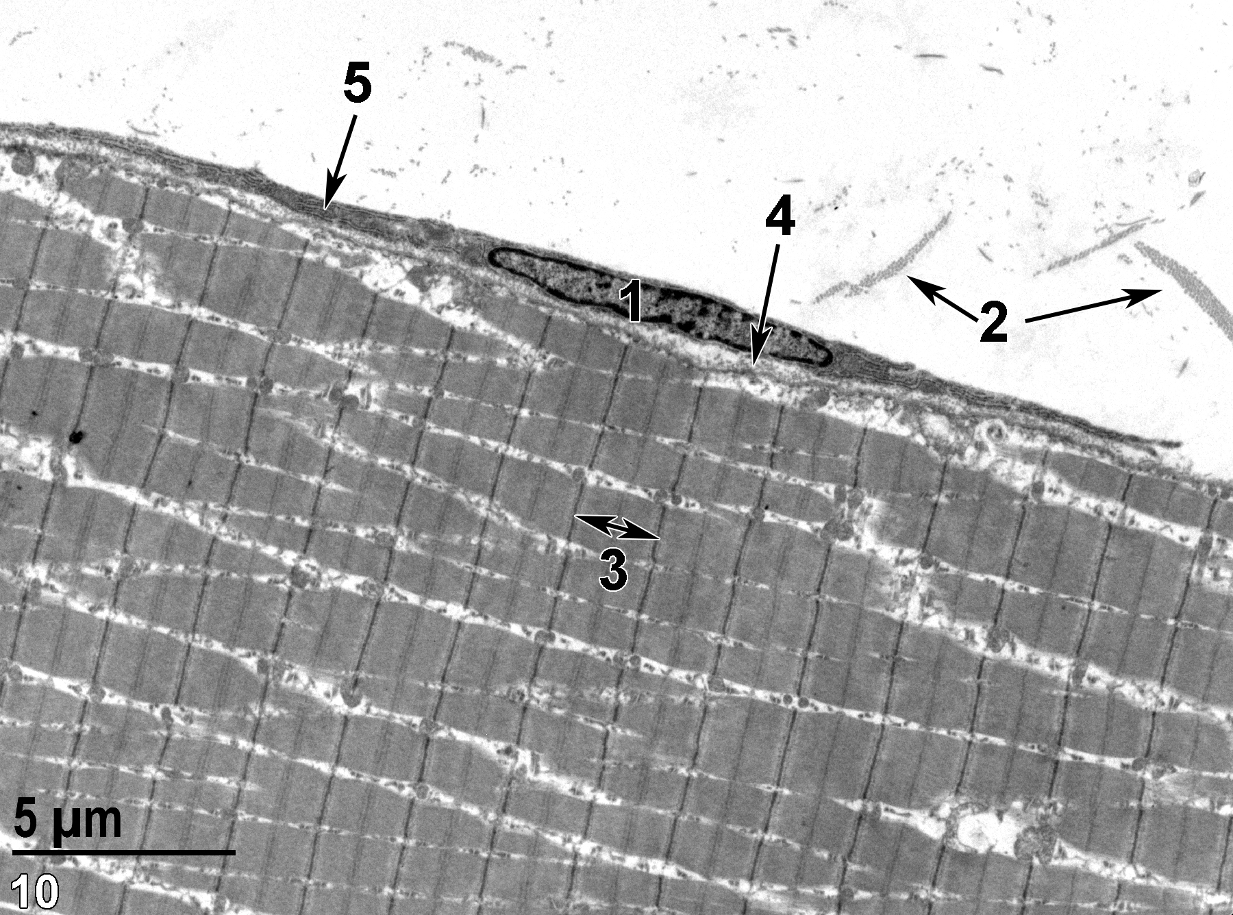
Figure 10. An ultrastructural image of a longitudinal section of skeletal muscle cell with a fibroblast (1) outside the sarcolemma. Collagen is located in the perimysium (2, double arrows) and also in the endomysium (4, arrow) beneath the fibroblast. The fibroblast contains an abundant rough endoplasmic reticulum (5, arrow). A single sarcomere (3, double-headed arrow) is shown. 4800x.
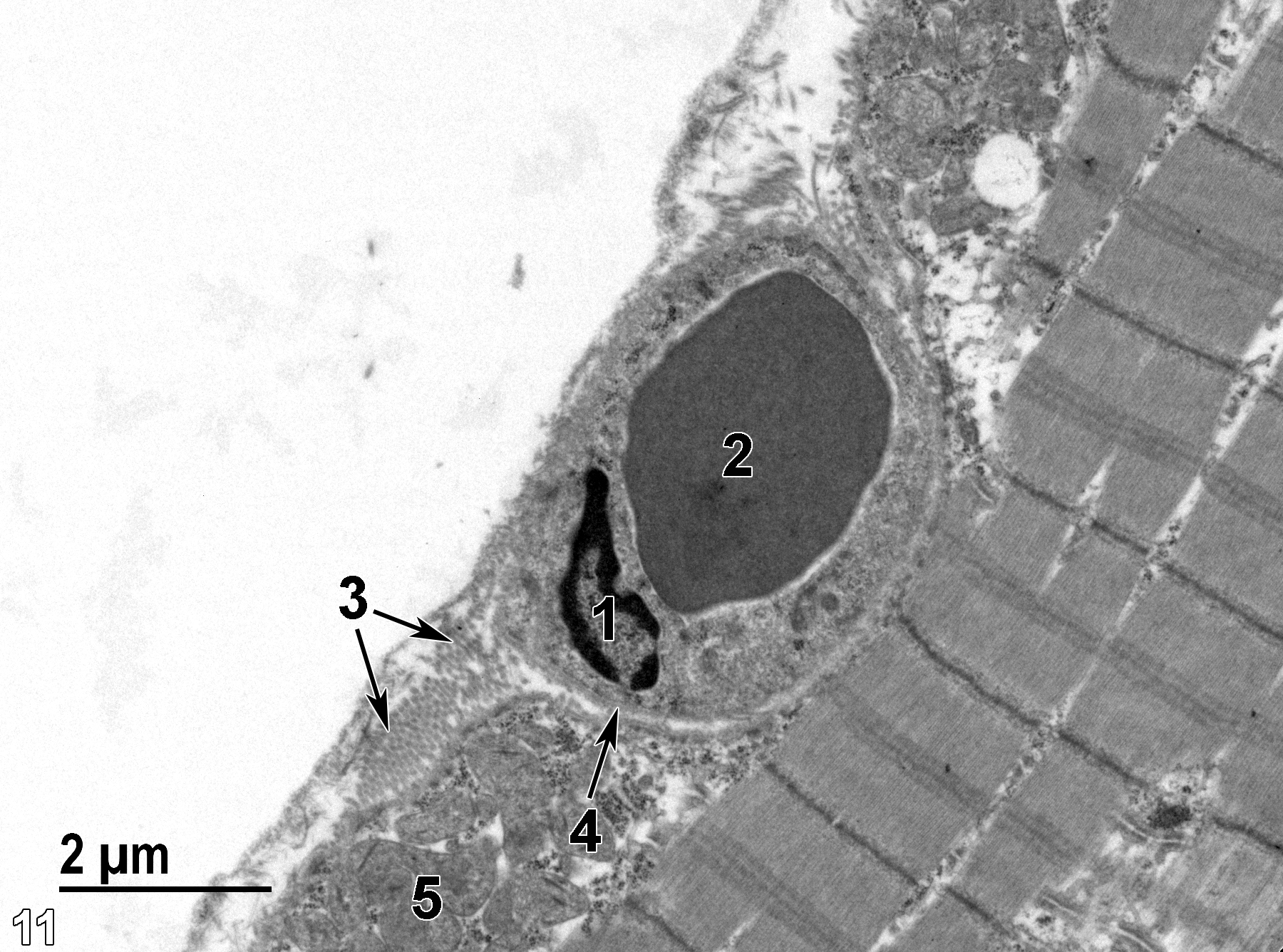
Figure 11. An ultrastructural image showing an endothelial nucleus (1) of a capillary containing an erythrocyte (2). The capillary is surrounded by a basal lamina (4, arrow). Clusters of collagen (3, double arrows) are evident. A group of mitochondria (5) is located at the periphery of the myocyte cytoplasm. 11000x.
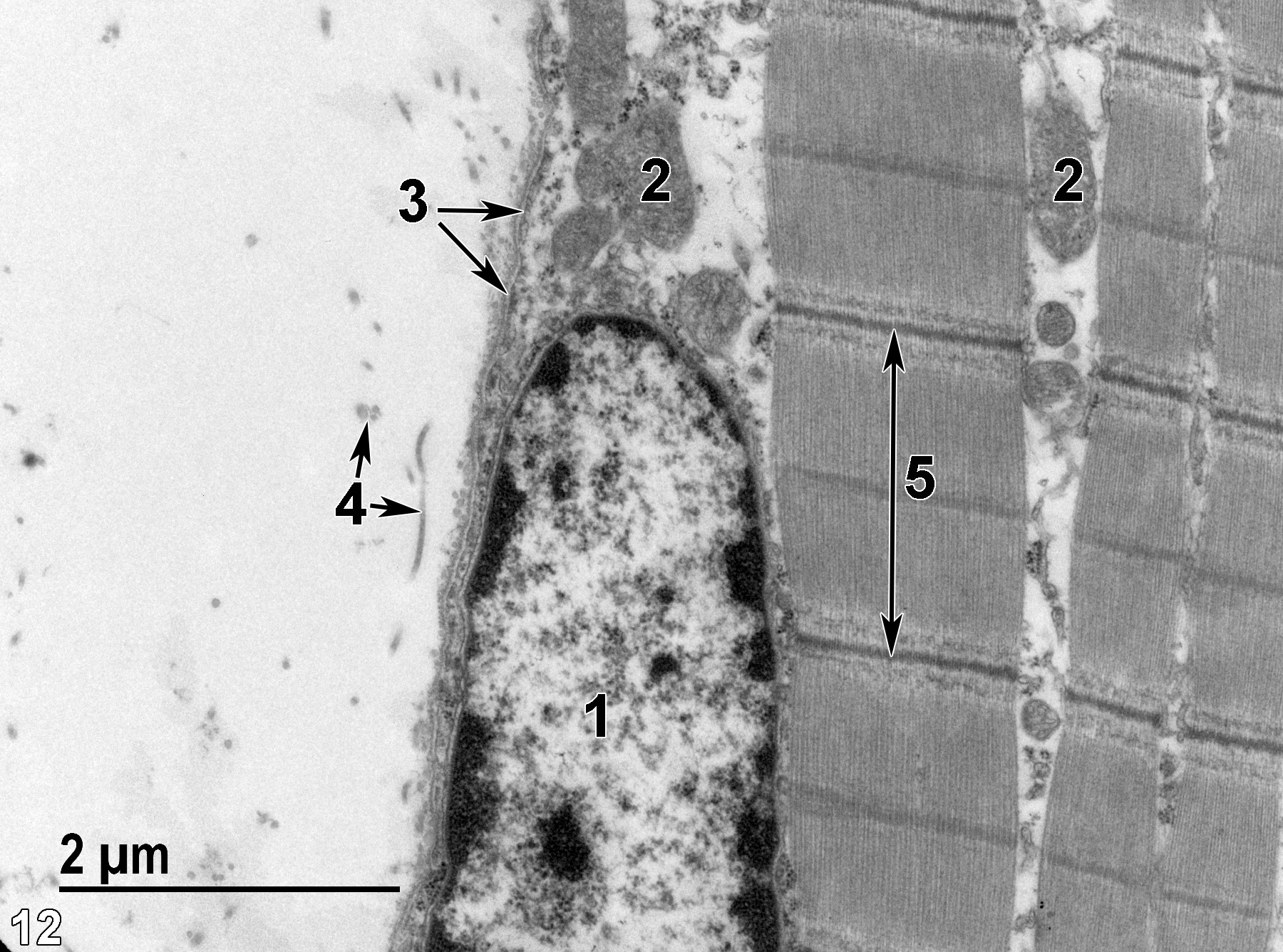
Figure 12. A higher magnification view of the periphery of a myocyte showing part of a myocyte nucleus (1) and mitochondria (2) between the myofibrils and just inside the myocyte sarcolemma and basal lamina (3, double arrows). Collagen (4, double arrows) is located just outside of the basal lamina. A single sarcomere (5) bounded by its Z lines (long double-headed arrow). 18500x.
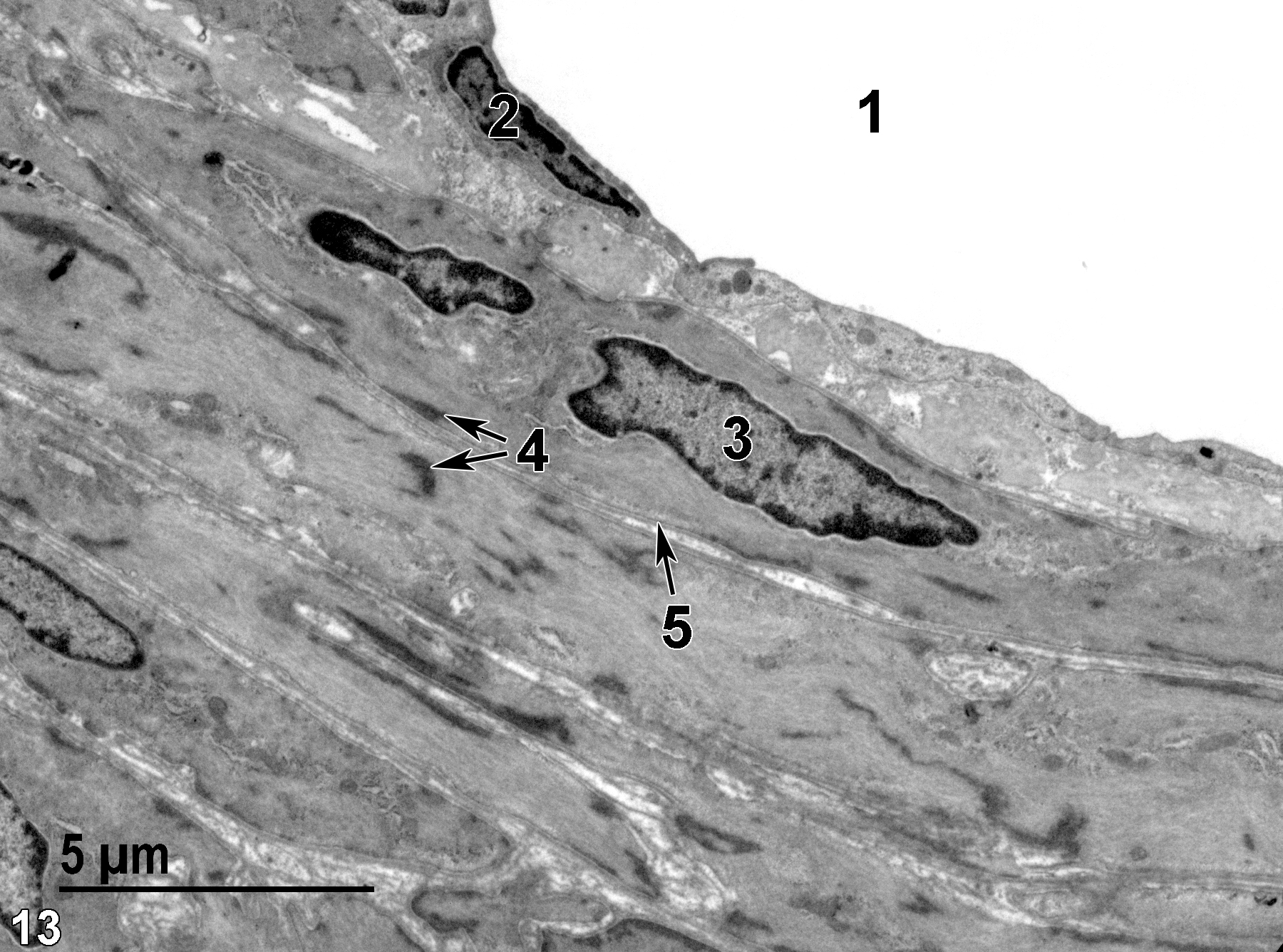
Figure 13. The lumen of an artery (1) lined with an endothelial cell layer, with one elongated endothelial nucleus (2). Beneath the endothelial cell layer are several layers of smooth muscle cells that also have elongated nuclei (3). The cytoplasm of the smooth muscle cells is filled with proteinaceous filaments and electron dense patches of alpha-actinin (4, double arrows). The individual smooth muscle cells (5) are surrounded by a basal lamina (arrow). 6800x.
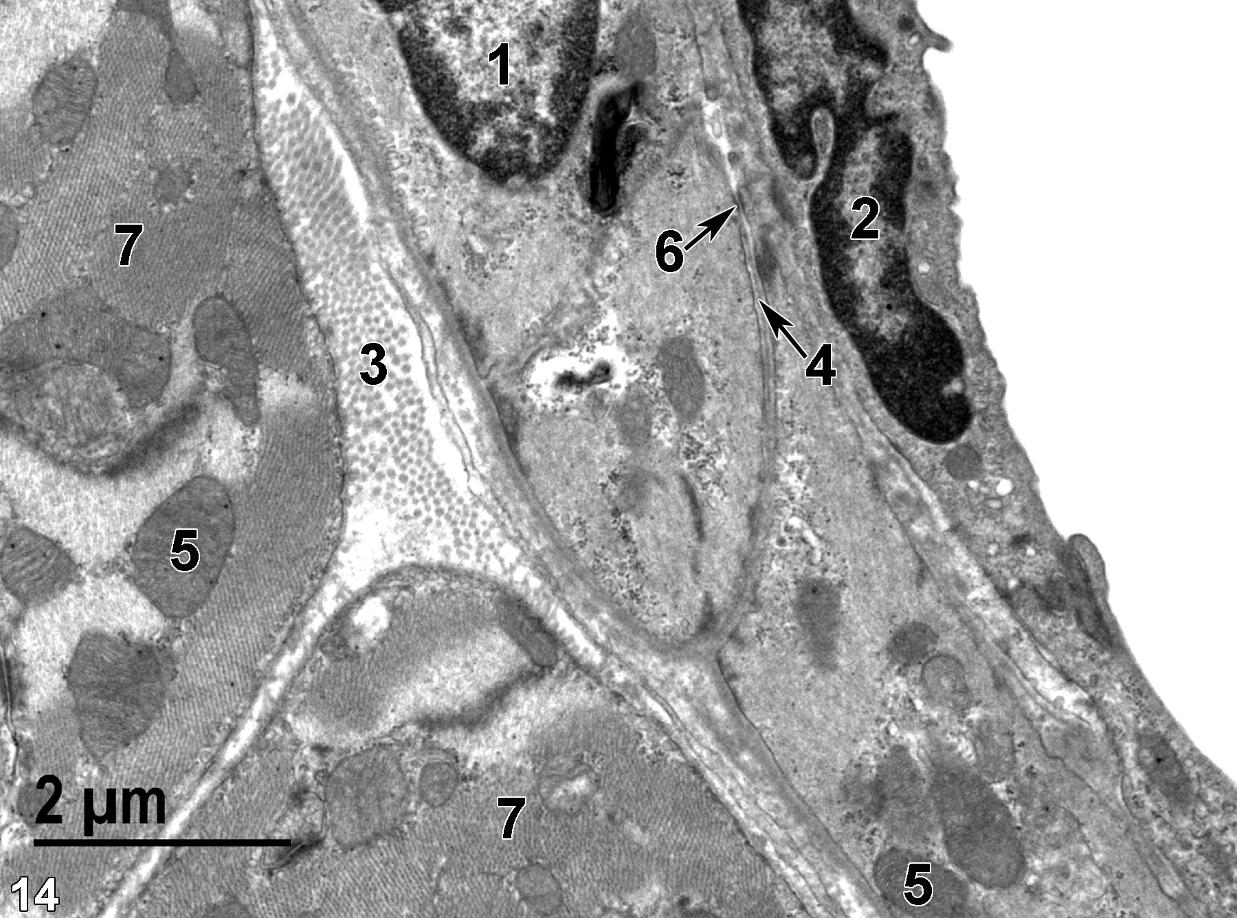
Figure 14. An image of a vein showing part of a nucleus (1) of a smooth muscle cell in a single layer of smooth muscle cells. The basal lamina (4, arrow) surrounds the smooth muscle cell, except where a desmosome (6, arrow) has formed with a thin extension of an adjacent smooth muscle cell. A pocket of collagen fibrils (3) lies between the smooth muscle cell layer and the adjacent striated cardiac muscle cells. The elongated nucleus (2) of an endothelial cell is shown lining the vein. Mitochondria (5) are less abundant in the smooth muscle cells than those in the adjacent striated cardiac myocytes (7). 11000x.
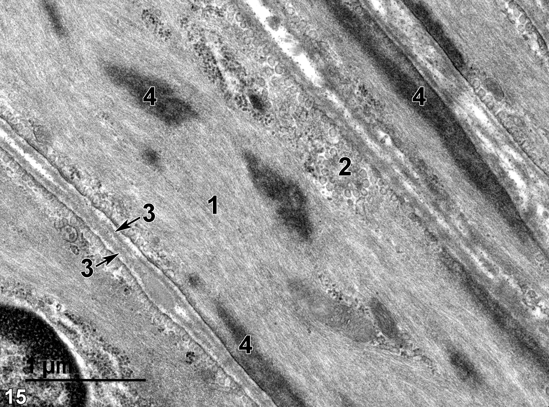
Figure 15. A high-magnification view of smooth muscle cells showing proteinaceous filaments (1) largely filling the cytoplasm of these cells. Clusters of vesicles (2) beneath the smooth muscle cell membrane are characteristic of smooth muscle cells. Each smooth muscle cell is enrobed with a basal lamina (3, arrows) and contains cytoplasmic densities (4) composed of alpha-actinin. 30000x.
| Eurell JA, Frappier BL, eds. 2006. Dellmann’s Textbook of Veterinary Histology. 6th ed. Ames, IA: Blackwell Publishing. |
| Pavelka M, Roth J. 2005. Functional Ultrastructure. An Atlas of Tissue Biology and Pathology. New York: Springer Wien. |
| Rhodin JAG. 1974. Histology: A Text and Atlas. New York: Oxford University Press. |
| Ross MH, Kaye GI, Pawlina W. 2003. Histology: A Text and Atlas. 4th ed. Philadelphia: Lippincott Williams & Wilkins. |
All Images


