

Reproductive System, Female
Vagina
Narrative
The vagina is a tubular organ lined with a vaginal wall with a well-vascularized mucous membrane that consists of a stratified epithelial cell layer, an intermediate epithelial cell layer, and a layer of basal epithelial cells. The vaginal epithelium undergoes changes depending on the stage of the estrous cycle observed (Dixon et al. 2018). The epithelial cells contain numerous tonofilaments and desmosomes that connect cellular projections into the intercellular spaces. Mitochondria, glycogen, and ribosomes are present. Below the epithelium is the lamina propria, which consists of collagenous connective tissue with fibroblasts, neutrophils, eosinophils, and mast cells. Below the lamina propria is a muscularis layer that consists of smooth muscle cells. The outermost layer of the vagina is the adventitia (Young and Heath 2000).
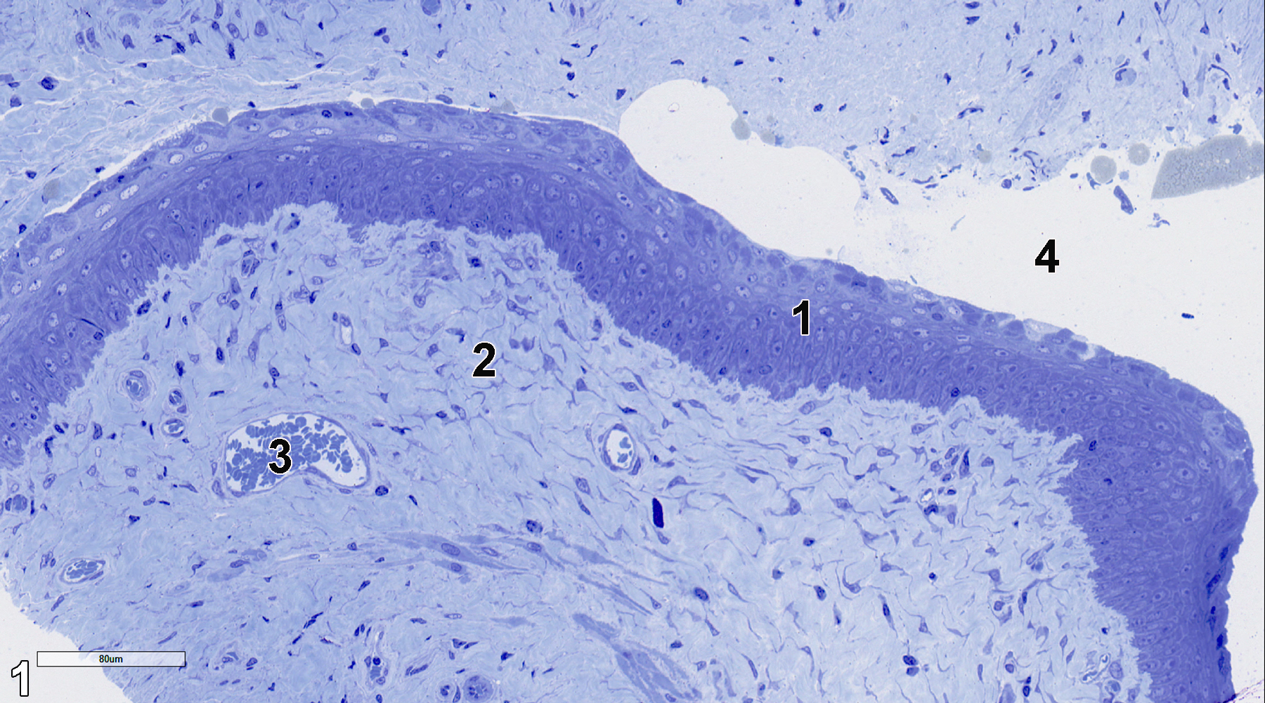
Figure 1. A 0.5-micron-thick semithin section stained with toluidine blue O that shows the epithelial cell layer (1), lamina propria (2), a blood vessel filled with erythrocytes (3), and the vagina lumen (4). 25x.
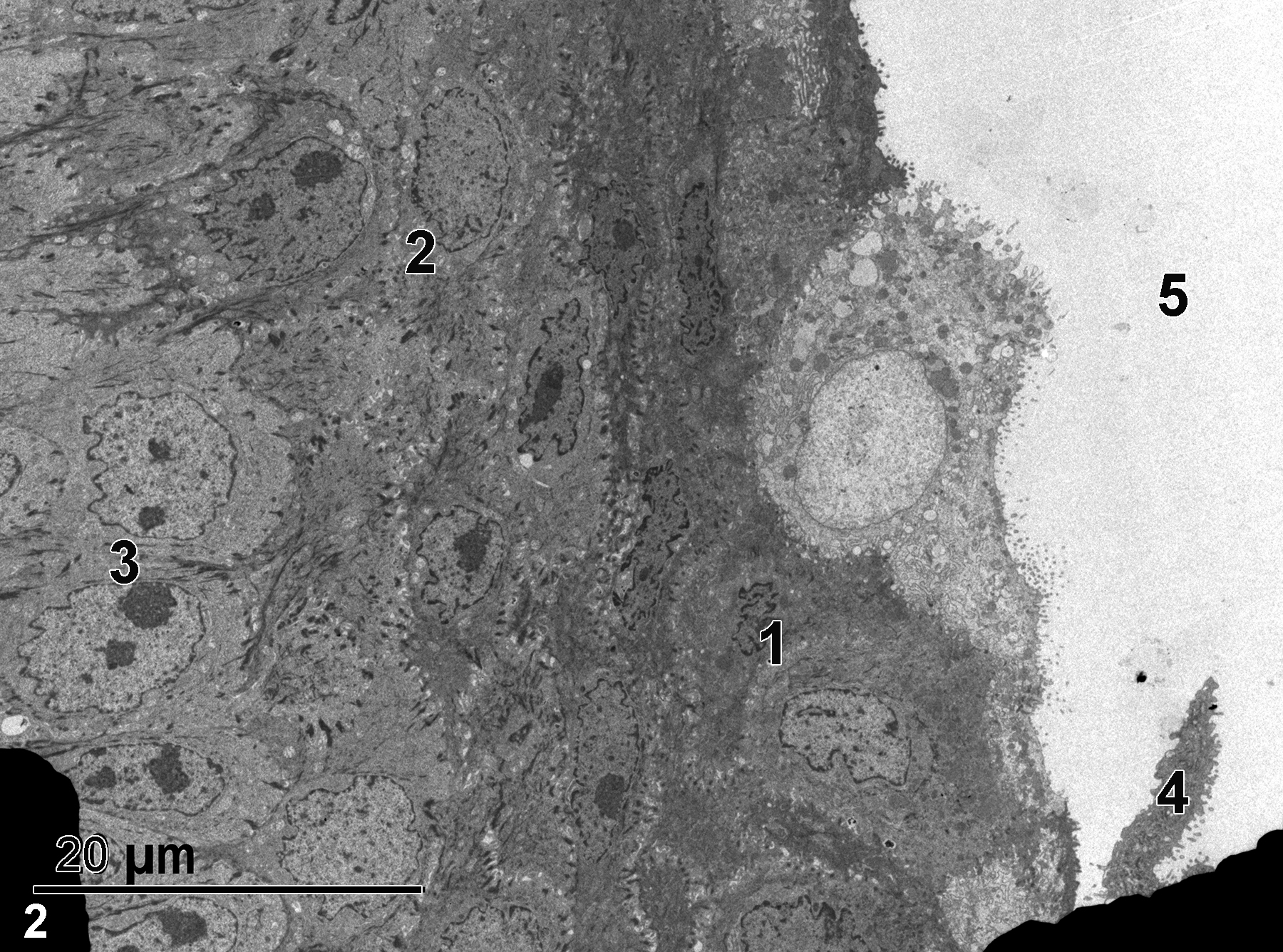
Figure 2. A low magnification view of the mucous membrane epithelial cells. The dark areas in the lower left and right of the image are grid bars. The lumen of the vagina (5) is lined with a superficial layer of stratified squamous epithelial cells (1), underlain with a layer of intermediate epithelial cells (2) that are more cuboidal, with a layer of cuboidal basal epithelial cells (3) beneath the intermediate layer. One superficial epithelial cell (4) is being shed. 1900x.
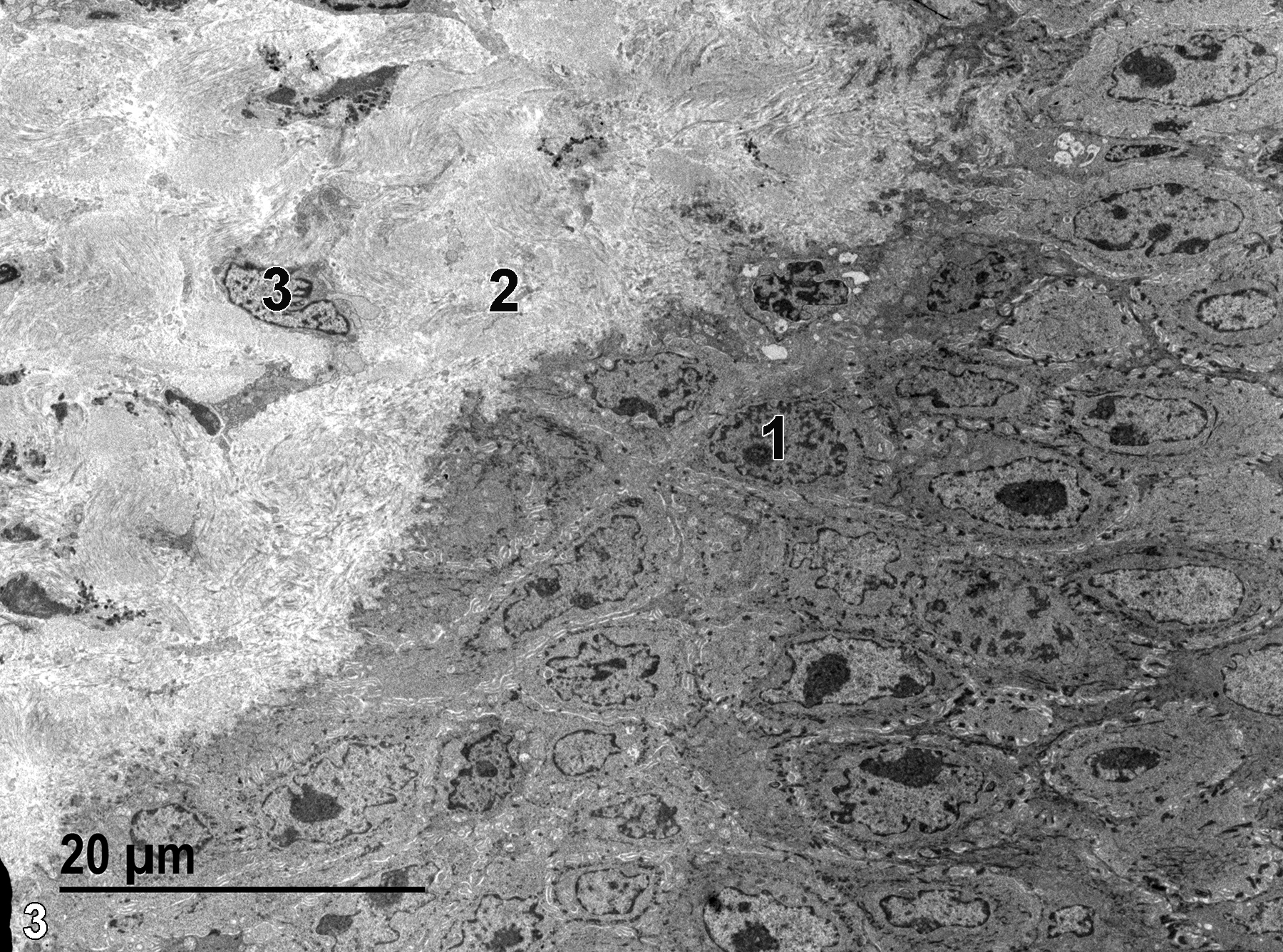
Figure 3. The nucleus (1) of a basal epithelial cell, part of the collagen layer comprising the lamina propria (2), and the nucleus of a fibroblast (3) in the lamina propria. 1900x.
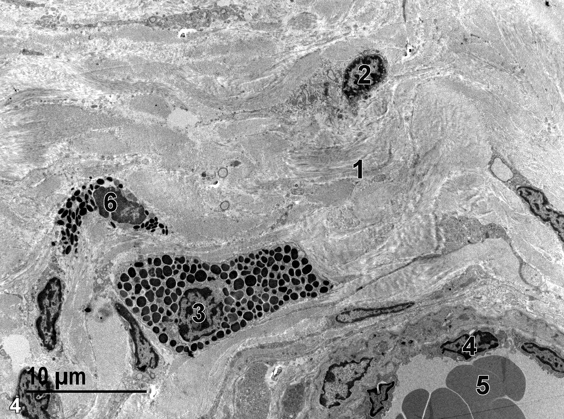
Figure 4. A higher magnification view of the lamina propria showing the bundles of collagen (1), the nucleus of a fibroblast (2), a single mast cell (3) that contains numerous mast cell granules of varying size and electron density, and the nucleus of an endothelial cell (4) lining a venule surrounded by a thin layer of smooth muscle cells and that contains erythrocytes (5). A single eosinophil (6) is present in the lamina propria. 2900x.
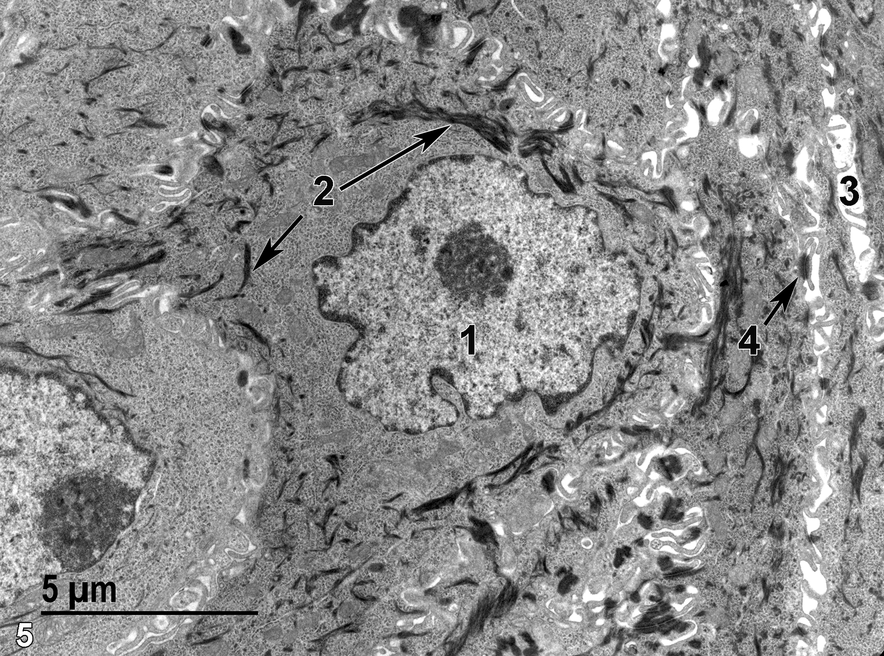
Figure 5. A high magnification view of epithelial cells of the mucous membrane. A nucleus of an epithelial cell (1) contains a single nucleolus, marginated chromatin, and has a slightly crenelated surface. Numerous bundles of tonofilaments (2, arrows) are present. The intercellular space (3) is filled with thin cytoplasmic extensions. Desmosomes (4, arrow) serve as attachment points between adjacent cells. 6800x.
| Dixon D, Vidal JD, Leininger JR, Jokinen MP. 2018. Chapter 27: Oviduct, uterus, and vagina. In Boorman’s Pathology of the Rat (Suttie AW, ed.). 2nd ed. London: Academic Press, 537−559. |
| Rhodin JAG. 1974. Histology: A Text and Atlas. New York: Oxford University Press. |
| Weiss L, ed. 1988. Cell and Tissue Biology: A Textbook of Histology. 6th ed. Baltimore: Urban & Schwarzenberg. |
| Young B, Heath JW. 2000. Wheater’s Functional Histology: A Text and Colour Atlas. 4th ed. Edinburgh, UK: Churchill Livingstone. |
All Images


