

Nervous System
Spinal Cord
Narrative
The spinal cord has a central canal with a butterfly-shaped gray matter area surrounded by white matter. There are three layers of connective tissue (meninges) surrounding the outer aspect of the white matter, starting from the innermost layer: pia mater, arachnoid, and dura mater. Gray matter of the brain and spinal cord consists of numerous nerve cells (perikaryon, nucleus, and dendrites), whereas the nerve fibers (axons) predominate in white matter. The perikarya are large polyhedral cells with central nuclei that have prominent nucleoli and little heterochromatin. They have abundant rough endoplasmic reticulum cisternae and large numbers of free ribosomes and intermediate filaments. In addition to the nerve cells, blood vessels and several types of microglial cells are found in both the white and gray matter of the spinal cord. Oligodendrocytes have fewer cytoplasmic processes than astrocytes and are often aligned in rows between axons. They have spherical nuclei and electron-dense cytoplasm with many microtubules, rough endoplasmic reticulum, and mitochondria. They produce myelin that surrounds myelinated axons. Astrocytes are the largest microglial cells, with large centrally located nuclei that lack nucleoli. Astrocytes provide structural support, supply energy, are involved in inflammatory responses, and are phagocytic. Protoplasmic astrocytes are found in gray matter and have numerous short branching cytoplasmic processes, whereas fibrous astrocytes are more common in white matter and have fewer, narrower, and straighter cytoplasmic processes than those found in protoplasmic astrocytes.
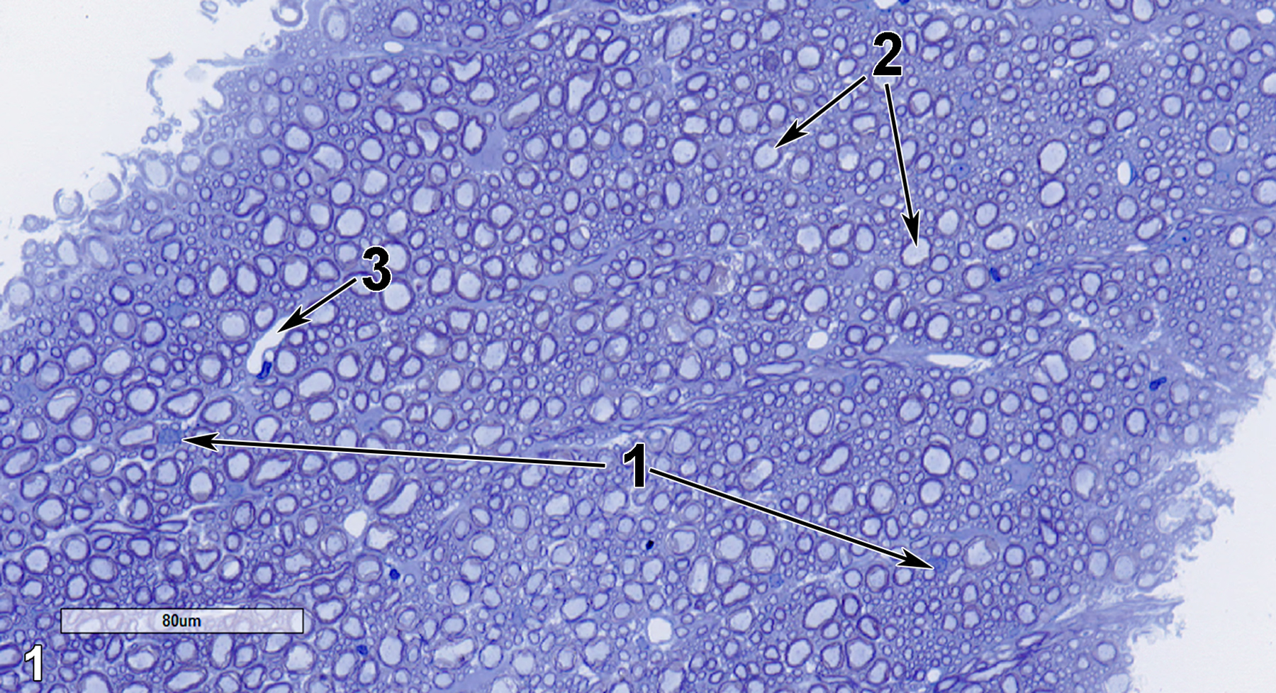
Figure 1. A semithin section (0.5 micrometer thick) of a toluidine blue O-stained transverse section of spinal cord white matter. The two large nuclei with nucleoli are consistent with nerve cell perikarya (1, arrows). Numerous sections of myelinated axons are present (2, arrows). A blood vessel is also shown (3, arrow). 25x.
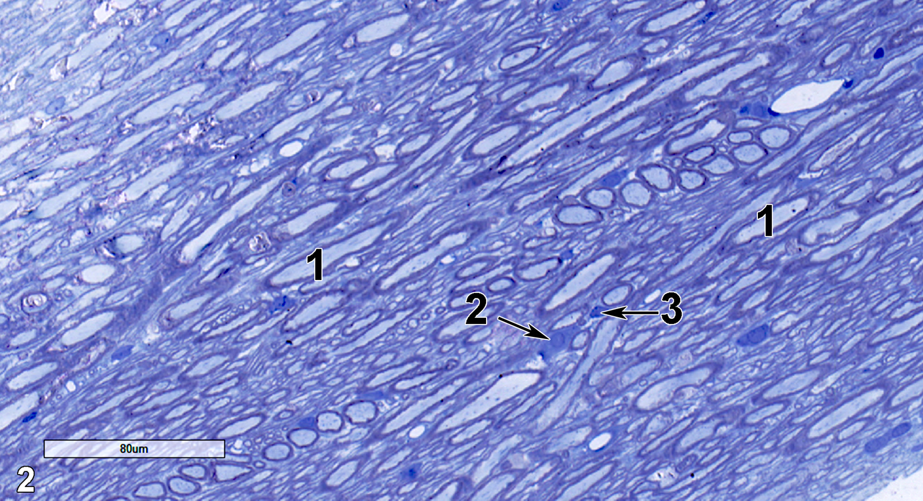
Figure 2. A semithin longitudinal section of spinal cord white matter. Large numbers of longitudinal sections of myelinated axons (1) are present. A round nucleus of a perikaryon is shown (2, arrow), along with a smaller, denser nucleus of a probable astrocyte (3, arrow). 25x.
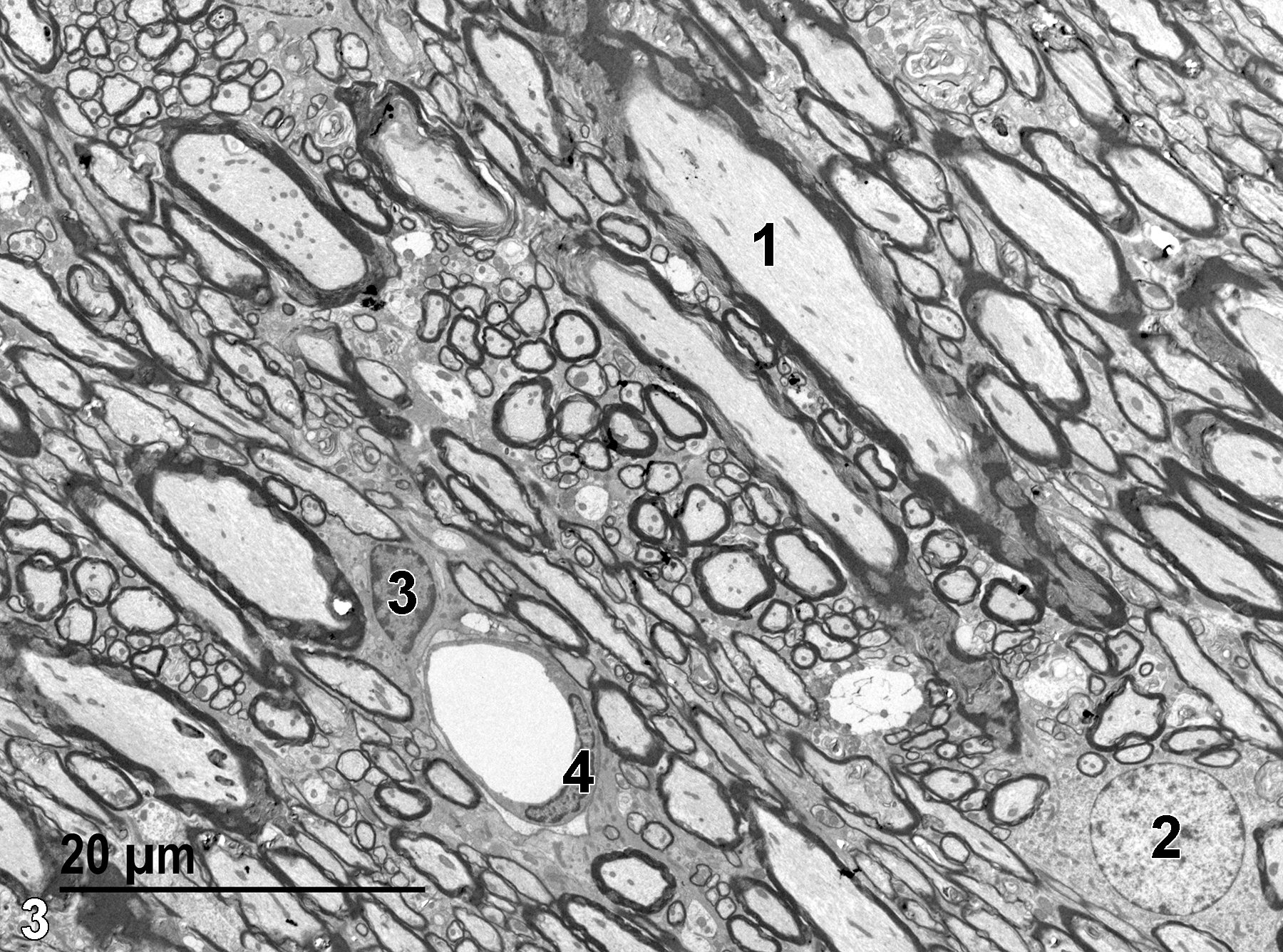
Figure 3. An electron micrograph of a longitudinal section of white matter showing a myelinated axon (1) with a number of electron-dense mitochondria and fibrillar material (microfilaments) in the axon cytoplasm. A single capillary (4) with an elongated endothelial nucleus is present. A large round nucleus (2) with little heterochromatin and lightly stained cytoplasm is characteristic of a perikaryon of a neuron. The smaller nucleus (3) with more marginated heterochromatin is consistent with an astrocyte. 1900x.
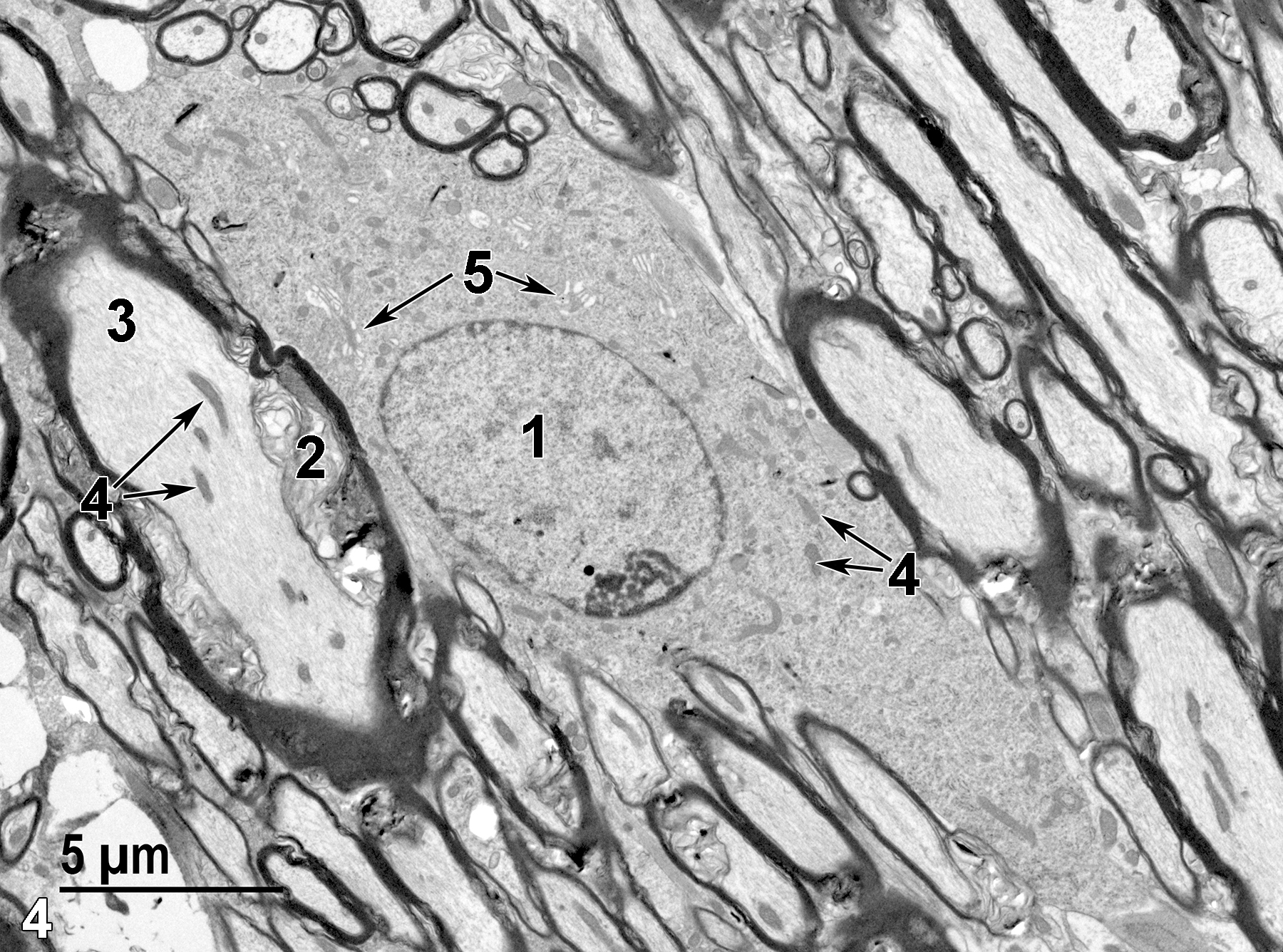
Figure 4. A higher magnification of a perikaryon, showing the round nucleus (1) with a single peripheral nucleolus and little heterochromatin. The cytoplasm has mitochondria (4, arrows) and Golgi bodies (5, arrows) that are visible at this magnification. The myelinated axon (3) has a few mitochondria (4, arrows). The disorganized membranous layers of the myelin sheath (2) are a fixation artifact. 4800x.
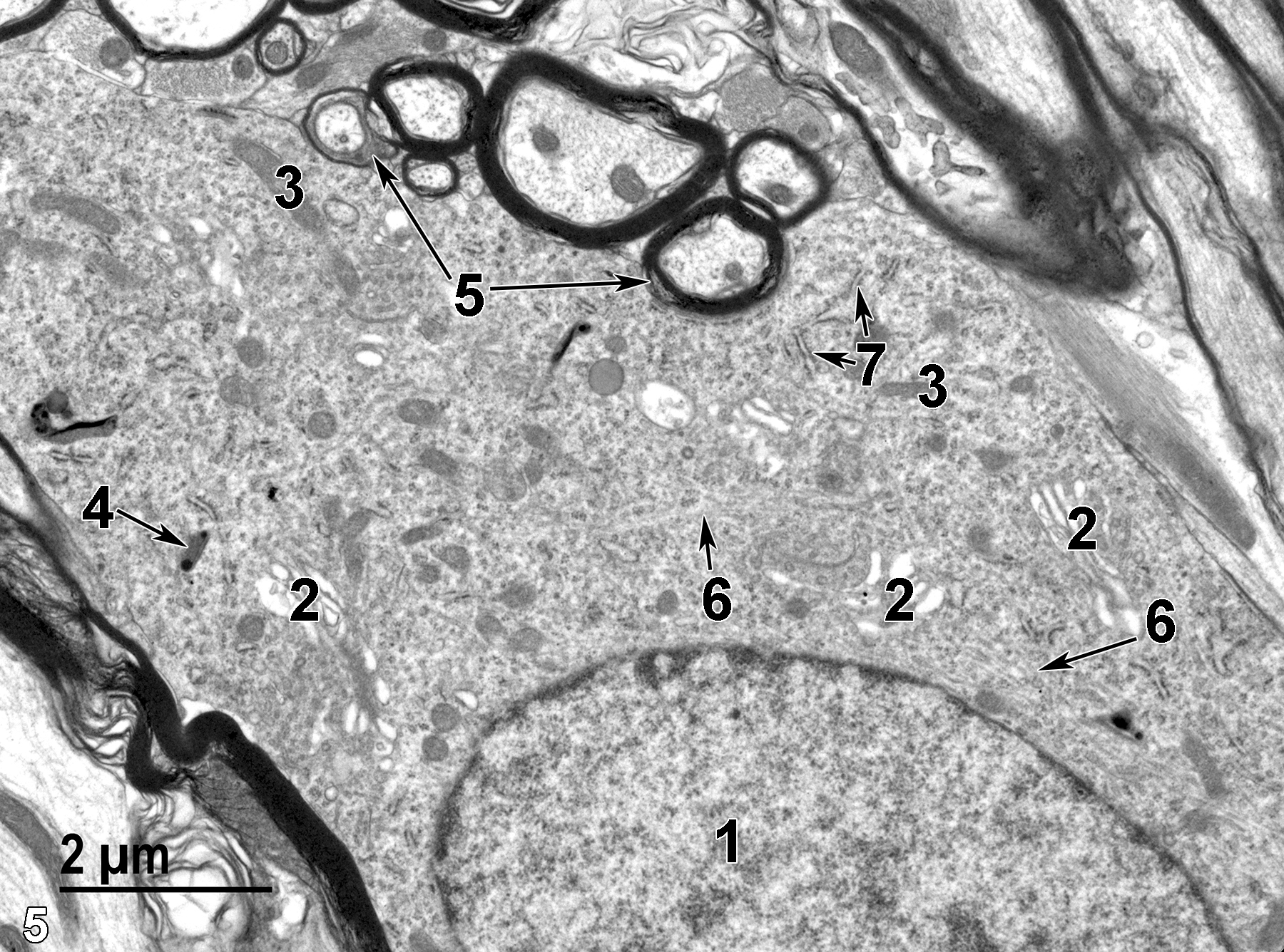
Figure 5. A higher magnification of the perikaryon seen in Figure 4. The nucleus (1) has closely associated Golgi bodies (2) and bundles of intermediate filaments (6, arrows). Mitochondria (3) are relatively numerous. One lysosome is present (4, arrow). Tubular cisternae of rough endoplasmic reticulum are scattered throughout the cytoplasm (7, arrows). Densely stained myelin surrounding axons is shown (5, arrows). 11000x.
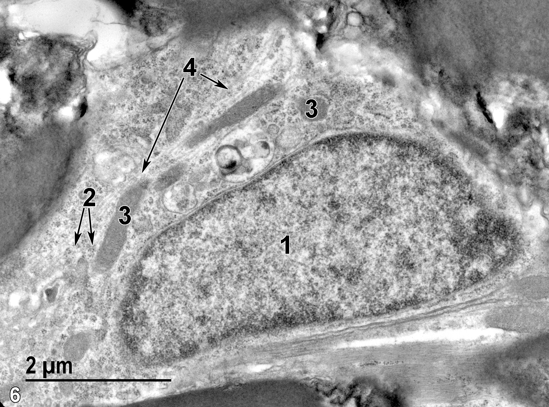
Figure 6. An astrocyte with an elongated nucleus (1), clusters of free ribosomes (2, arrows), mitochondria (3), and bundles of intermediate filaments (4, arrows). 18500x.
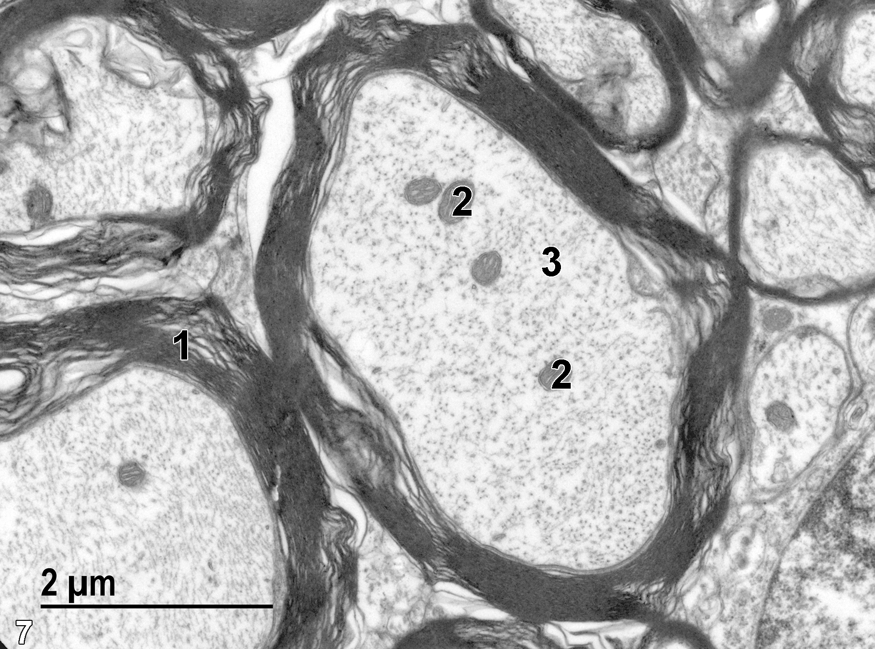
Figure 7. A high magnification of transverse sections of myelinated axons. They have myelin consisting of whorls of membranes (1) surrounding the axon that contains mitochondria (2) and numerous microfilaments (3) in cross section. 18500x.
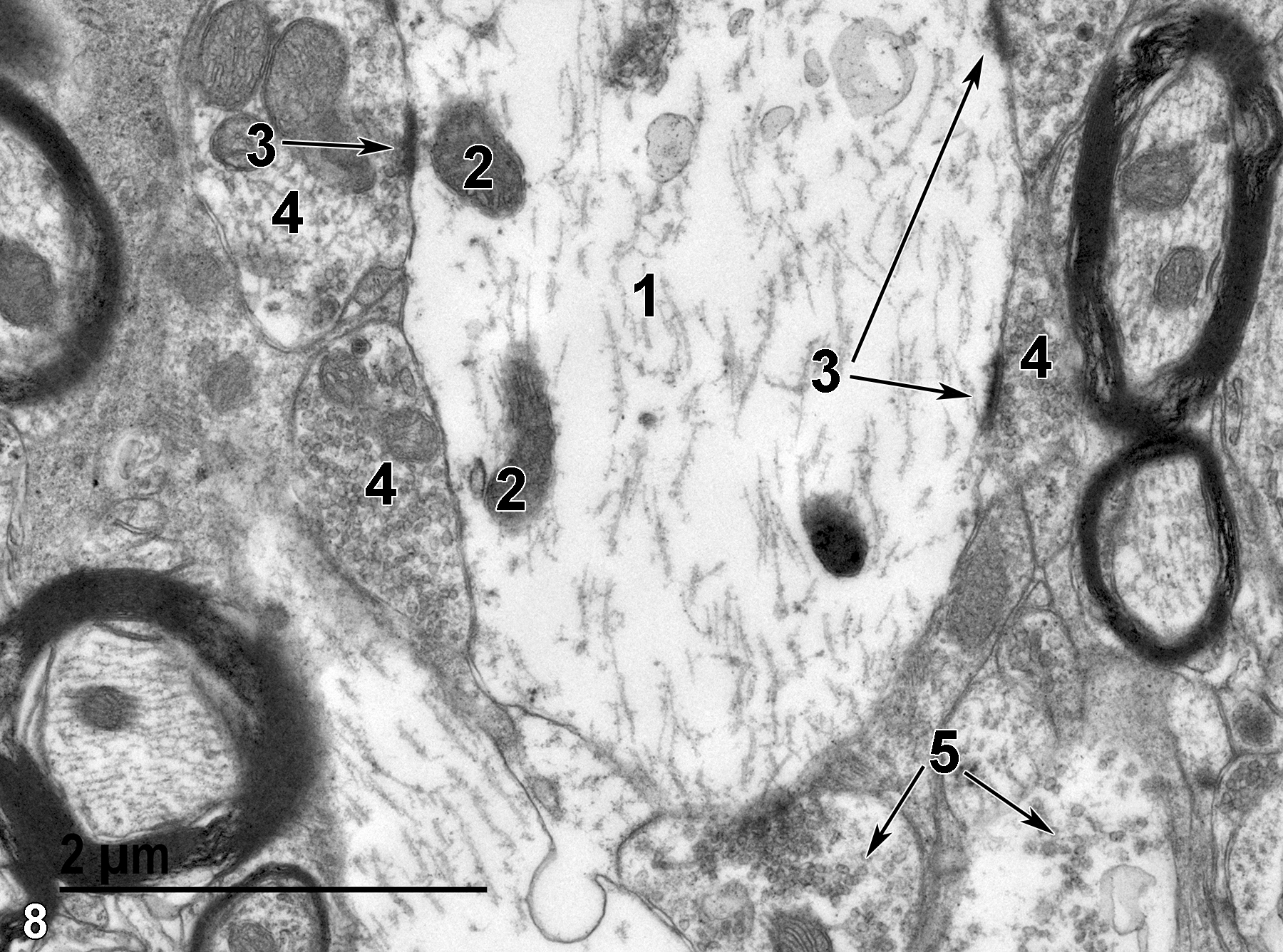
Figure 8. An unmyelinated axon with numerous microfilaments (1), mitochondria (2), and axo-dendritic synapses (3, arrows). Surrounding the axon are a number of dendritic boutons (4) containing synaptic vesicles (5). 23000x.
| Boorman GA, Eustis SL, Elwell MR, Montgomery CA, Jr., MacKenzie WF, eds. 1990. Pathology of the Fischer Rat: Reference and Atlas. New York: Academic Press. |
| Dellmann HD, Eurell J, eds. 1998. Textbook of Veterinary Histology. 5th ed. Philadelphia: Lippincott Williams & Wilkins. |
| Rhodin JAG. 1974. Histology: A Text and Atlas. New York: Oxford University Press. |
| Ross MH, Kaye GI, Pawlina W. 2003. Histology: A Text and Atlas. 4th ed. Philadelphia: Lippincott Williams & Wilkins. |
| Weiss L, ed. 1988. Cell and Tissue Biology: A Textbook of Histology. 6th ed. Baltimore: Urban & Schwarzenberg. |
All Images


