

Reproductive System, Female
Cervix
Narrative
The cervix of the rat can be divided histologically into an endocervix (upper/anterior) and an exocervix (lower/posterior). The endocervix is a transitional zone that leads into each horn of the uterus (Picut and Remick 2016). The exocervix is non-glandular and continuous with the vaginal mucosa. It has a superficial layer of mucus cells subtended by stratified non-keratinized squamous epithelial cells. Similar to the vagina, the cervical epithelium undergoes changes depending on the stage of the estrous cycle observed, but according to Dixon et al. (2018), the changes can be less distinct and more variable. The lamina propria is a broad fibrous layer consisting of a collagenous matrix, fibroblasts, and fairly sparse smooth muscle cells.
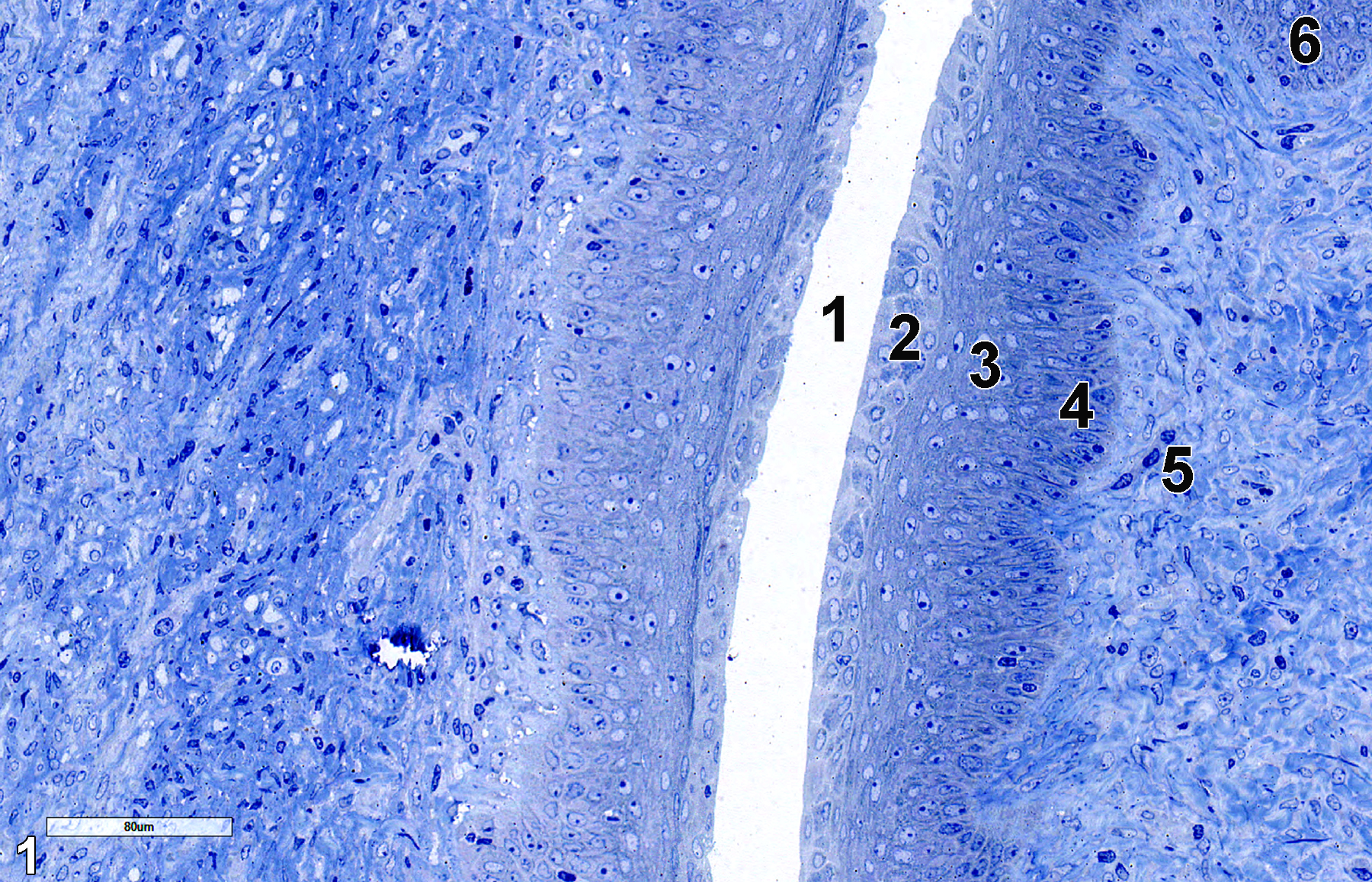
Figure 1. A semithin section (0.5 micrometer thick) of a toluidine blue O-stained section of the exocervix showing the cervical lumen (1), the mucus cell layer (2), the stratified non-keratinized epithelial cells (3), the basal cells of the stratified epithelial cell layer (4), the lamina propria (5), which consists of a collagenous matrix with smooth muscle cells and fibroblasts, and a single tubular gland (6). 25x.
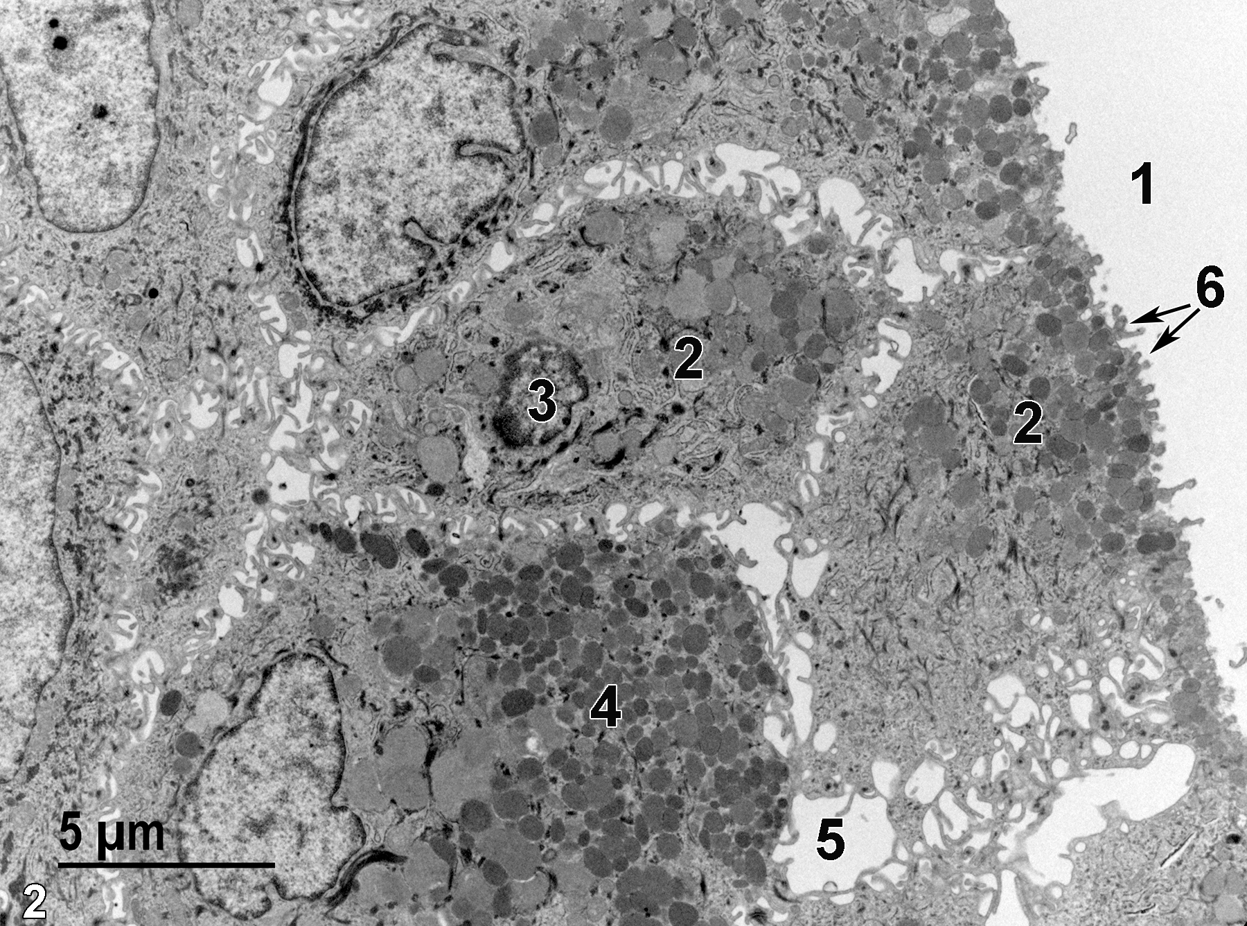
Figure 2. An electron micrograph of an area similar to Figure 1 showing the cervical lumen (1), a mucus cell (2) with a single nucleus (3), many mucigen granules (4) of varying size and electron density, intercellular space (5), and microvilli of the mucus cells protruding into the cervical lumen (6, arrows). 4800x.
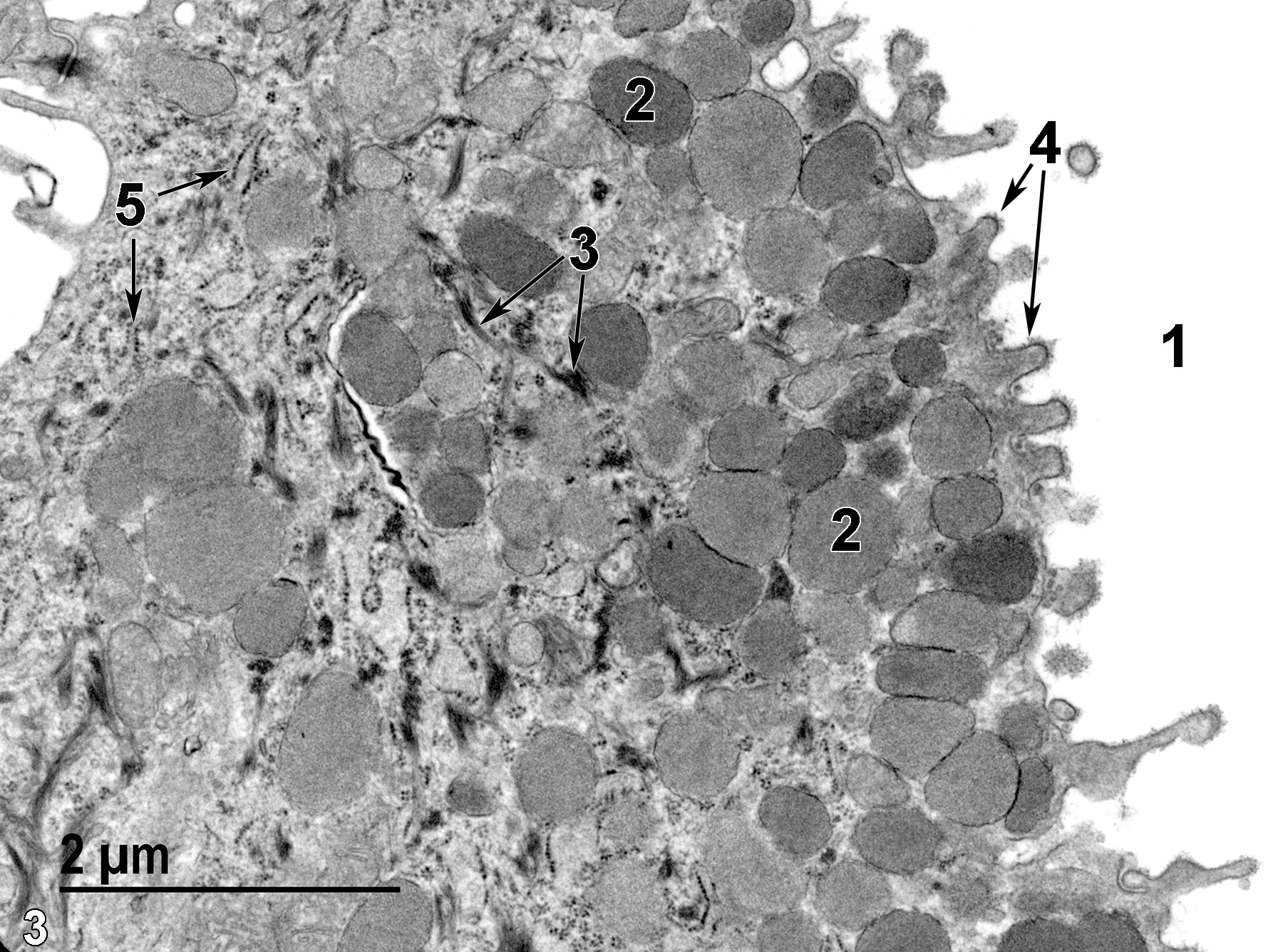
Figure 3. A higher magnification view of a single mucus cell showing the cervical lumen (1), mucigen granules (2) of varying density, bundles of tonofilaments (3, arrows), microvilli with glycocalyx materials associated with their plasma membranes (4, arrows), and rough endoplasmic reticulum elements (5, arrows). 8500x.
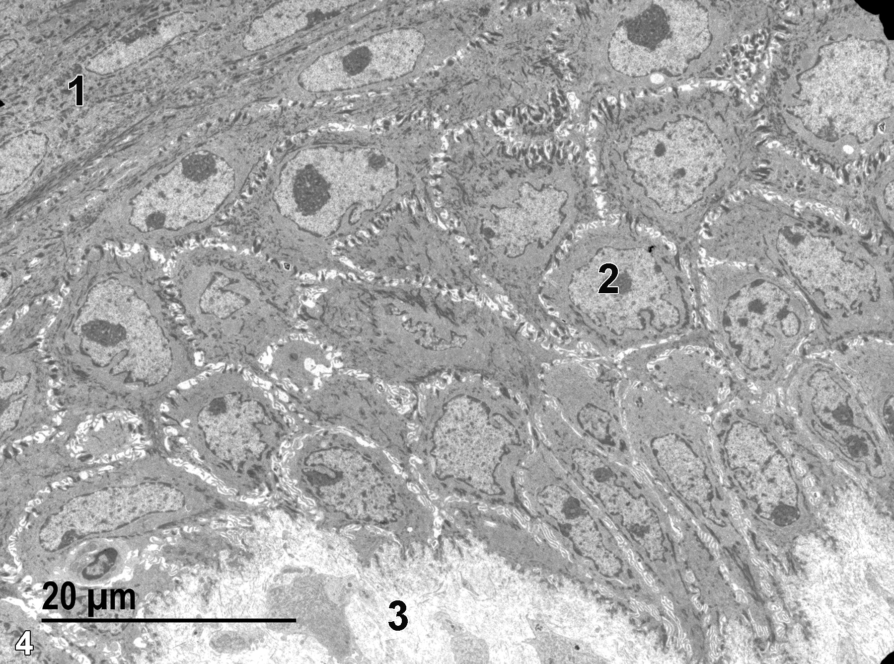
Figure 4. Stratified squamous epithelial cells (1) that underlay the more cuboidal basal stratified epithelial cells (2), which are, in turn, underlain by the connective tissue of the lamina propria (3). 1900x.
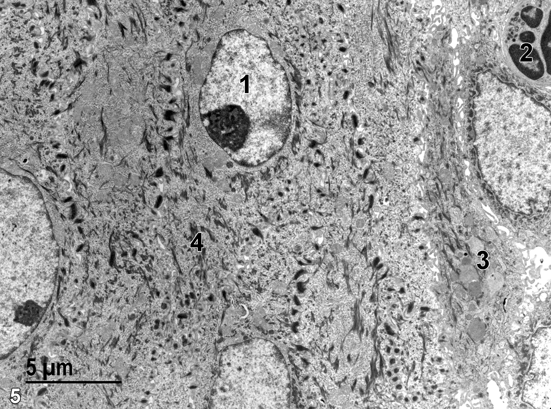
Figure 5. A higher magnification view of the stratified squamous epithelial cells. A single nucleus (1) with a prominent nucleolus is present. The upper right corner of the image contains part of a neutrophil (2). Mucigen granules (3) can be seen in a single mucus cell adjacent to the squamous epithelium. Numerous bundles of tonofilaments (4) are visible. 4800x.
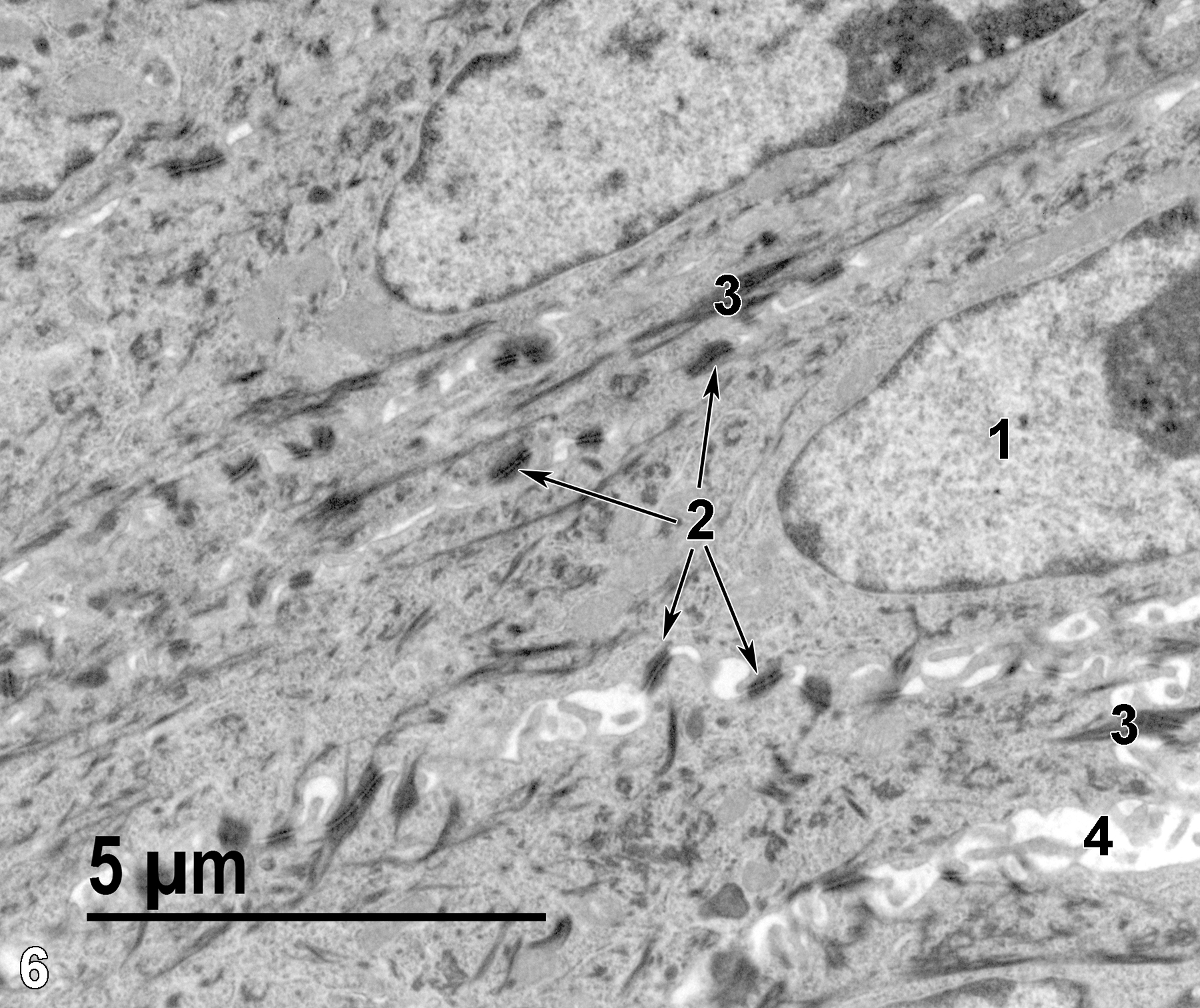
Figure 6. An even higher magnification view of squamous epithelial cells showing a nucleus (1) with a prominent nucleolus, a number of desmosomes (2, arrows) binding adjacent cell surfaces together, bundles of tonofilaments (3), and an intercellular space (4). 6800x.
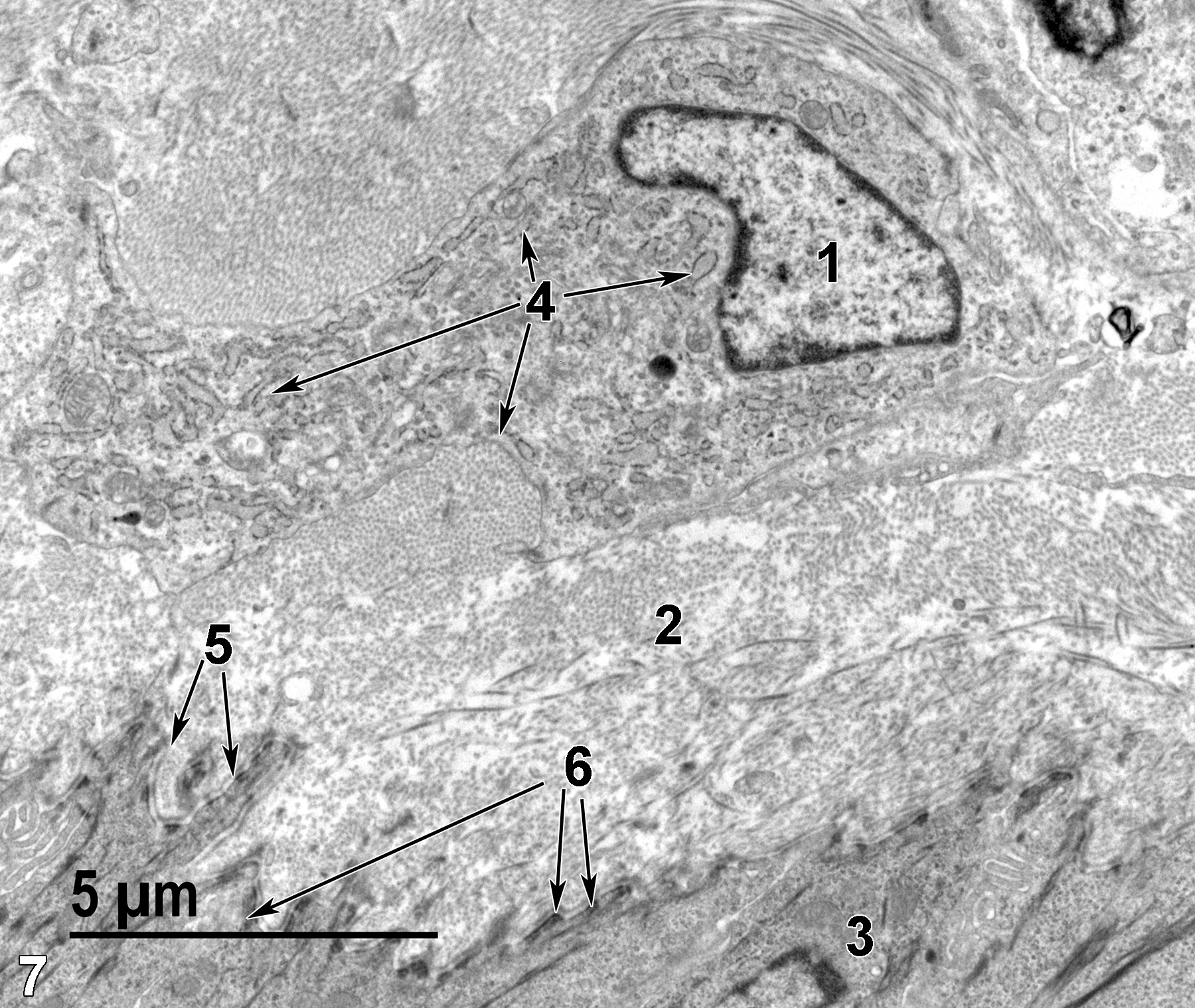
Figure 7. A portion of the lamina propria is shown. A fibroblast nucleus (1) is shown. The cytoplasm of the fibroblast is filled with a large quantity of rough endoplasmic reticulum (4, arrows). Collagen fibrils (2) are the predominant element of the lamina propria. At the bottom of the image are the most basal cells (3) of the stratified epithelium. The electron densities scattered along the junction of the cell membrane and the basal lamina (5, arrows) are hemidesmosomes (6, arrows). 6800x.
| Dellmann HD, Eurell J, eds. 1998. Textbook of Veterinary Histology. 5th ed. Philadelphia: Lippincott Williams & Wilkins. |
| Dixon D, Vidal JD, Leininger JR, Jokinen MP. 2018. Chapter 27: Oviduct, uterus, and vagina. In Boorman’s Pathology of the Rat (Suttie AW, ed.). 2nd ed. London: Academic Press, 537−559. |
| Picut CA, Remick AK. 2016. Chapter 7: Female reproductive system. In Atlas of Histology of the Juvenile Rat (Parker GA, Picut CA, eds.). London: Academic Press, London, 203-226. |
| Rhodin JAG. 1974. Histology: A Text and Atlas. New York: Oxford University Press. |
| Weiss L, ed. 1988. Cell and Tissue Biology: A Textbook of Histology. 6th ed. Baltimore: Urban & Schwarzenberg. |
All Images


