

Musculoskeletal System
Bone
Narrative
Compact (cortical) bone forms the exterior of all bones, whereas spongy (trabecular) bone is found in the ends of long bones and in the interior of short bones. Compact bone has Haversian and Volksmann’s canals, whereas trabecular bone lacks central blood vessels and Haversian and Volksmann’s canals.
Lamellar bone is composed of the bone matrix, which contains collagen, amorphous organic ground substance [protein polysaccharides and glycoproteins, such as sialoprotein (Rhodin, 1974), and inorganic materials (calcium phosphate, calcium carbonate, and sodium). Osteoblasts form the bone matrix and osteoclasts resorb bone. Within the lamellar bone, the Haversian canals are arranged parallel to the long axis of the bone and contain small blood vessels. The smaller Volkmann’s canals are perpendicular to the long axis of the bone. Finally, osteocytes are found within Howship’s lacunae within the lamellar bone matrix.
The bone marrow at the center of the lamellar bone consists of hemopoietic (myeloid) tissue responsible for the production of erythrocytes, leukocytes, osteoblasts, osteoclasts, reticular cells (fixed macrophages), and megakaryocytes.
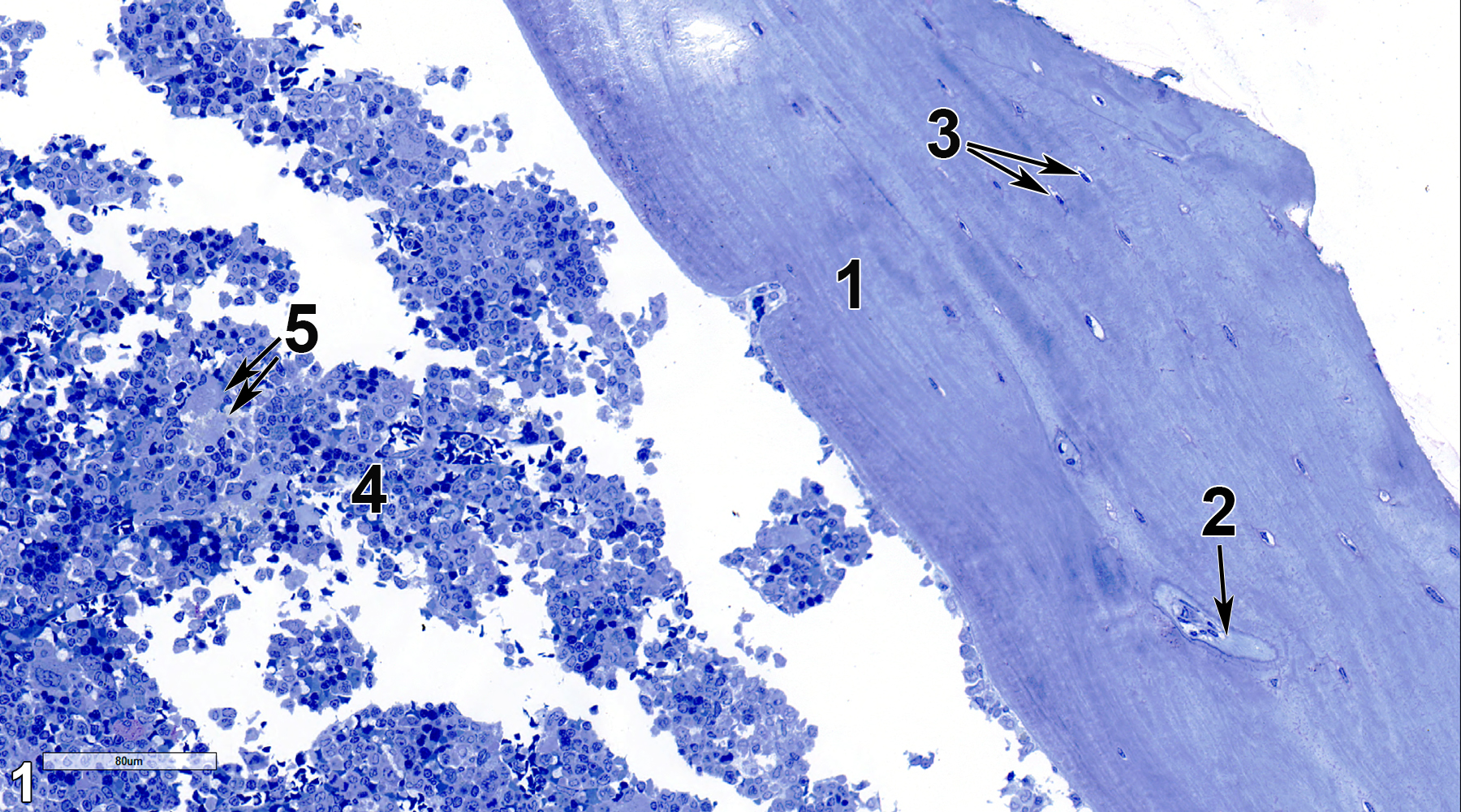
Figure 1. A semithin section (0.5 micrometer thick) of a toluidine blue O-stained section showing cortical bone (1) with a Haversian canal (2, arrow), lacunae containing osteocytes (3, double arrows), and bone marrow (4), containing a variety of progenitor cells, erythrocytes, leukocytes, and megakaryocytes (5, double arrows). 25x.
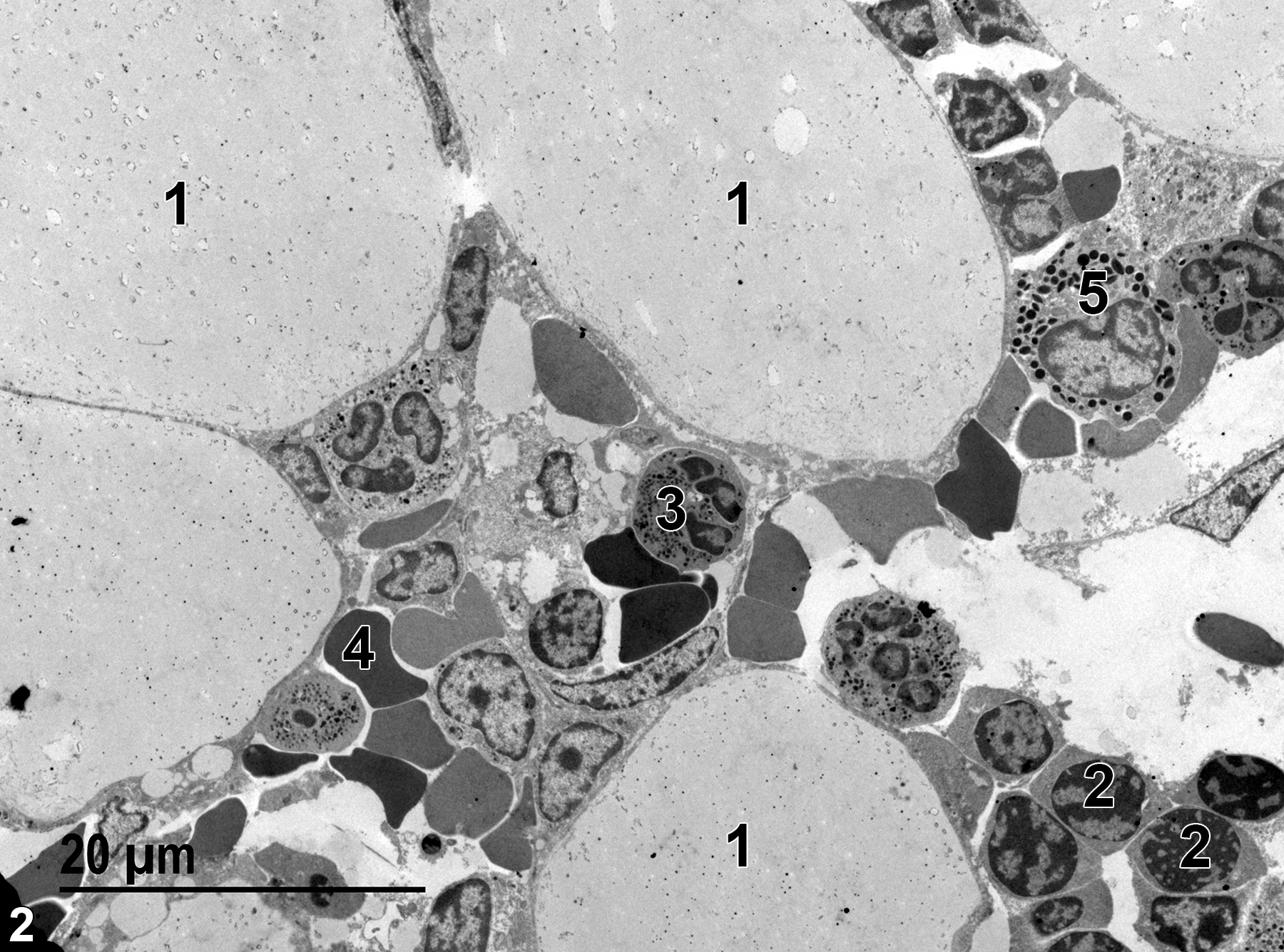
Figure 2. A low magnification view of the bone marrow, containing adipocytes (1), erythroblasts (2), a neutrophil (3), an erythrocyte (4), and an eosinophil (5). 1900x.
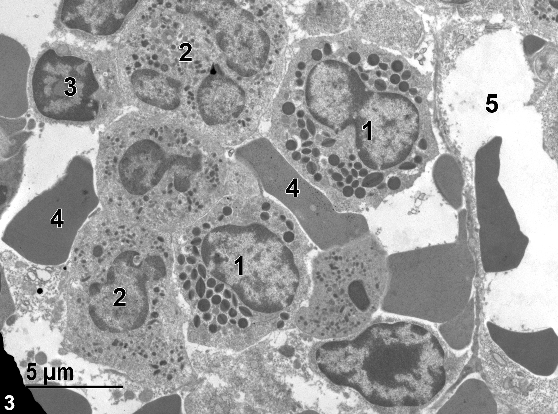
Figure 3. A higher magnification view of marrow structures, including eosinophils (1) with characteristic elliptical granules with linear inclusions, neutrophils (2) with prominent lobed nuclei and three different populations of specific granules, an erythroblast (3) with characteristic nuclei with large amounts of heterochromatin and relatively sparse cytoplasm, erythrocytes (4), and a capillary (5) that contains erythrocytes. 4800x.
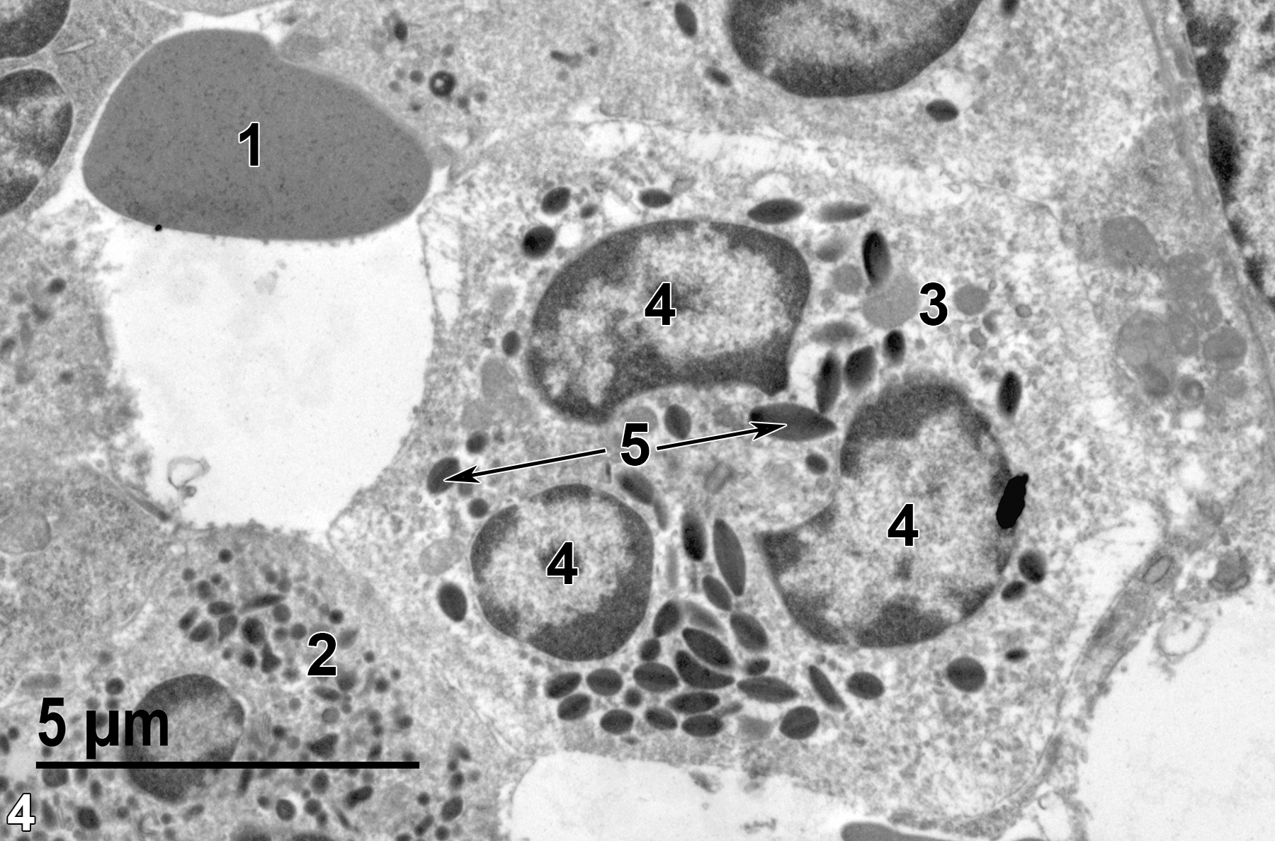
Figure 4. An even higher magnification image showing an erythrocyte (1), a neutrophil (2) with a population of small specific granules, an eosinophil (3) with three pieces of a lobed nucleus (4), and characteristic elliptical granules with linear inclusions (5, arrows). 6800x.
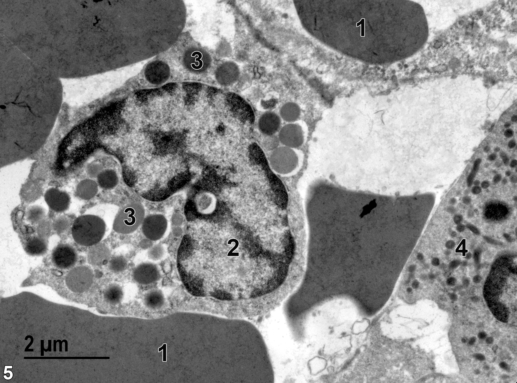
Figure 5. Several erythrocytes (1) and a relatively rare basophil (2) with relatively large and variably sized basophilic granules of varying electron density (3). A neutrophil with elongated granules (4), spherical electron-dense granules, and smaller more electron-lucent granules is located at the lower right of the image. 11000x.
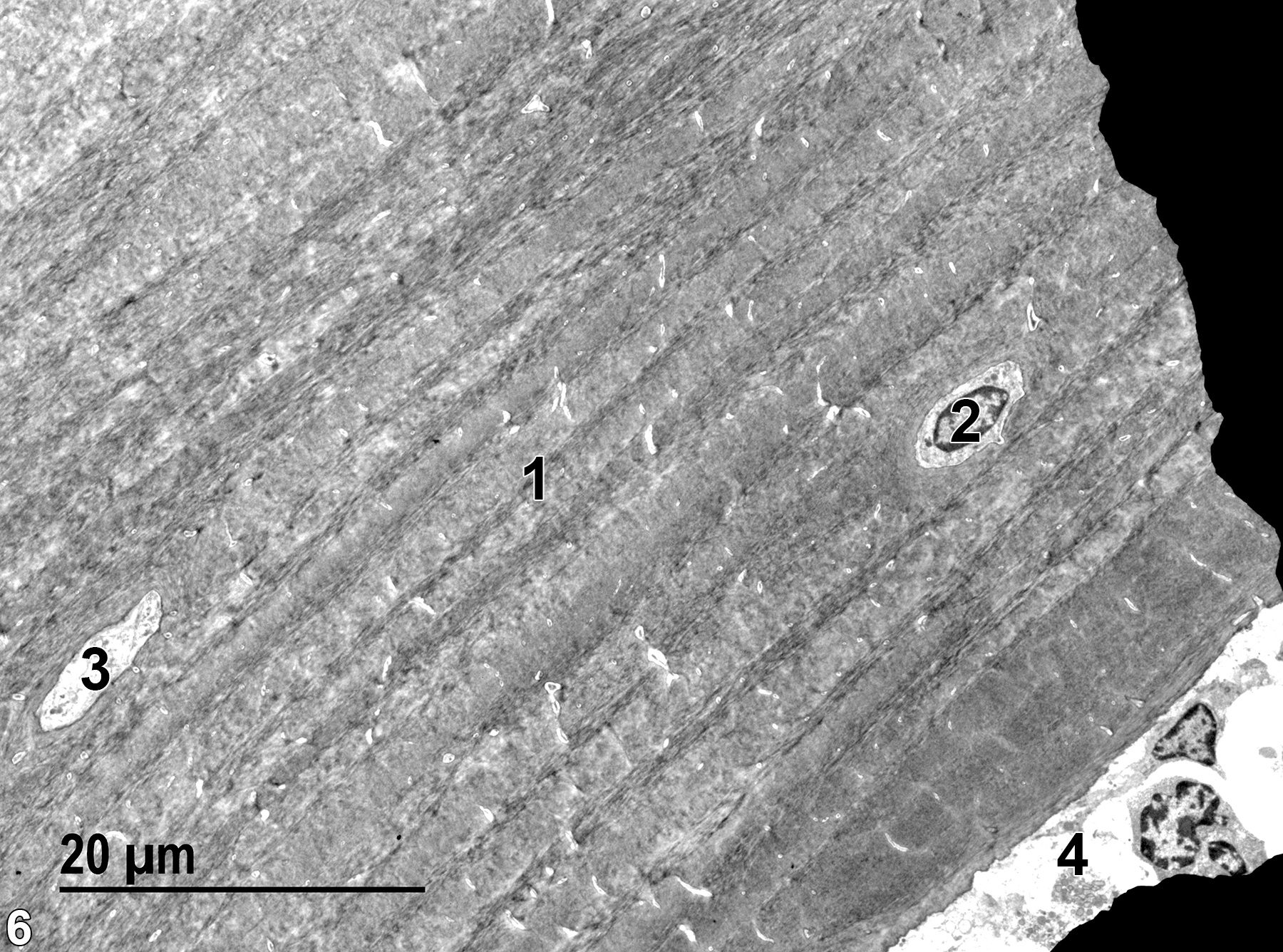
Figure 6. A low magnification view of cortical bone, showing the lamellar structure of the bone matrix (1), with a single Howship’s lacuna (2) containing an osteocyte. The dark areas at the right of the image are grid bars blocking the electron beam. A portion of a Haversian canal (3) is oriented parallel to the long axis of the bone. The edge of the bone marrow (4). 1900x.
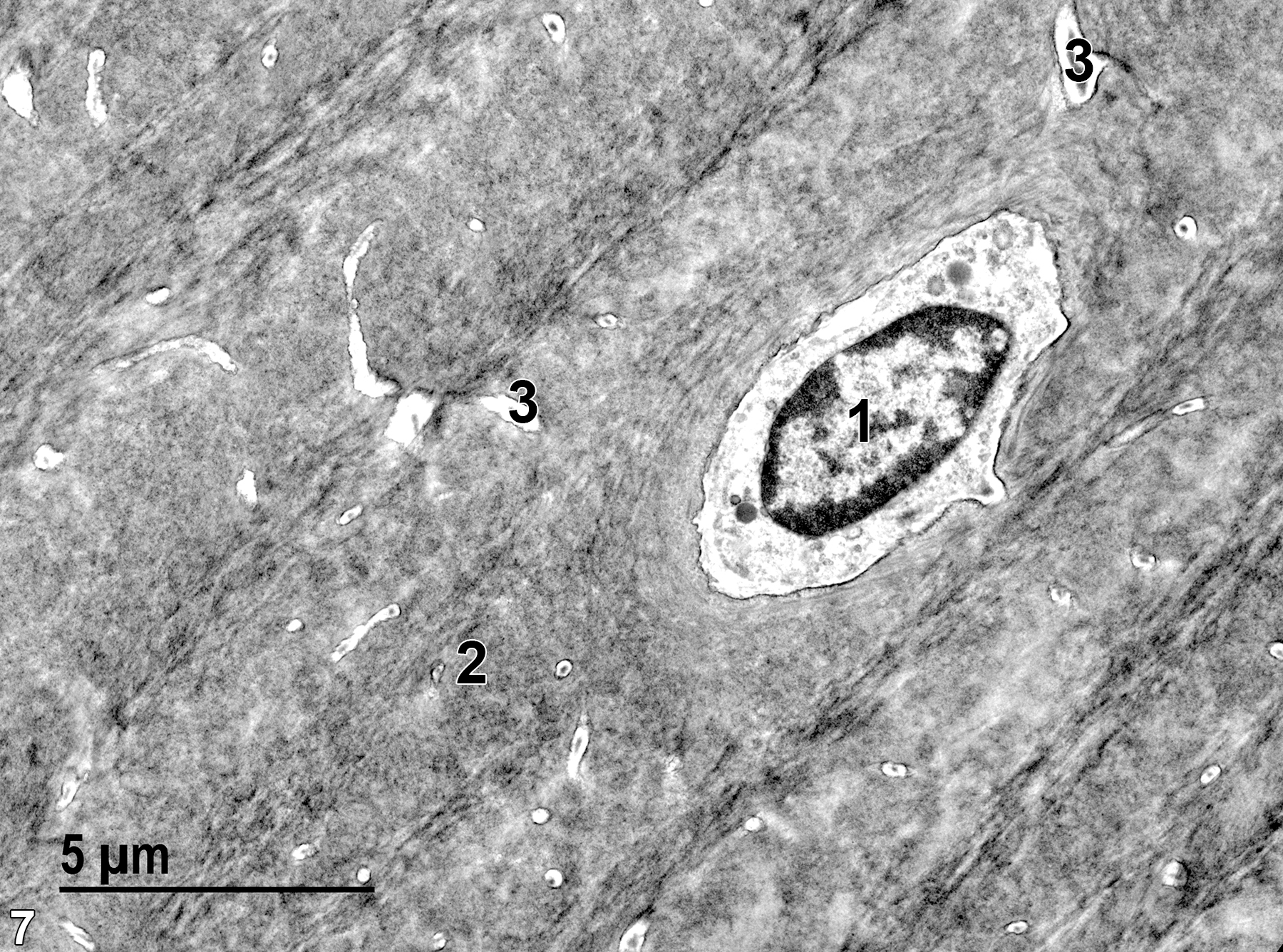
Figure 7. An enlarged view of Figure 6. The nucleus of an osteocyte (1) is surrounded by poorly preserved cytoplasm because of the decalcification procedure necessary to allow sectioning of the bone sample. The lamellar bone matrix (2) is largely parallel to the long axis of the bone, except for the concentric lamellae surrounding Howship’s lacuna containing the osteocyte. Small Volksmann’s canals (3) that are perpendicular to the long axis of the bone are more easily seen at this magnification than at the magnification in Figure 6. 6800x.
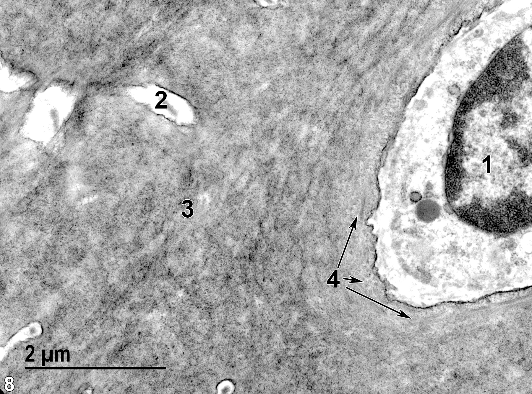
Figure 8. An even higher magnification view of Figure 6, showing the osteocyte nucleus (1), a Volksmann’s canal (2), and both longitudinal and cross-sectional views (4, arrows) of the collagen fibrils within the bone matrix (3). 18500x.
| Boorman GA, Eustis SL, Elwell MR, Montgomery CA, Jr., MacKenzie WF, eds. 1990. Pathology of the Fischer Rat: Reference and Atlas. New York: Academic Press. |
| Dellmann HD, Eurell J, eds. 1998. Textbook of Veterinary Histology. 5th ed. Philadelphia: Lippincott Williams & Wilkins. |
| Rhodin JAG. 1974. Histology: A Text and Atlas. New York: Oxford University Press. |
| Weiss L, ed. 1988. Cell and Tissue Biology: A Textbook of Histology. 6th ed. Baltimore: Urban & Schwarzenberg. |
| Young B, Heath JW. 2000. Wheater’s Functional Histology: A Text and Colour Atlas. 4th ed. Edinburgh, UK: Churchill Livingstone. |
All Images


