

Alimentary System
Small Intestine
Narrative
The small intestine is comprised of the duodenum, the jejunum, and the ileum. The surface is covered with absorptive villi. The villi have absorptive columnar epithelial cells (enterocytes) with microvilli, mucous (goblet) cells, and enteroendocrine cells with membrane-bound electron dense granules. At the base of villi are Peyer’s patches composed of lymphoid tissue. Beneath the epithelium is a lamina propria composed of connective tissue with lymphatic and blood capillaries. A thin smooth muscle layer (muscularis mucosae) separates the lamina propria from the underlying submucosa, which is comprised of nerve fibers, loose connective tissue, and blood vessels. The submucosa is subtended by the tunica muscularis. The outermost layer of the intestines is the serosal (tunica adventitia) tissue, that is a mesothelium consisting of squamous epithelial cells.
The duodenum is attached to the glandular stomach and contains broad or irregular folds or ridges and has the largest villi of the small intestine. The villi are broad and tongue shaped. The jejunum is the next segment and has shorter villi that are conical or rounded. The final segment of the small intestine, the ileum, has club-like villi.
Duodenum
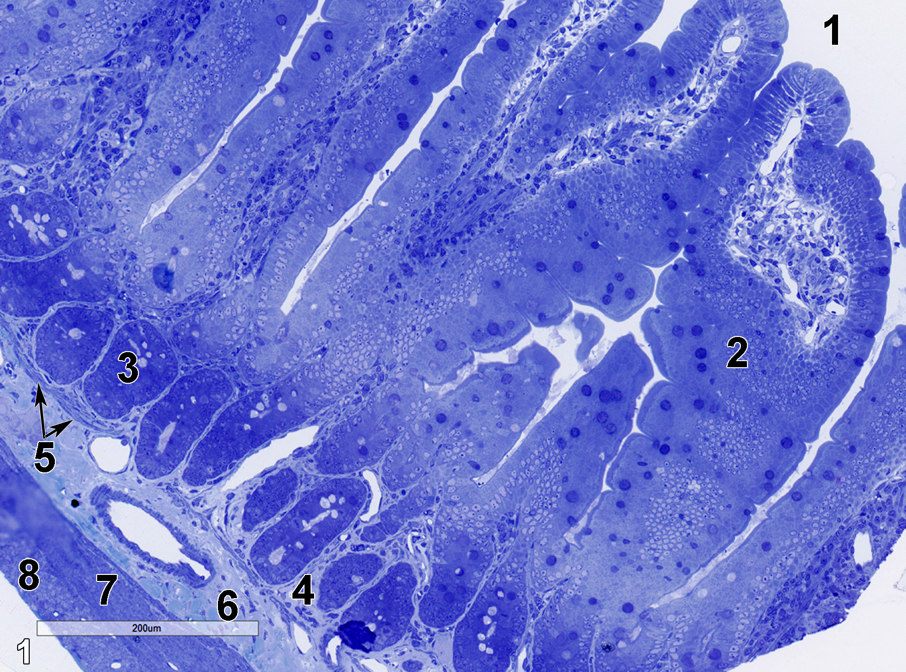
Figure 1. A semithin section (0.5 micrometer thick) of a toluidine blue O-stained portion of the duodenum, the first segment of the small intestine after the glandular stomach. The villi (2) extend into the intestinal lumen (1). The lamina propria (4) surrounds numerous Brunner’s glands (3). The lamina propria is subtended by a thin muscularis mucosae (5), below which is the submucosa (6) consisting of a collagenous matrix with vessels and fibroblasts as major components. An inner circular layer of smooth muscle tissue (7), along with an outer longitudinal layer of smooth muscle tissue (8) comprise the tunica muscularis. 15x.
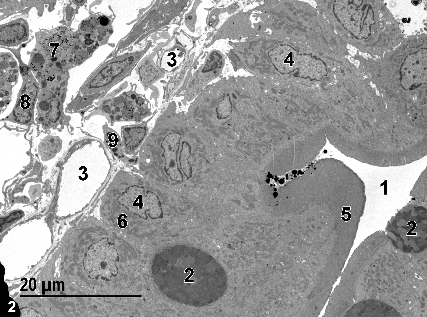
Figure 2. The duodenal lumen (1) lined with absorptive columnar epithelial cells with basal nuclei (4), surface microvilli (5), and numerous mitochondria (6). Occasional mucous (goblet) cells (2) containing mucous droplets are present. Capillaries (3) are present in the lamina propria, along with macrophages (7), fibroblasts (8), and an eosinophil (9). 1900x.
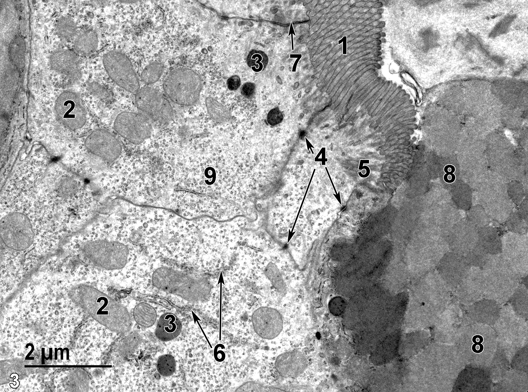
Figure 3. A high magnification view of the duodenal epithelial cells. The surface of the less electron-dense absorptive cells is covered with densely packed microvilli (1). Mitochondria (2) are pleomorphic. A number of small lysosomes (3) are present, along with desmosomes (4, arrows) and other elements of junctional complexes (7, arrow) binding epithelial cells to each other. Rootlets of the terminal web (5) can be seen below the microvilli. Elements of rough endoplasmic reticulum (6, double arrows) and numerous polyribosomes (9) are present. The adjacent mucous cell is more electron-dense because of the closely packed mucous granules (8). 11000x.
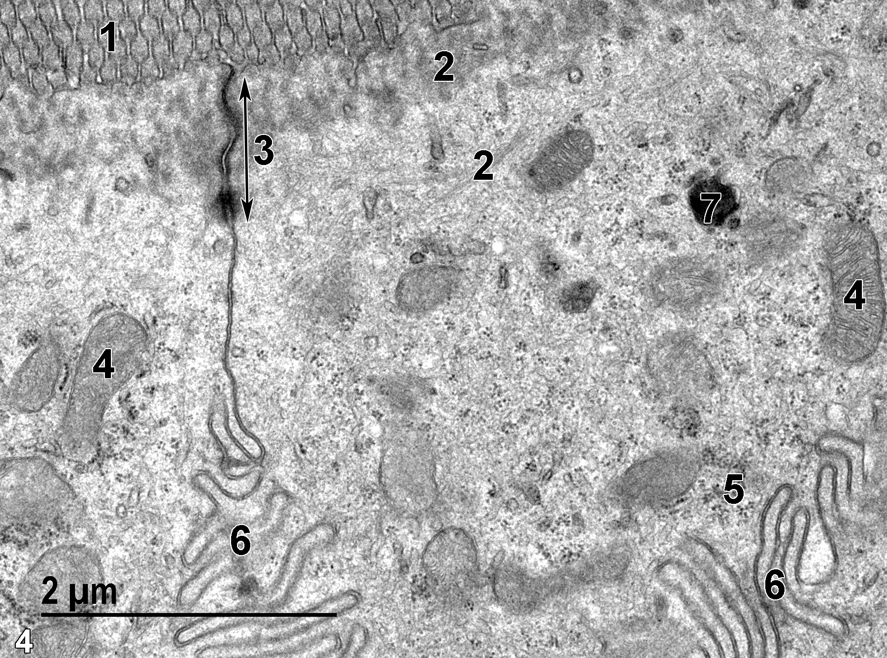
Figure 4. A higher magnification view of an absorptive cell. The microvilli at the cell surface (1) have fibrous terminal web material (2) at their base, with some of the proteinaceous fibrils extending some distance into the cytoplasm below the terminal web. A junctional complex (3) terminating in a desmosome (double-headed arrow) is present above the infoldings of adjacent epithelial cells (6). Free ribosome clusters (5) are present in the cytoplasm, along with mitochondria (4) and a lysosome (7). 23000x.
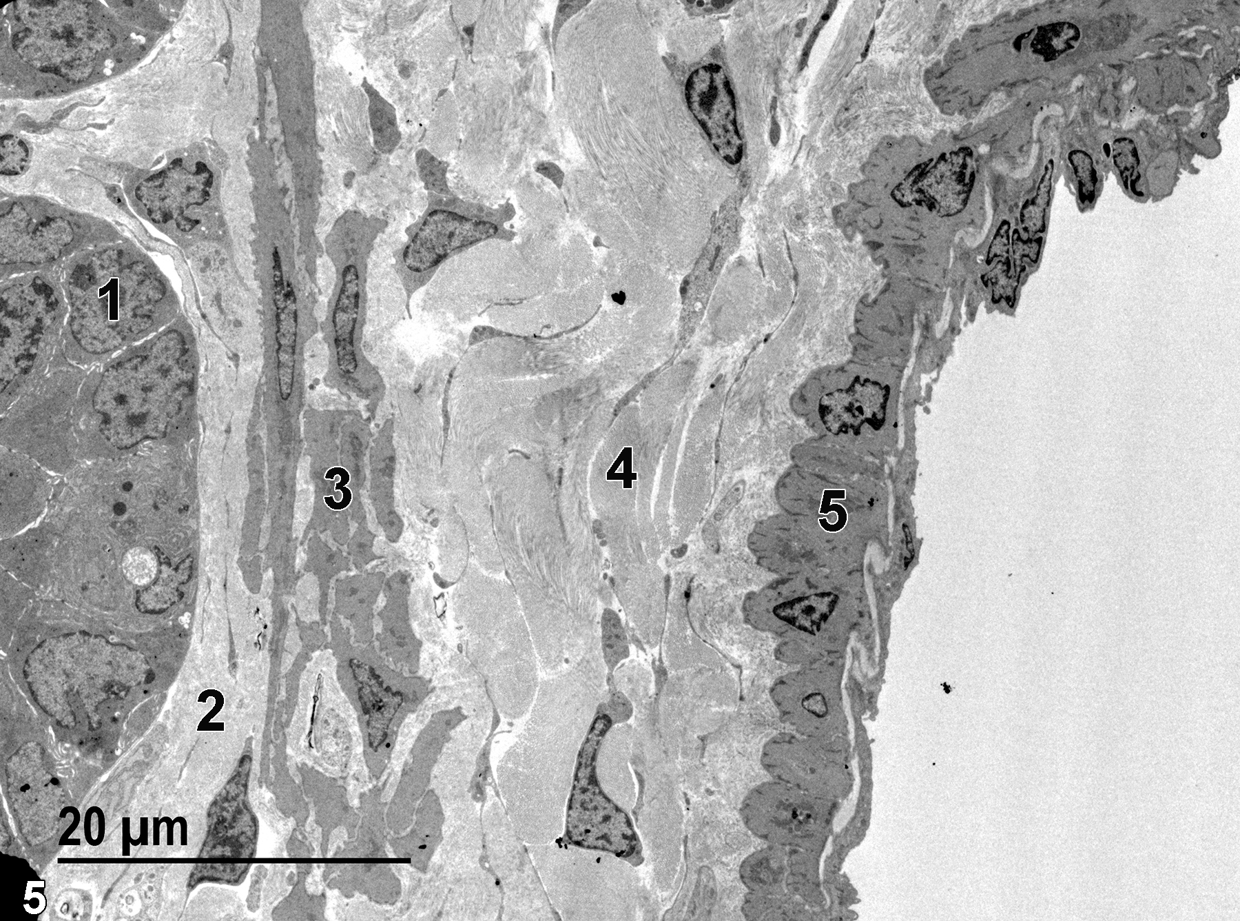
Figure 5. The base of a villus with a single epithelial cell nucleus labeled (1). Below the villus cells is the lamina propria (2) with primarily collagen fibrils and fibroblasts. Directly below the lamina propria is the muscularis mucosae (3) composed of smooth muscle cells and collagen fibrils between the cells. Below that layer is the submucosa (4), which consists of large amounts of collagen and sparse fibroblasts. The final layer to the right in the image is composed of smooth muscle cells (5) of the tunica muscularis. 1900x.
Jejunum
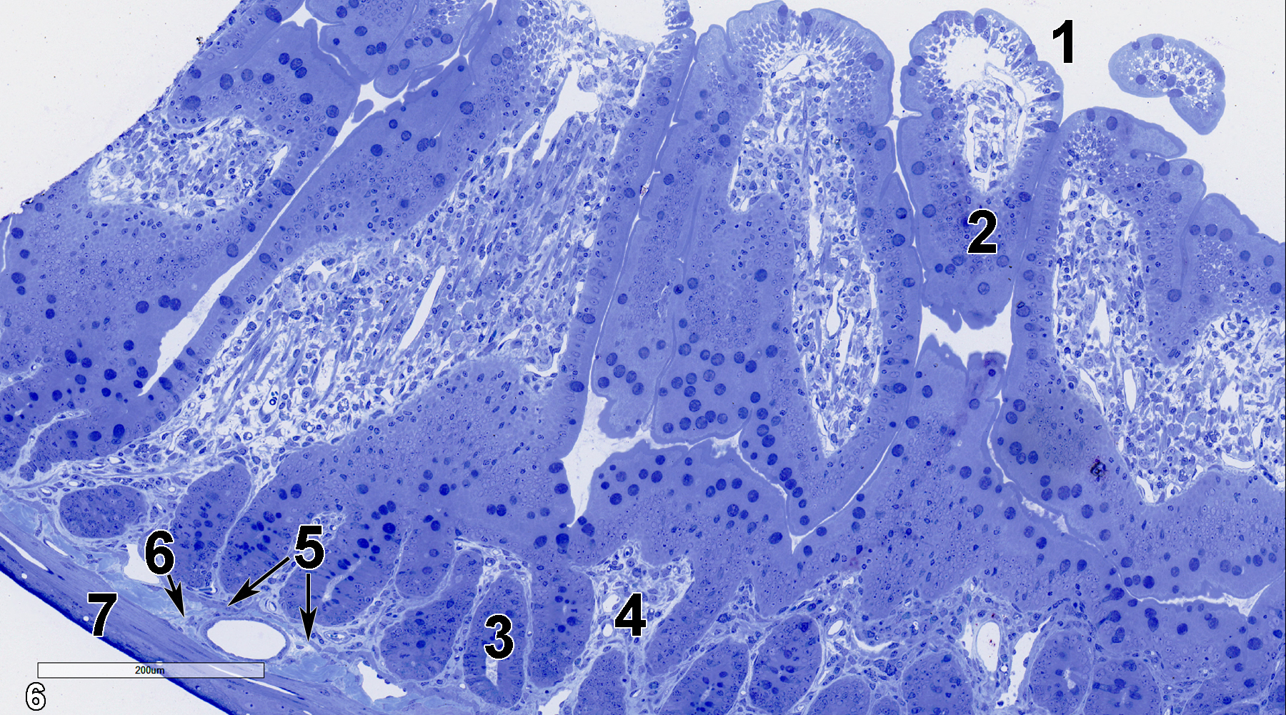
Figure 6. A semithin section (0.5 micrometer thick) of a toluidine blue O-stained portion of the jejunum. The intestinal lumen (1) is lined with villi with absorptive cells (2) covering their surfaces. At the basal portion of the villi is a lamina propria (4). Glands (3) are present within the lamina propria. A thin muscularis mucosae (5, double arrows) separates the lamina propria from the submucosal layer (6, arrow), which contains connective tissue and vessels. Directly below the submucosal layer is the tunica muscularis (7). 15x.
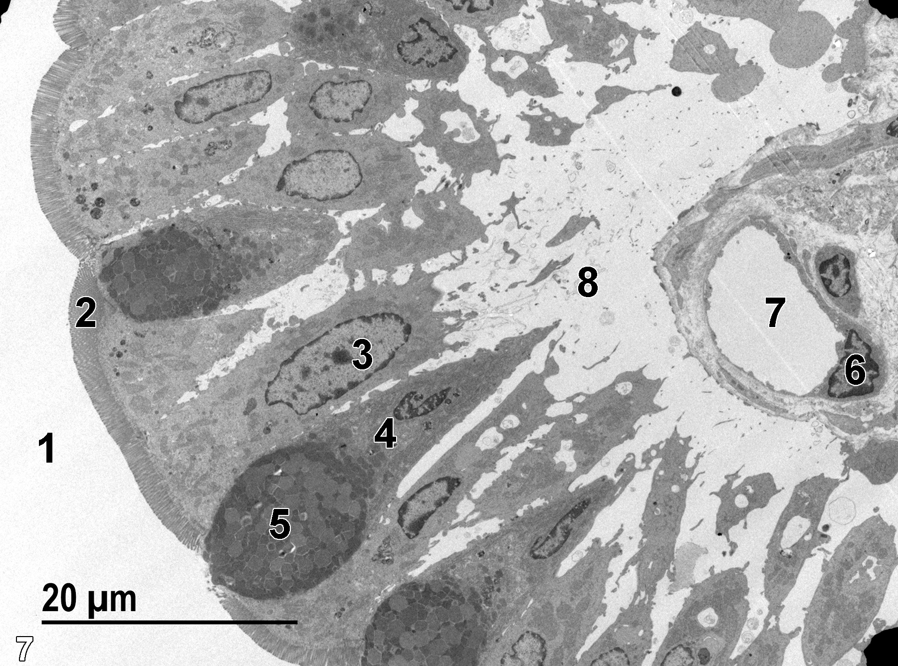
Figure 7. A low magnification electron micrograph of a villus protruding into the intestinal lumen (1). A nucleus of an absorptive cell (3) is located basally in the epithelial cell, whereas the surface of the cell has a dense covering of microvilli (2). A mucous cell (4) contains a mass of mucous droplets (5) near the luminal surface. The interior of the villus (8) contains a capillary bed with surrounding connective tissue. A single capillary lumen (7) with a thin endothelial lining and a single nucleus (6) are shown. 1900x.
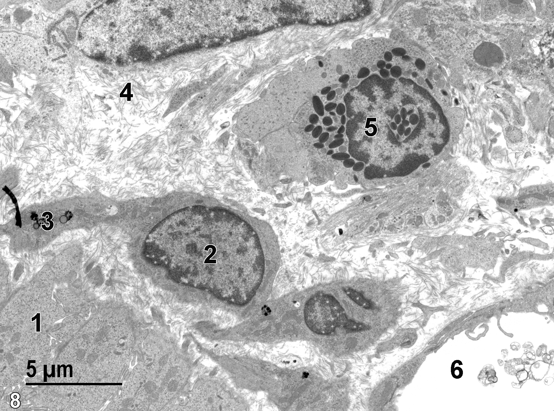
Figure 8. A higher magnification view of the lamina propria (1) beneath the epithelial cells of a villus, showing a fibroblast nucleus (2) and lysosomes (3) in the fibroblast cytoplasm. The lamina propria also contains numerous collagen fibrils (4) and a single eosinophil (5) with characteristic specific granules that have a lenticular shape. A single blood vessel (6) containing membranous debris is shown. 4800x.
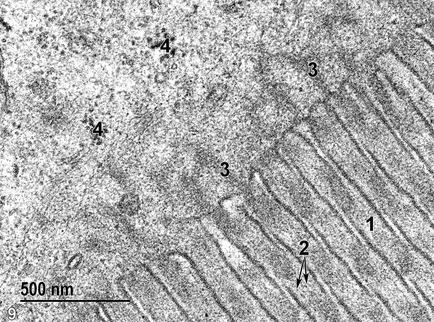
Figure 9. A high magnification view of the surface of an absorptive epithelial cell of a villus. The microvilli (1) contain a core of proteinaceous filaments (2, double arrows) that are connected with the rootlets of the terminal web (3) below the microvilli. Clusters of free ribosomes (4) are present in the cytoplasm. 68000x.
Ileum
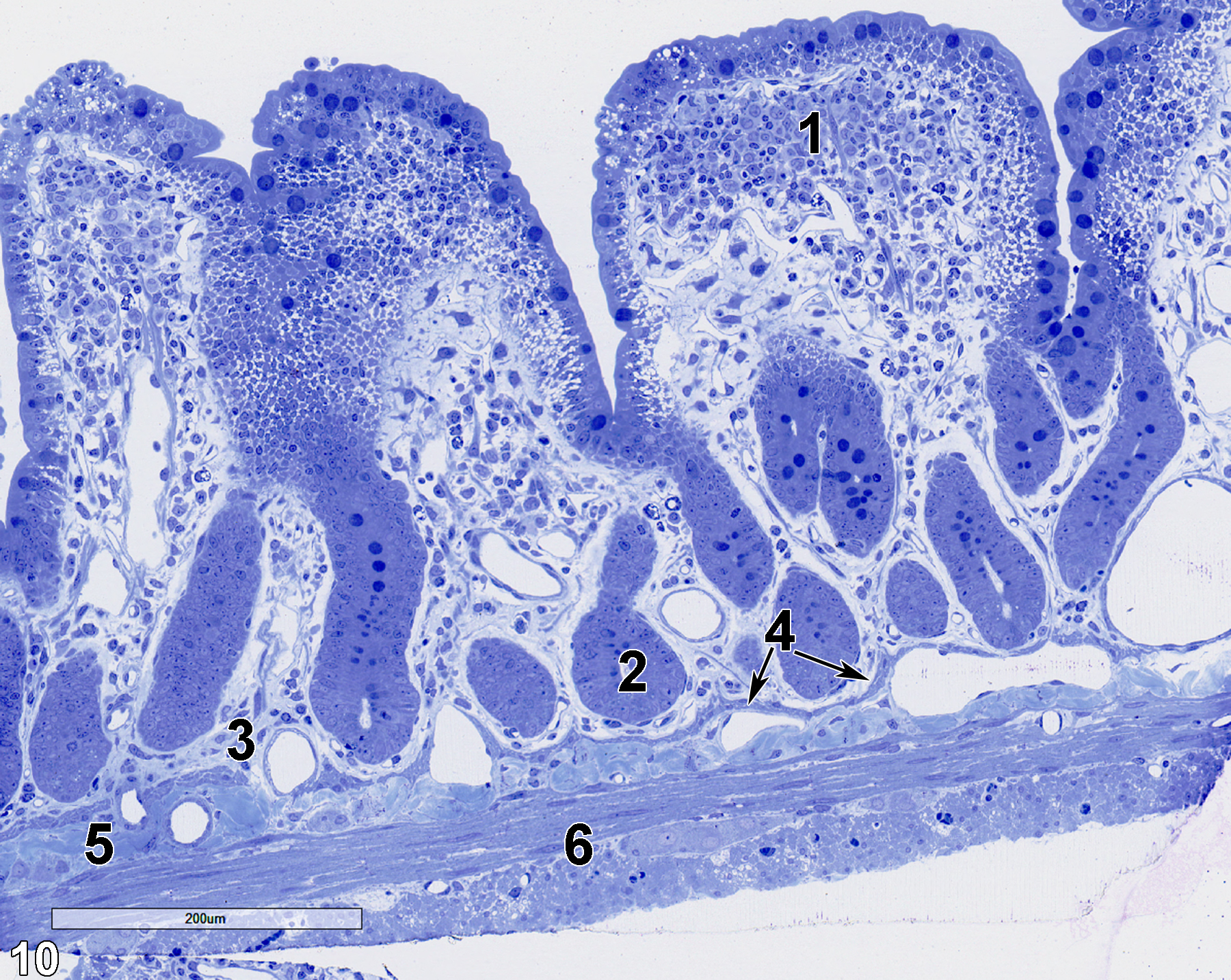
Figure 10. A semithin section (0.5 micrometer thick) of a toluidine blue O-stained portion of the ileum. It has relatively short villi (1) with basal glands (2) embedded in the lamina propria (3), subtended by a thin muscularis mucosae of smooth muscle cells (4, double arrows) that are overlaying the submucosa (5), which is composed of connective tissue, fibroblasts, and vessels. The muscle cells of the tunica muscularis (6) are shown. 15x.
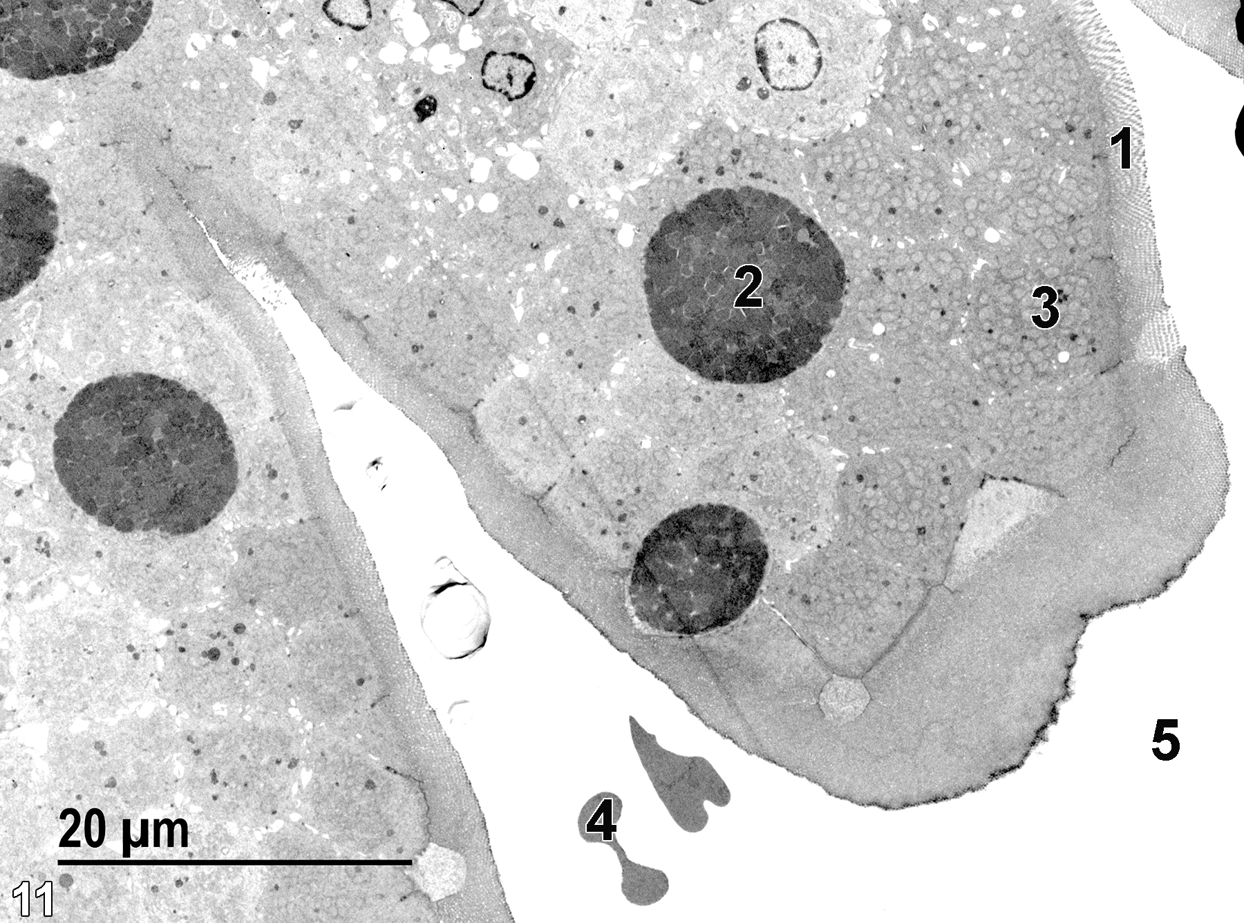
Figure 11. The tips of two villi, covered with microvilli (1). Intermixed with the absorptive epithelial cells (3) are mucous cells (2) with more electron-dense content, primarily mucous droplets. The intestinal lumen (5) contains a few erythrocytes (4), probably introduced during sample collection. 1900x.
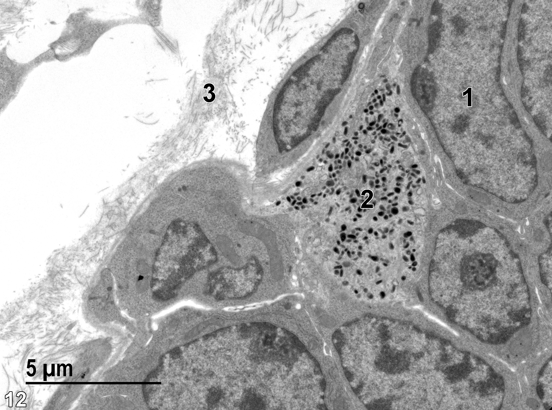
Figure 12. A view of the base of an ileum villus showing a nucleus of an absorptive cell (1). An enteroendocrine cell (2) contains numerous electron-dense secretory granules. Collagen fibrils (3) are a major component of the lamina propria. 6800x.
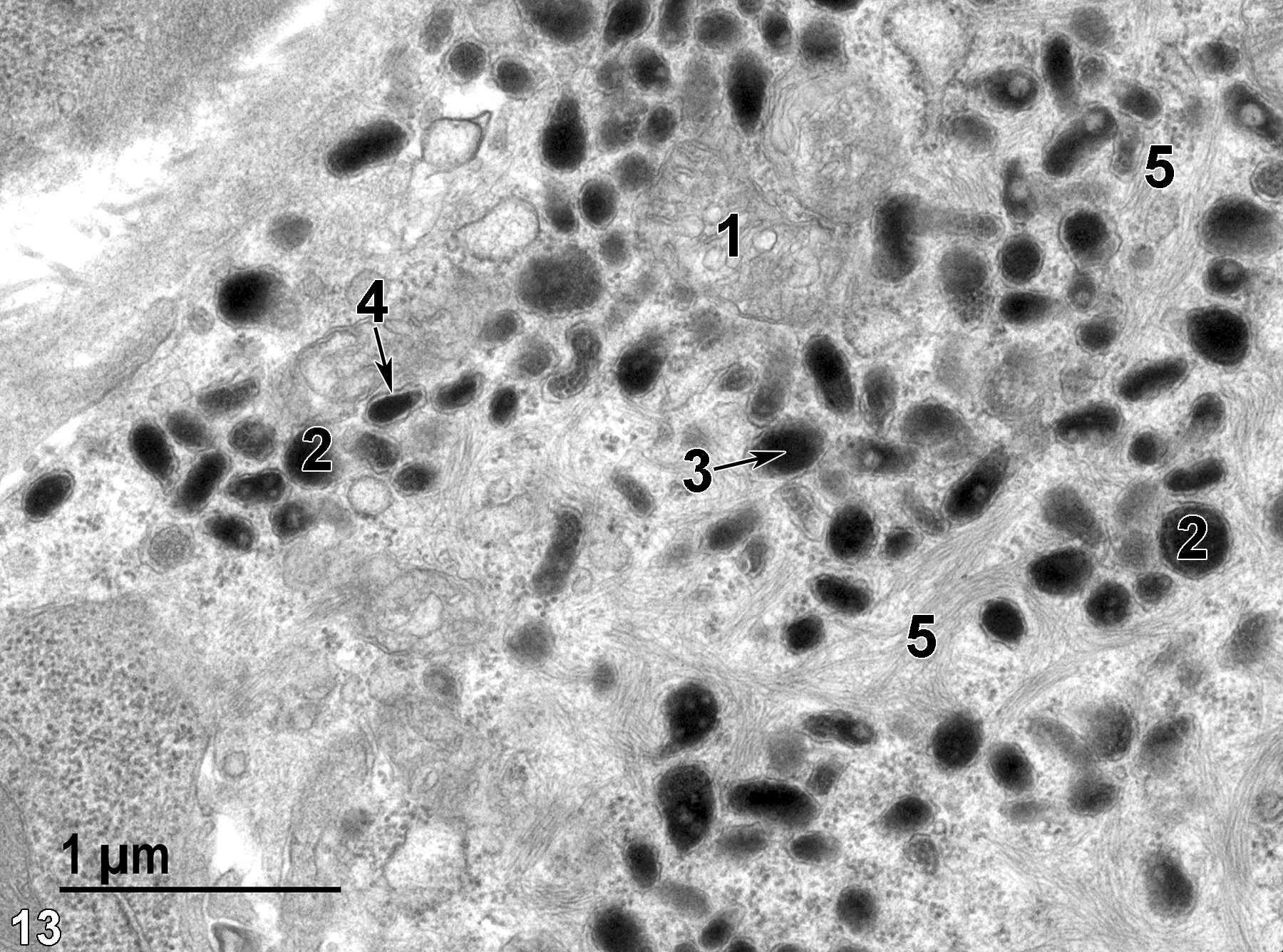
Figure 13. A high magnification view of an enteroendocrine cell. A mitochondrion (1) is surrounded by secretory granules (2) that vary somewhat in size and shape. The electron-dense core (3, arrow) is frequently separated from the membrane surrounding the granule (4). The cell also contains bundles of tonofilaments (5). 30000x.
| Boorman GA, Eustis SL, Elwell MR, Montgomery CA, Jr., MacKenzie WF, eds. 1990. Pathology of the Fischer Rat: Reference and Atlas. New York: Academic Press. |
| Cross PC, Mercer KL. 1993. Cell and Tissue Ultrastructure: A Functional Perspective. New York: W.H. Freeman and Company. |
| Dellmann HD, Eurell J, eds. 1998. Textbook of Veterinary Histology. 5th ed. Philadelphia: Lippincott Williams & Wilkins. |
| Rhodin JAG. 1974. Histology: A Text and Atlas. New York: Oxford University Press. |
| Ross MH, Kaye GI, Pawlina W. 2003. Histology: A Text and Atlas. 4th ed. Philadelphia: Lippincott Williams & Wilkins. |
| Uehara T, Elmore SA, K.A. Szabo KA. 2017. Chapter 6: Esophagus and stomach. In Boorman’s Pathology of the Rat (Suttie AW, ed). 2nd ed. London: Academic Press, 35-50. |
| Weiss L, ed. 1988. Cell and Tissue Biology: A Textbook of Histology. 6th ed. Baltimore: Urban & Schwarzenberg. |
All Images


