

Alimentary System
Pancreas
Narrative
The pancreas is surrounded by a thin capsule of loose connective tissue and is composed of exocrine (acinar) tissue and endocrine (Islets of Langerhans) tissue. The exocrine tissue is primarily glandular parenchymal cells separated into lobules by septa of connective tissue with ducts, blood vessels, neuronal cells, and lymphatics. The parenchymal epithelial cells are organized into acini surrounded by basal laminae. Acini have tall columnar or pyramidal epithelial cells with basally located nuclei and apical regions filled with secretory (zymogen) granules surrounding the acinar lumen. The epithelial cell surface bordering the acinar lumen has short microvilli. The endocrine tissue (Islets of Langerhans) can be found throughout the pancreas but is more prevalent in the tail region. There is no capsule separating the Islets of Langerhans cells from the acinar tissue, but there are collagen fibrils and a thin basal lamina between these two types of tissue. The epithelial cells of the Islets are subdivided into four major types of cells. The alpha cells make up approximately 20% of the Islets and are located primarily at the periphery of the Islets. These cells have numerous fairly uniform secretory granules and mitochondria that tend to be elongated and to be smaller than those found in the beta cells. Glucagon is produced by alpha cells. The most common cell is the beta cell. These cells are found more at the interior of the Islets and produce insulin. They tend to have more mitochondria that are larger and more spherical than those of the alpha cells and are reported to have less rough endoplasmic reticulum (Rhodin 1974). The secretory granules of these cells tend to be more polymorphic than those of the alpha cells, with prominent spaces between the granule surface and the electron-dense core. Two other cell types, delta cells and c cells are less common and have also been described by Rhodin (1974).
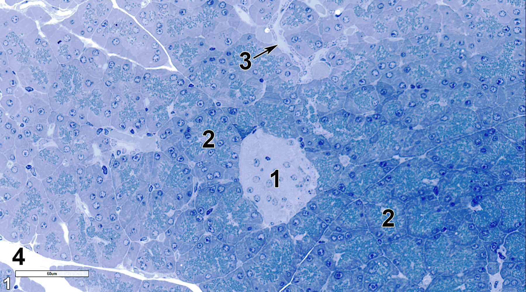
Figure 1. A semithin section (0.5 micrometer thick) of a toluidine blue O-stained section showing a single Islet of Langerhans (1), which is composed of endocrine cells, and a large number of acini (2), which consist of epithelial cells surrounding the acinar lumens. A septum (3), primarily composed of collagenous tissue, separates lobules of the acinar tissue. Part of an interlobular duct (4) is shown. 40x.
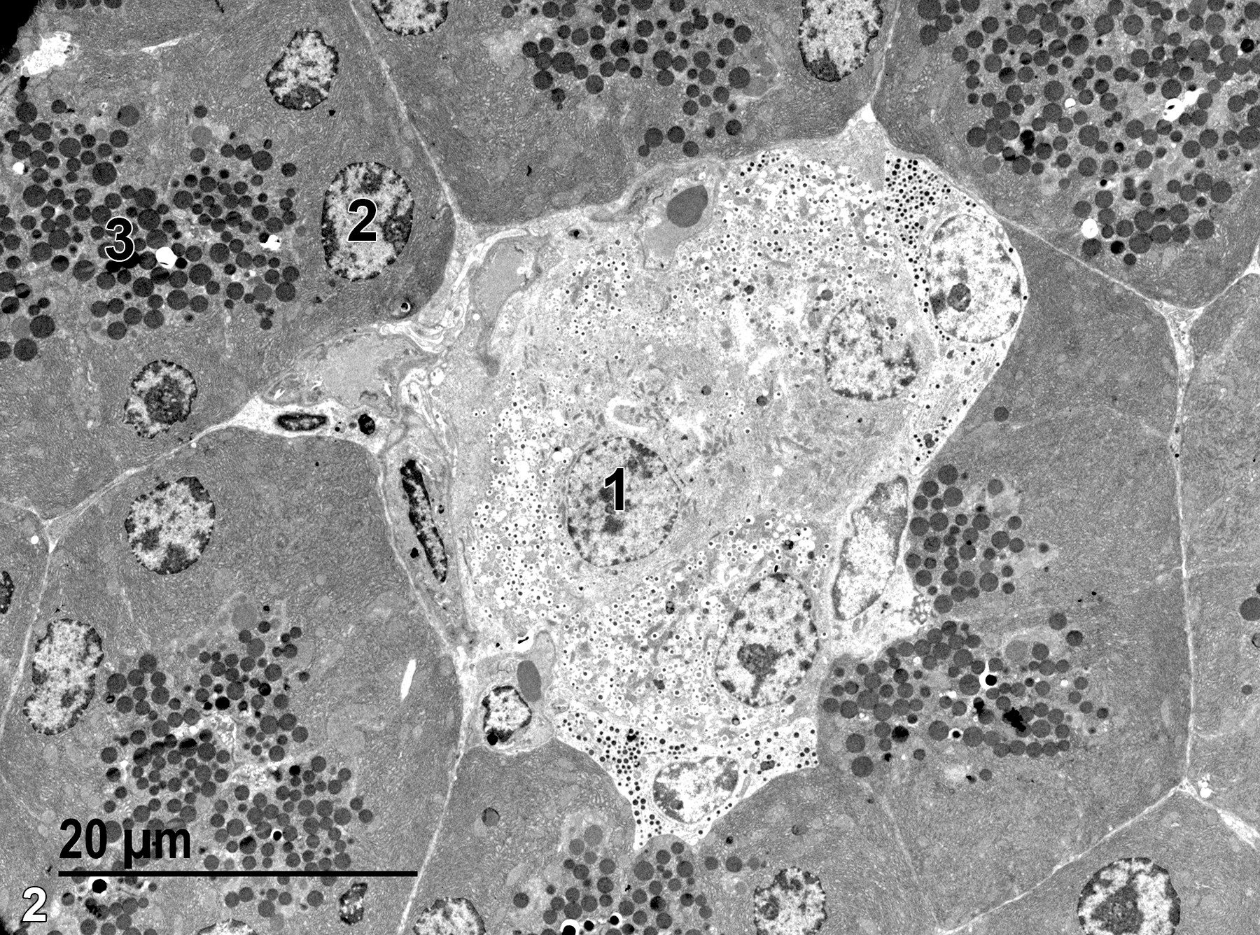
Figure 2. A low magnification electron microscope image of the same area shown in Figure 1. The less electron-dense area in the center is an Islet of Langerhans, with one of the endocrine cell nuclei (1) shown. A nucleus located near the base of an acinar epithelial cell (2) is located beneath the more apical zymogen granules (3). 1900x.
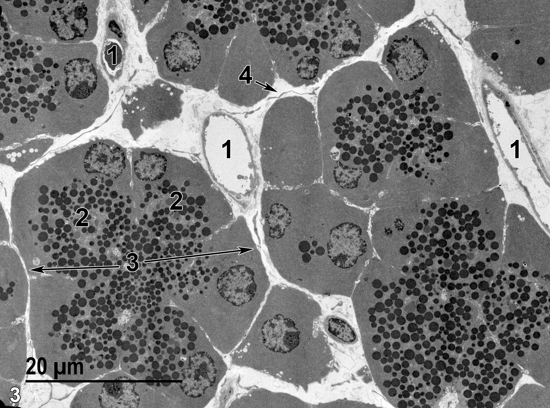
Figure 3. A higher magnification view of the parenchymal tissue showing capillaries (1) in the septal areas separating the acini (3, double arrows). Individual acinar glandular epithelial cells (2) have basal nuclei and apical zymogen granules and are arranged around acinar lumens. Some elements of connective tissue of an intralobular septum (4) can be seen between the lobules, which are composed of collections of acini. 1900x.
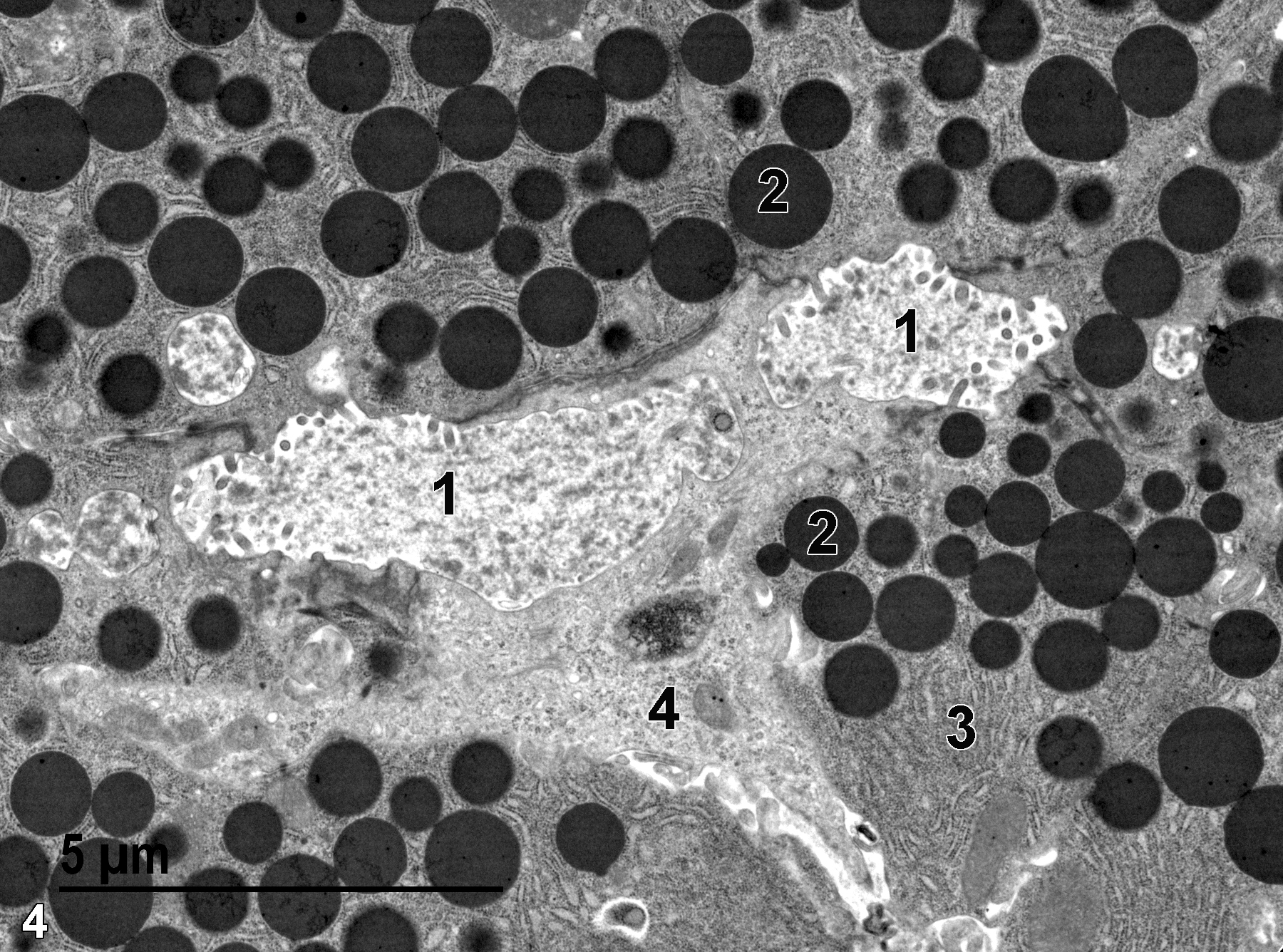
Figure 4. An even higher magnification of acinar epithelial cells showing the short microvilli (1) protruding into the acinar lumens, zymogen granules (2), accumulations of rough endoplasmic reticulum (3), and a pale staining centroacinar cell (4). 9300x.
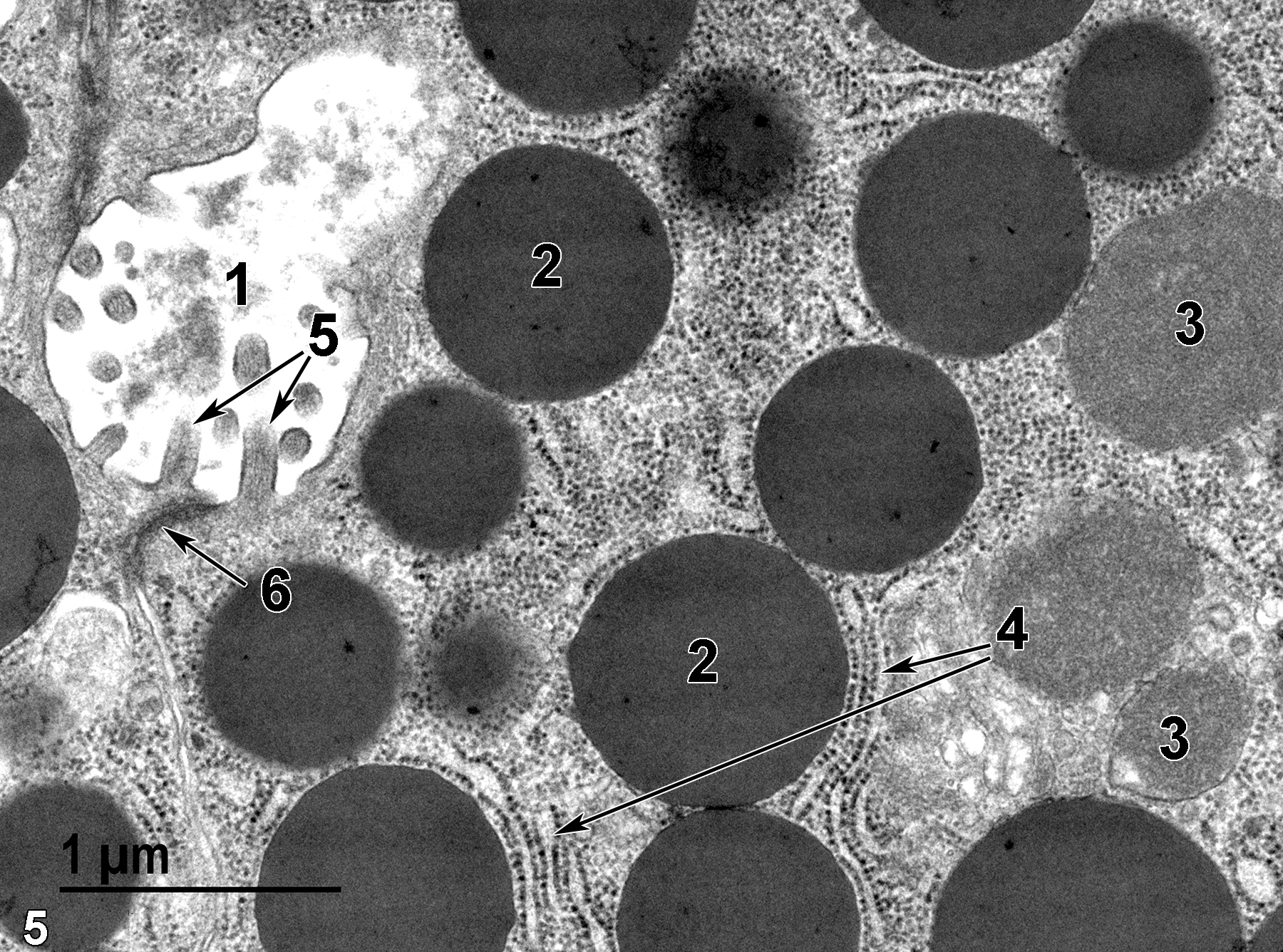
Figure 5. The highest magnification view of the apical region of parenchymal epithelial cells showing the acinar lumen (1), zymogen granules (2), mitochondria (3), rough endoplasmic reticulum (4, short and long arrows), microvilli of the epithelial cells extending into the acinar lumen (5, double arrows), and a junctional complex between adjacent epithelial cells (6, arrow). 30000x.
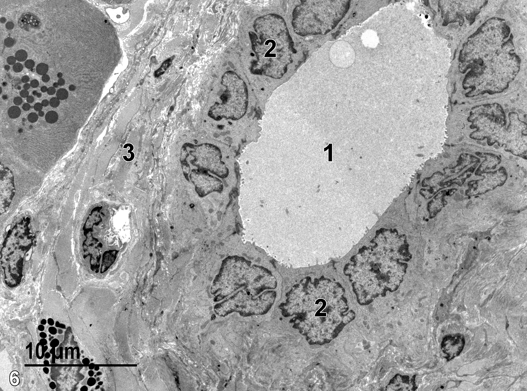
Figure 6. The intralobular duct lumen (1) contains some finely flocculent material and has short microvilli from the duct cells protruding into the lumen. The duct is composed of a single layer of low cuboidal epithelial cells, each with a single nucleus (2). An interlobular septum (3) contains bundles of collagen that appear as homogeneous gray areas at this magnification. 2900x.
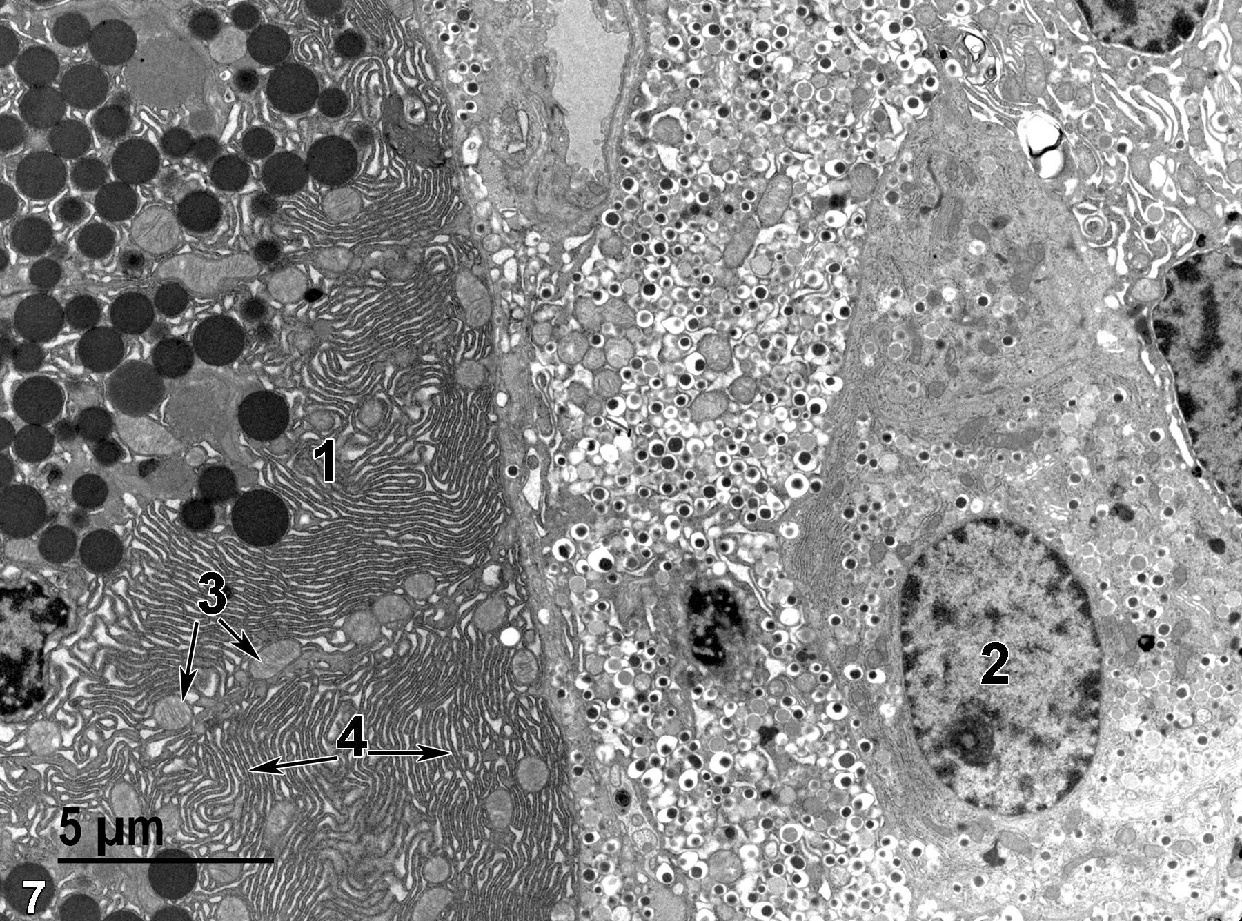
Figure 7. A glandular epithelial cell of the parenchyma (1) to the left, containing masses of rough endoplasmic reticulum (4, double arrows) and mitochondria (3, double arrows) in addition to the electron dense zymogen granules. To the right is the edge of the less electron-dense endocrine cells of an Islet of Langerhans. The nucleus (2) of one of the endocrine cells is labeled. 4800x.
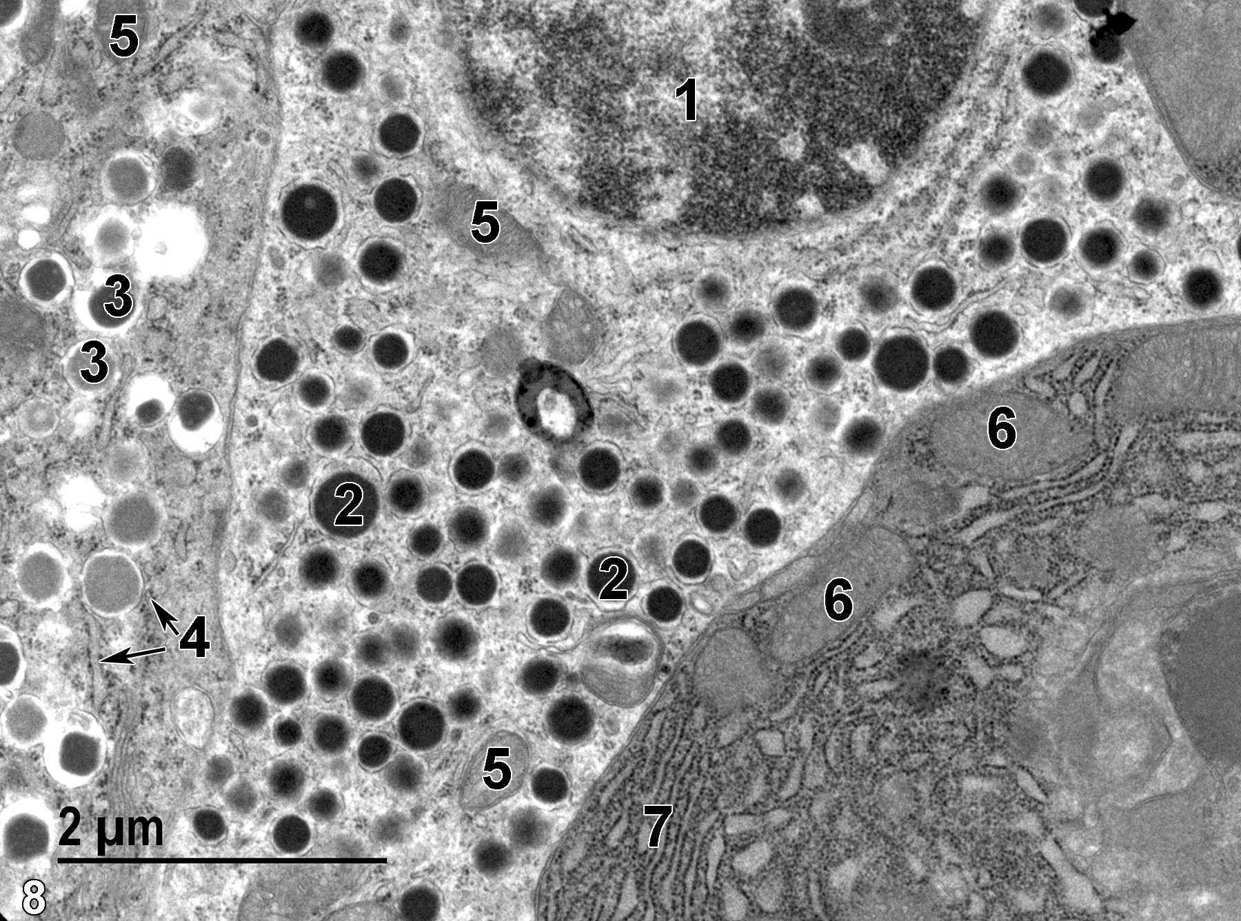
Figure 8. An alpha cell (glucagon cell) nucleus (1). Note the characteristic electron-dense granules (2) with little space between the granule membrane and contents, and the mitochondria (5). The cell to the lower right of the image is part of an exocrine cell containing large amounts of rough endoplasmic reticulum (7) and a few mitochondria (6). The typical zymogen granules and nucleus of the cell are out of the plane of section. The cell to the far left of the image is a beta cell (insulin cell) with variably electron-dense granules (3) with a core typically separated from the membrane by a clear space. Elements of rough endoplasmic reticulum (4, double arrows) are evident. 18500x.
| Cross PC, Mercer KL. 1993. Cell and Tissue Ultrastructure: A Functional Perspective. New York: W.H. Freeman and Company. |
| Dellmann HD, Eurell J, eds. 1998. Textbook of Veterinary Histology. 5th ed. Philadelphia: Lippincott Williams & Wilkins. |
| Rhodin JAG. 1974. Histology: A Text and Atlas. New York: Oxford University Press. |
| Weiss L, ed. 1988. Cell and Tissue Biology: A Textbook of Histology. 6th ed. Baltimore: Urban & Schwarzenberg. |
All Images


