

Immune System
Lymph Node, Mesenteric
Narrative
A mesenteric lymph node is covered with a capsule that consists of connective tissue with some smooth muscle cells and elastic fibers. Some capsular material extends into the cortex, forming the trabecula (reticular meshwork) of the stroma. Directly beneath the capsule is a subcapsular sinus that is continuous with cortical sinuses, with associated dendritic cells and connective tissue that includes the trabeculae. This reticular meshwork supports lymphocytes, macrophages, and other leukocytes. The outer cortex parenchyma contains lymphoid follicles with dense populations of lymphocytes and germinal centers surrounded by more diffuse lymphatic tissue. The diffuse lymphocytic parenchyma extends into the deep cortex (paracortex). The medullary portion of the lymph node consists of cords that contain lymphocytes and plasma cells, as well as sinuses that contain macrophages, plasma cells, and lymphocytes.
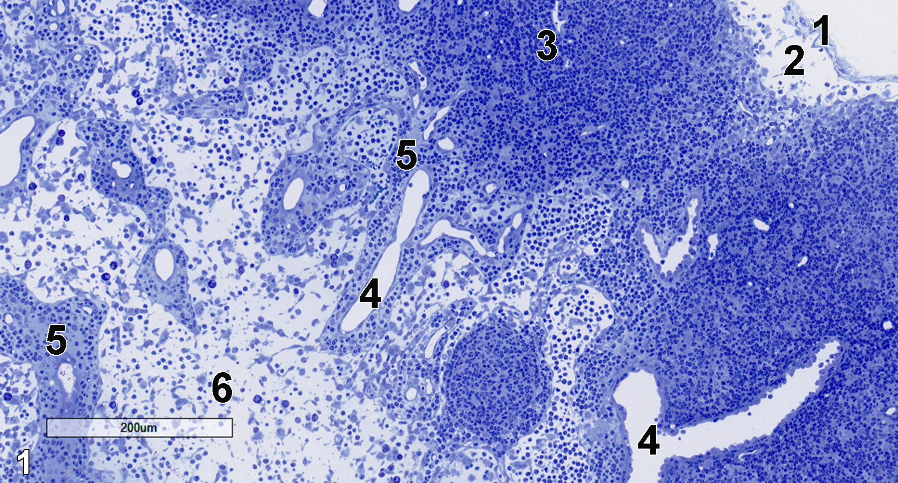
Figure 1. A semithin section (0.5 micrometer thick) of a toluidine blue O-stained portion of a mesenteric lymph node. A thin capsule (1) is underlain by the subcapsular sinus (2), which contains lymphocytes. The cortical parenchyma below the subcapsular sinus shows the dense accumulation of lymphocytes of a lymphoid follicle (3). Cortical sinuses (4) are surrounded by the connective tissue of the trabeculae (5). The medullary sinuses are composed of loose lymphatic tissue (6). 10x.
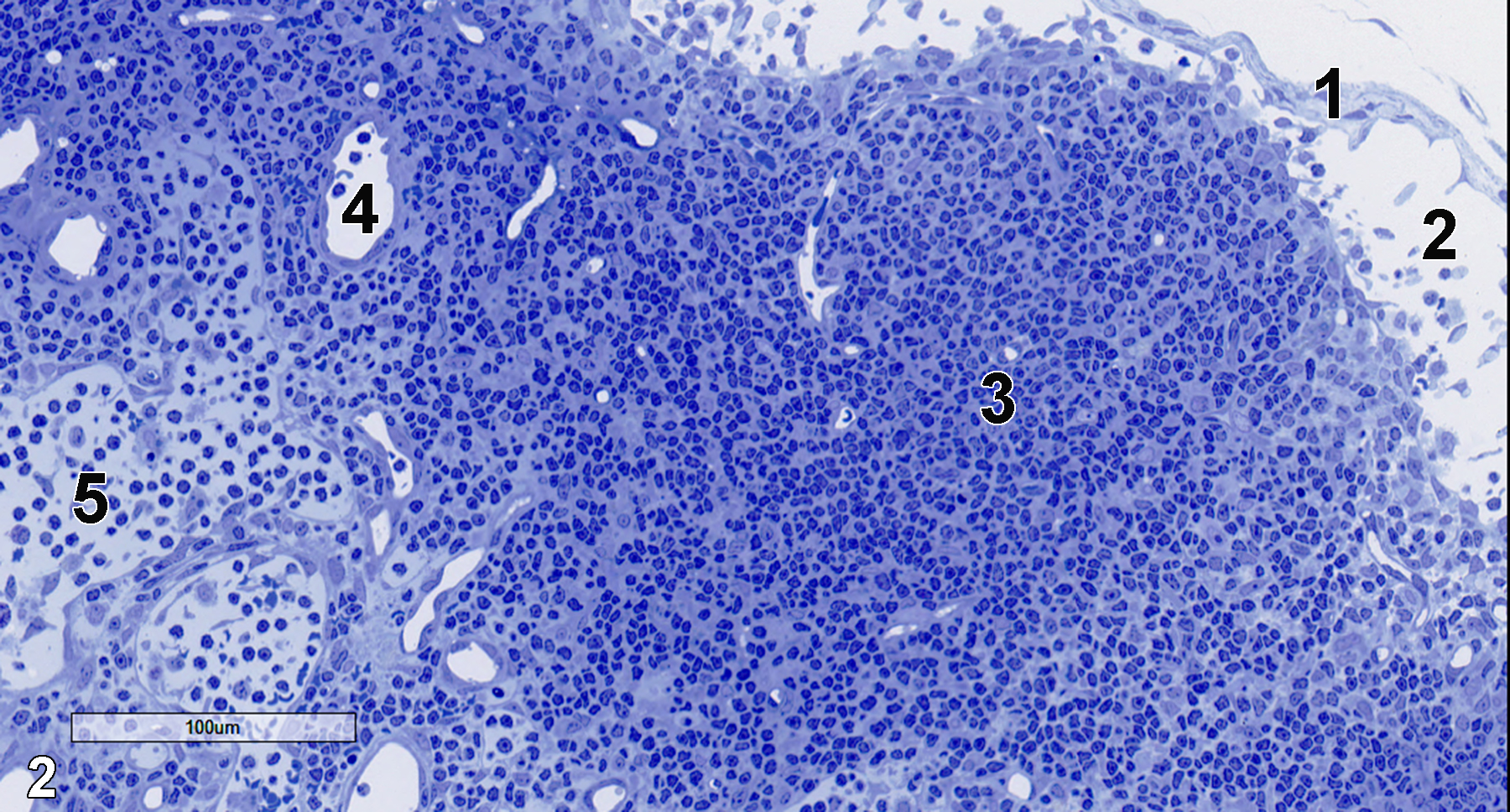
Figure 2. Another semithin section at slightly higher magnification showing the capsule (1), subcapsular sinus (2), and cortical parenchyma, which consist of lymphocytes (3) and cortical sinuses (4). Medullary sinuses (5) are also visible. 20x.
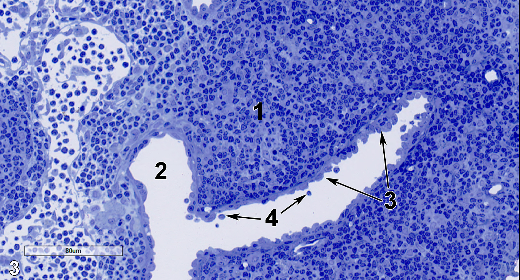
Figure 3. A portion of the cortical parenchyma (1) with a lymphatic sinus (2) lined by endothelial cells (3, arrows) and that contains lymphocytes in the sinus lumen (4, arrows). 25x.
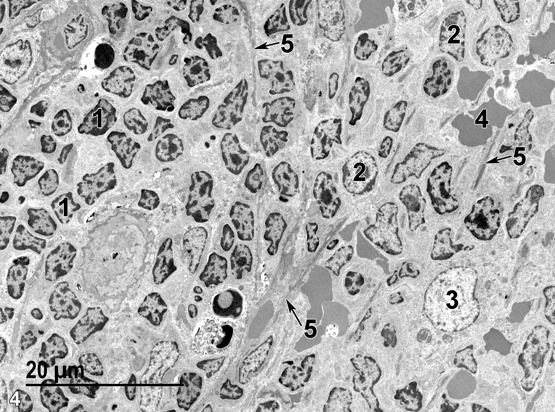
Figure 4. An ultrastructural view of a portion of the cortical parenchyma, which consists of small lymphocytes (1), medium-sized lymphocytes (2), a large lymphocyte (3), and erythrocytes (4). Portions of the trabecular meshwork are present (5, arrows). 1900x.
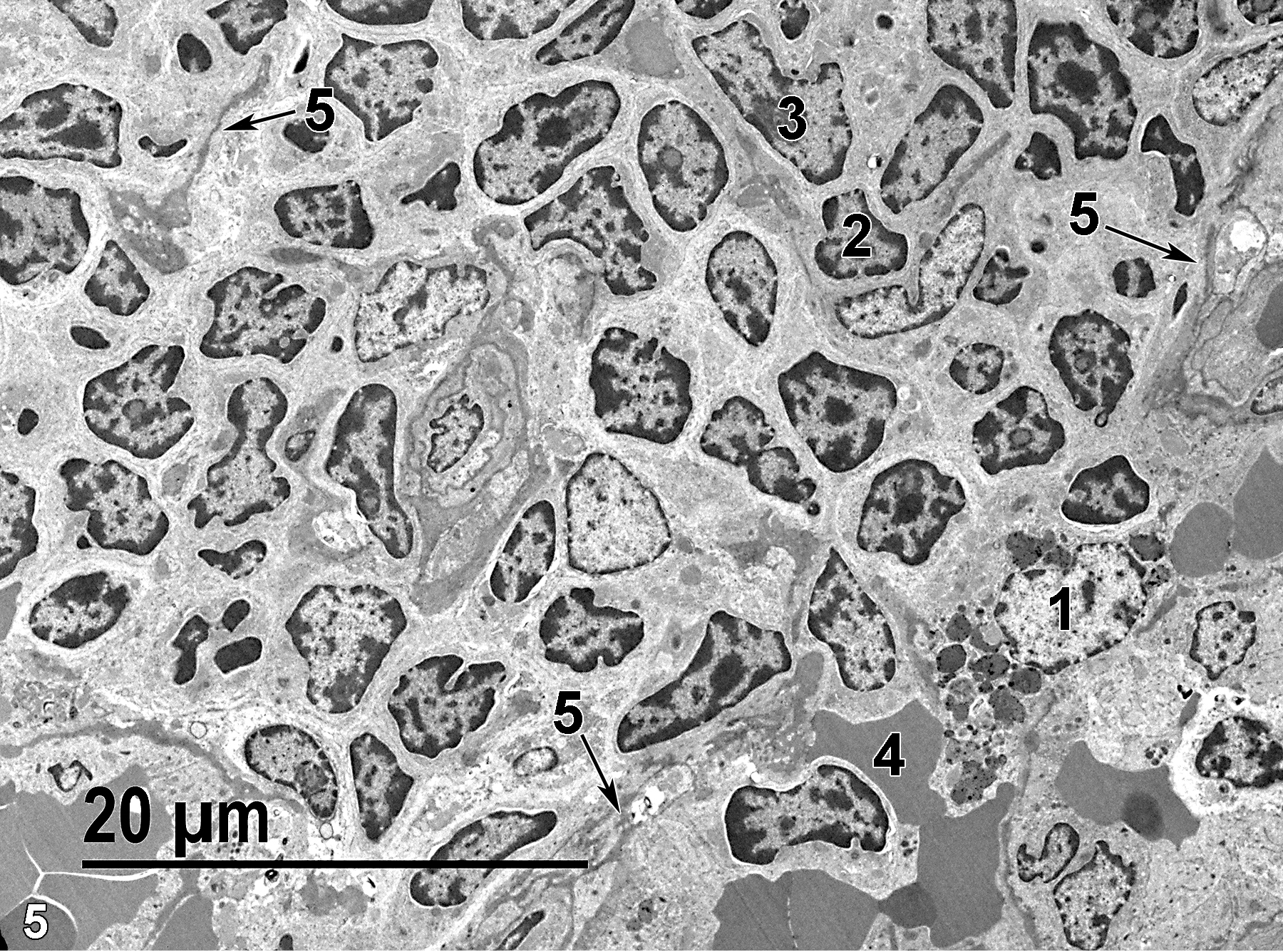
Figure 5. Another portion of the cortical parenchyma, which contains a macrophage (1), a small lymphocyte (2), a medium-sized lymphocyte (3), and an erythrocyte (4). Portions of the trabecular meshwork are evident (5, arrows). 1900x.
| Dellmann HD, Eurell J, eds. 1998. Textbook of Veterinary Histology. 5th ed. Philadelphia: Lippincott Williams & Wilkins. |
| Rebelatto MC. 2018. Chapter 24: Spleen, lymph nodes, and thymus. In Boorman’s Pathology of the Rat (Suttie AW, ed.). 2nd ed. London: Academic Press, 469-491. |
| Rhodin JAG. 1974. Histology: A Text and Atlas. New York: Oxford University Press. |
| Ross MH, Kaye GI, Pawlina W. 2003. Histology: A Text and Atlas. 4th ed. Philadelphia: Lippincott Williams & Wilkins. |
| Weiss L, ed. 1988. Cell and Tissue Biology: A Textbook of Histology. 6th ed. Baltimore: Urban & Schwarzenberg. |
All Images


