

Special Senses System
Eye
Narrative
The eye consists of an anterior portion that captures light (cornea and pupil), regulates light (iris and ciliary body), and focuses the light on the retina (lens). The posterior aspect of the eye consists of the retina, which has ten layers, including photoreceptors (rods and cones) that are stimulated by light and nerve cells that conduct signals to the optic nerve and from there to the brain.
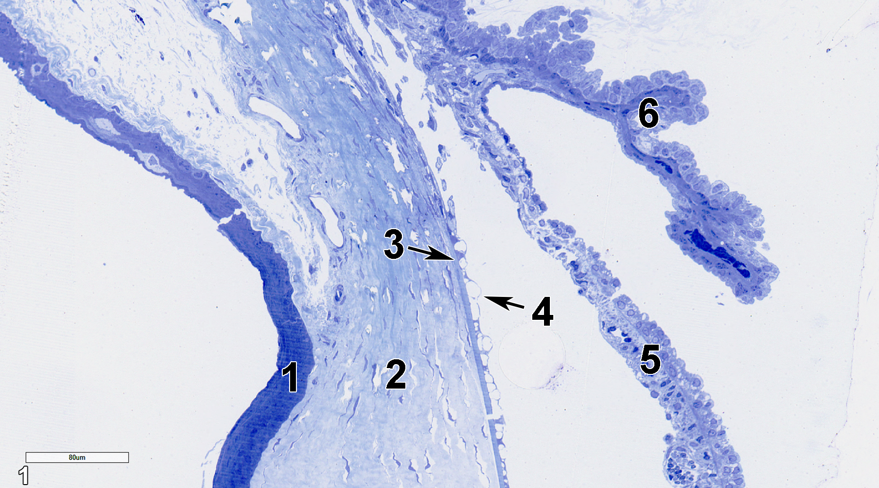
Figure 1. A toluidine blue O-stained semithin section (0.5 micrometer thick) of the anterior region of the eye showing the corneal epithelium (1), corneal stroma (2), Descemet’s membrane (3, arrow), corneal endothelium (4, arrow), iris (5), and ciliary body (6). 25x.
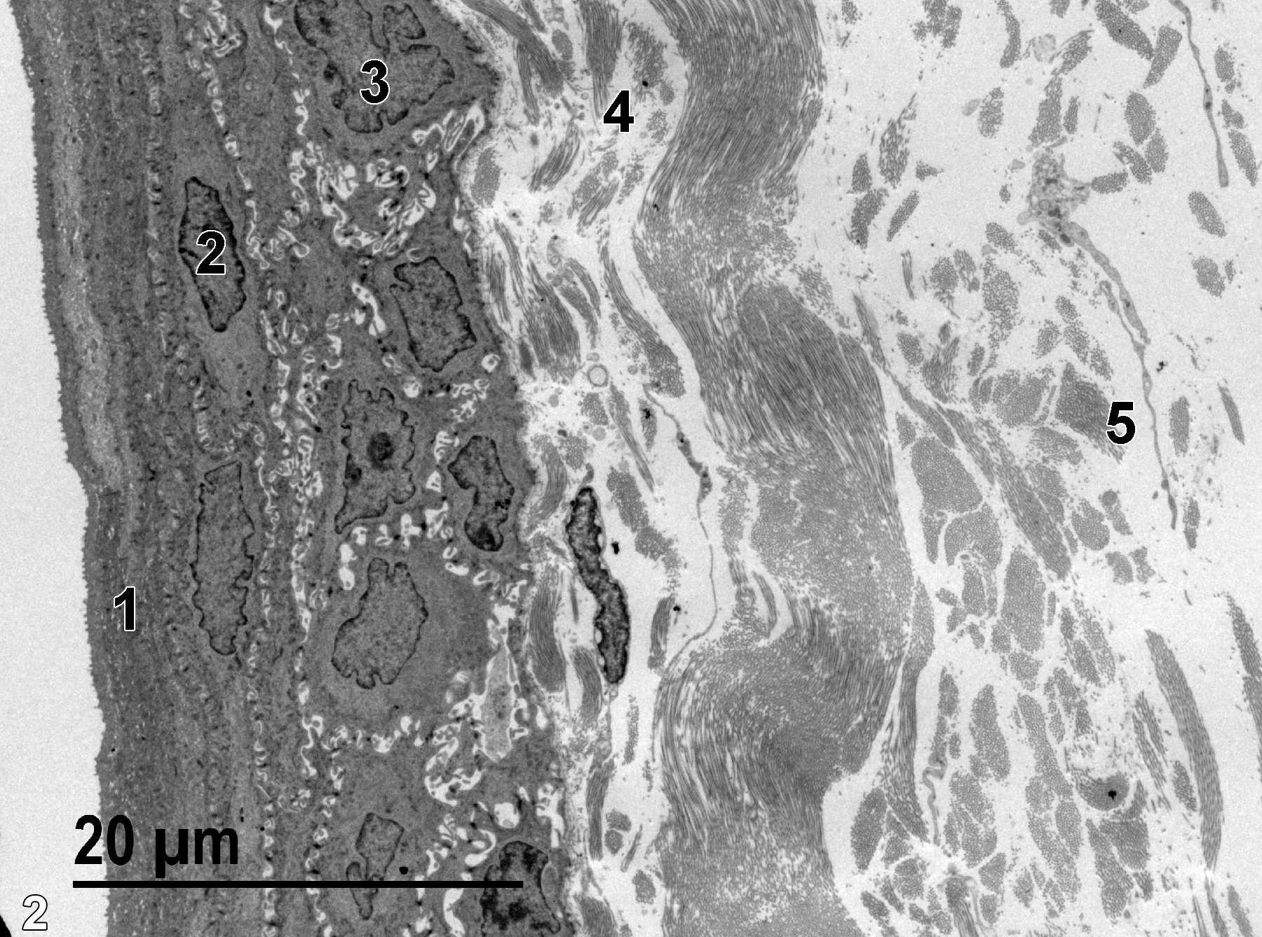
Figure 2. An ultrathin section (80-90 nm thick) of the cornea. The outermost layer of the cornea is composed of flattened squamous epithelial cells (1) with short vermiform ridges that resemble short microvilli in section (Weiss 1988, p. 1076). The nuclei (2) in this layer are elongated with peripheral heterochromatin. Below the squamous epithelial layer are several layers of polygonal cells and columnar cells (3). The first narrow layer of the corneal stroma (4) consists of randomly distributed collagenous fibrils. This layer is known as Bowman’s membrane in human eyes (Dunn et al 2018). The much wider corneal stroma layer (5) is composed of bundles of collagenous fibrils of uniform diameter, running in different directions, with matrical materials between them, as well as sparse populations of fibroblasts. 1900x.
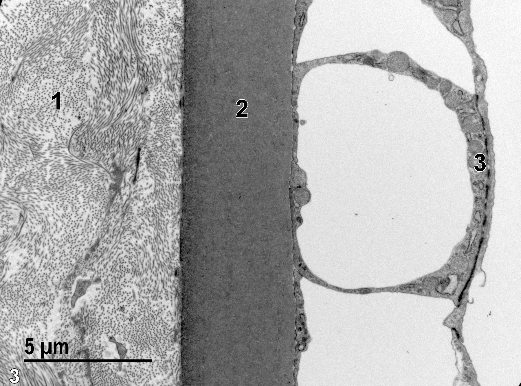
Figure 3. A higher magnification view of the basal area of the corneal stroma (1) with bundles of collagen fibrils and small cytoplasmic extensions of fibroblasts. Deep to the stromal layer is the homogeneous elastic membrane (2) composing Descemet’s membrane. This layer, in turn, underlays the corneal endothelium (3), which is highly vacuolated in the area imaged. 6800x.
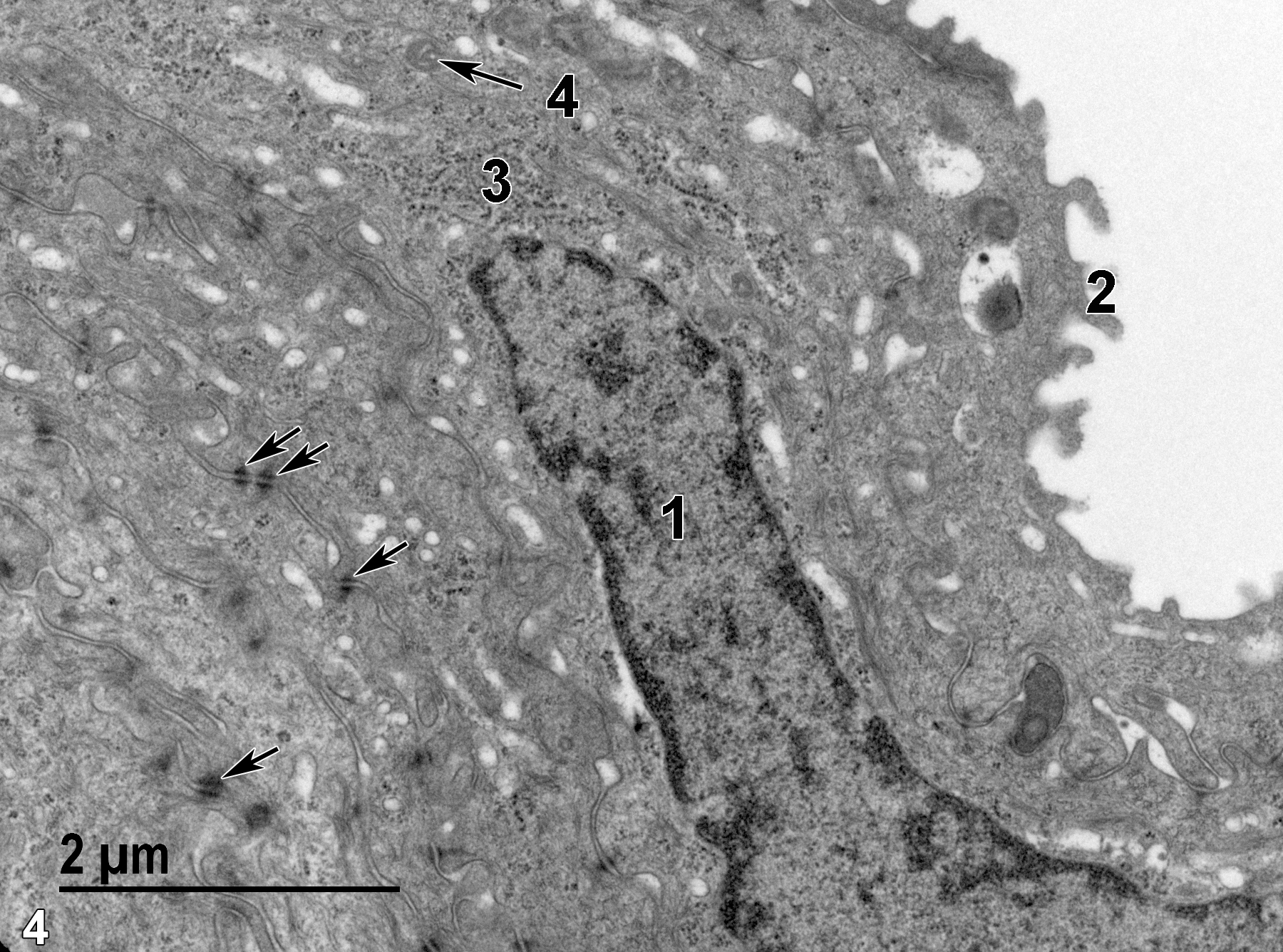
Figure 4. A high magnification view of squamous epithelial cells of the cornea, showing a portion of a typical elongate squamous epithelial cell nucleus (1) with marginated heterochromatin, transverse sections of the vermiform ridges (2) of the superficial epithelial cells, desmosomes between adjacent cells (arrows), polysomes and rough endoplasmic reticulum (3), and relatively scarce small mitochondria (4, arrow). 18500x.
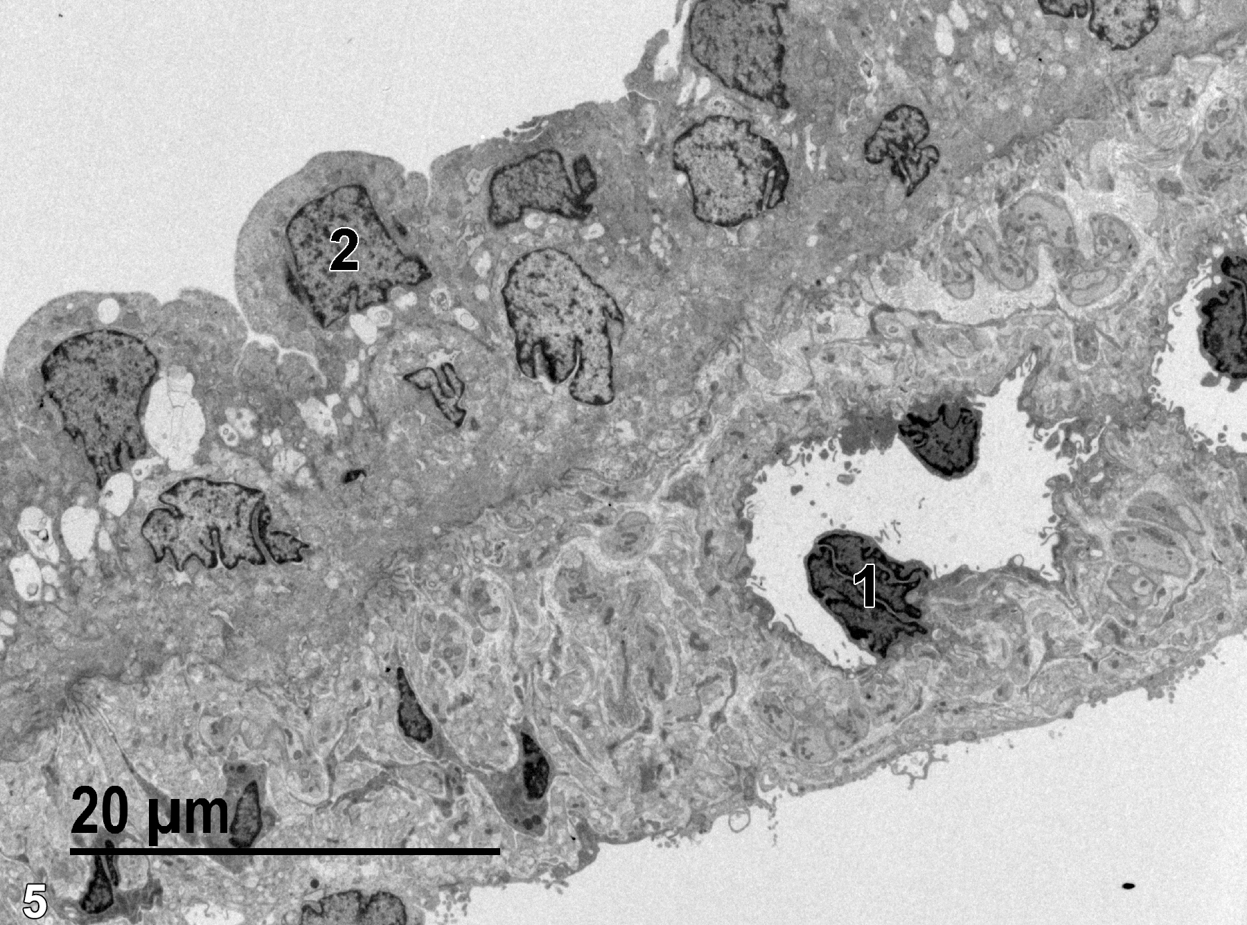
Figure 5. A low magnification view of a portion of the iris. An endothelial nucleus (1) of a capillary in the anterior surface of the iris is shown, surrounded by neural elements and connective tissue. The posterior surface of the iris is characterized by cuboidal epithelial cells containing a single nucleus (2). 1900x.
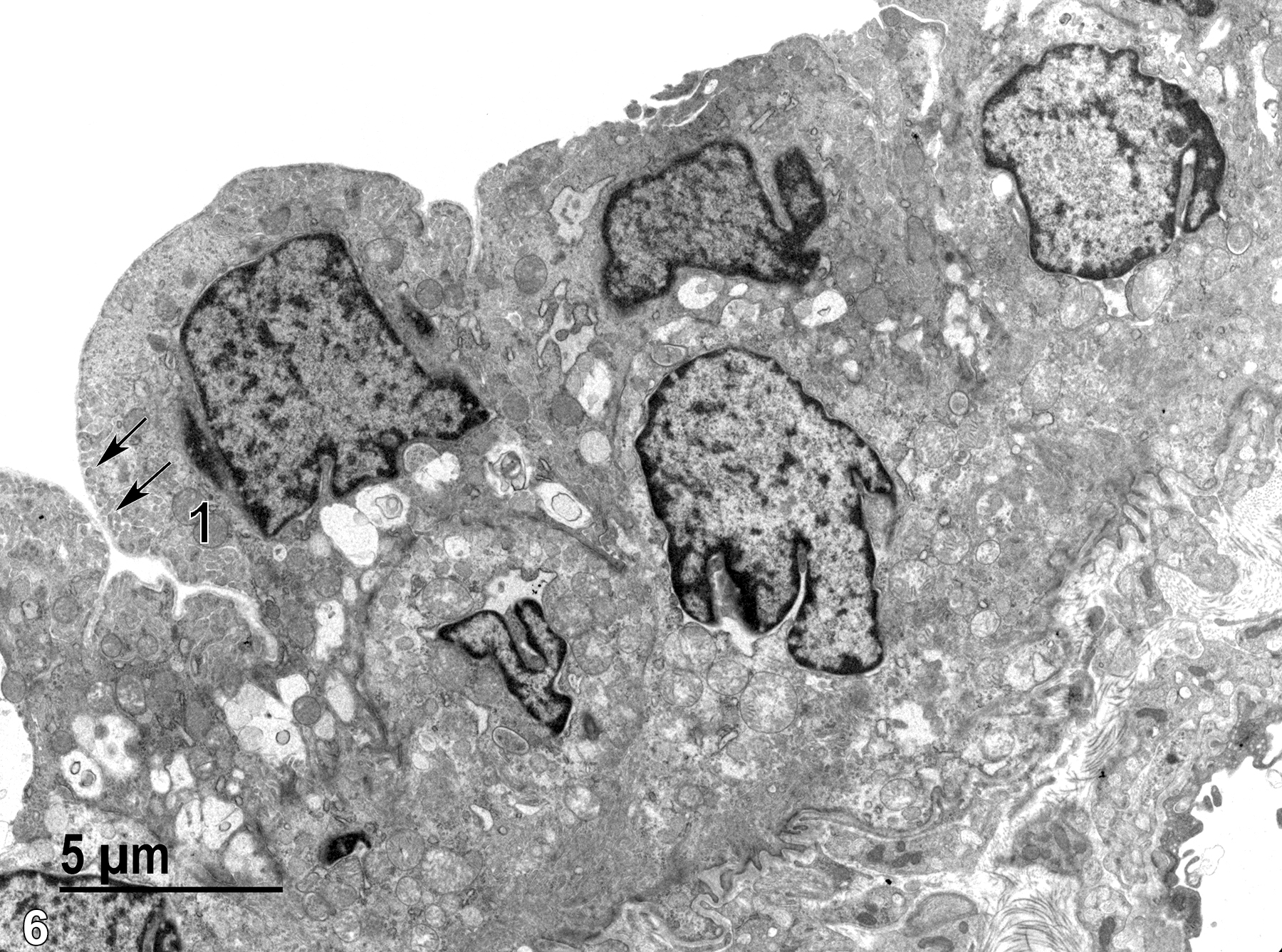
Figure 6. A higher magnification view of the epithelial cells of the posterior iris surface with infoldings of the cell surface (arrows) overlaid by a thin basal lamina. Mitochondria (1) are mostly round with relatively dense matrical content. Vacuolated areas are seen near the basal aspect of the cells. 4800x.
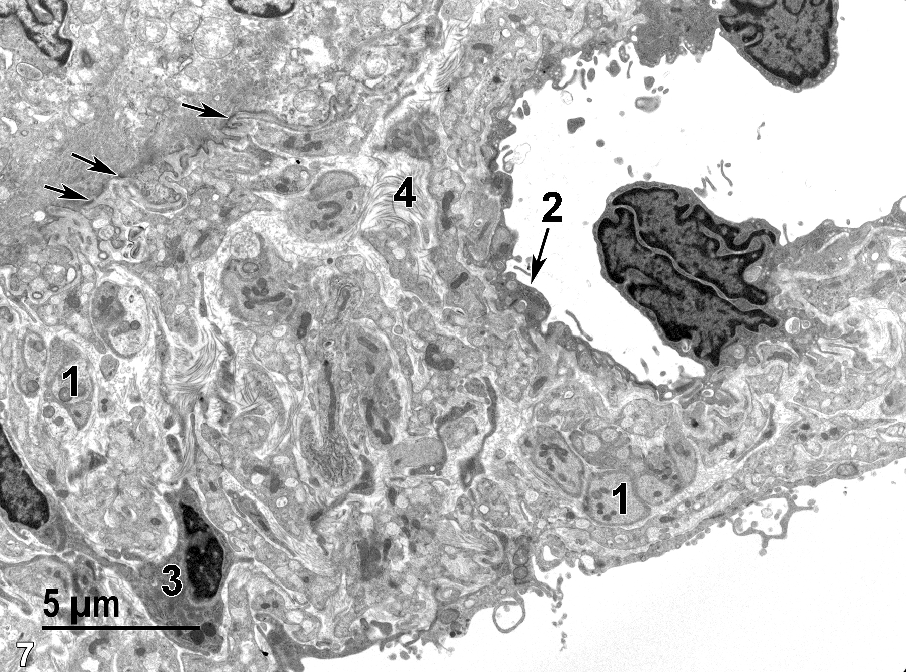
Figure 7. A higher magnification view of the anterior surface of the iris shown in Figure 5. A number of neural elements (1) containing nuclei and barely discernable fibrous elements (microtubules and microfilaments) are present. The thin endothelial lining (2, arrow) of a capillary is evident. A fibroblast (3) with an elongated nucleus is present. The fibroblastic population of this layer is responsible for the production of the collagenous matrix (4). The border between the collagenous stromal layer that consists of the anterior iris surface and the epithelial cells of the posterior iris surface is demarcated by a basal lamina and electron-dense hemidesmosomes (arrows). 4800x.
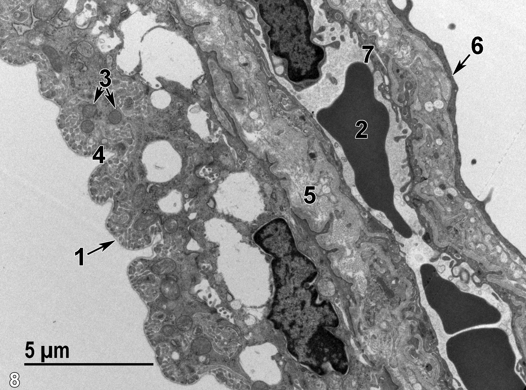
Figure 8. A low magnification view of the ciliary process, with cuboidal epithelial cells facing the posterior chamber with markedly infolded surfaces (4) beneath a basal lamina (1). Numerous mitochondria (3, double arrows) show mostly rounded profiles. There are two layers of epithelial cells, and the elongated nucleus (6, arrow) represents the pigmented layer of epithelial cells in the rat, although the albino rat shown here has few cells with melanin granules (none shown here). The ciliary stroma (5) contains large amounts of collagen produced by fibroblasts. A large capillary containing erythrocytes (2) is lined with a thin endothelial layer (7). 6800x.
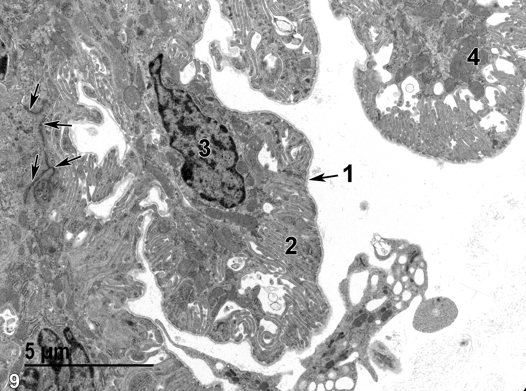
Figure 9. Another view of the ciliary process clearly showing the basal lamina (1) overlying the markedly infolded cell surface (2) of an epithelial cell with a single nucleus (3). Mitochondria (4) are plentiful and elongated junctional complexes (arrows) can be seen between adjacent epithelial cells. 6800x.
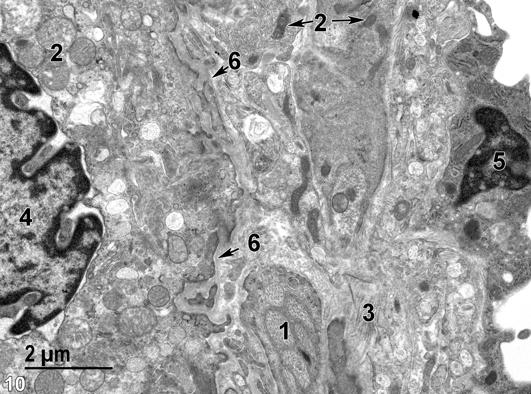
Figure 10. A higher magnification view of the stromal area of the ciliary process in the right half of the image with neural processes containing microtubules (1) and collagen bundles (3) produced by fibroblasts (5). An epithelial cell that has a nucleus (4) with irregular borders and marginated heterochromatin is separated from the stromal layer by a basal lamina (6, arrow). Note the difference in size, morphology, and matrical density of the mitochondria (2) of the epithelial cell versus the stromal cells (double arrows). 11000x.
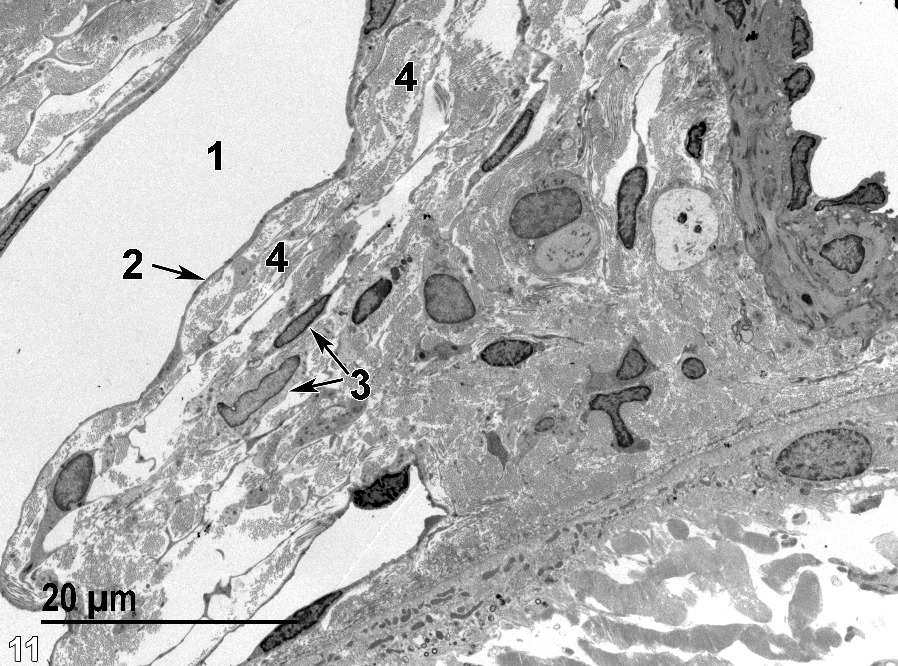
Figure 11. One end of Schlemm’s canal (1) lined with a thin endothelium (2, arrow) and underlaid by several layers of collagen fibrils (4) separated by thin bands of cytoplasm of the fibroblasts (3, double arrows) of this trabecular meshwork. 1900x.
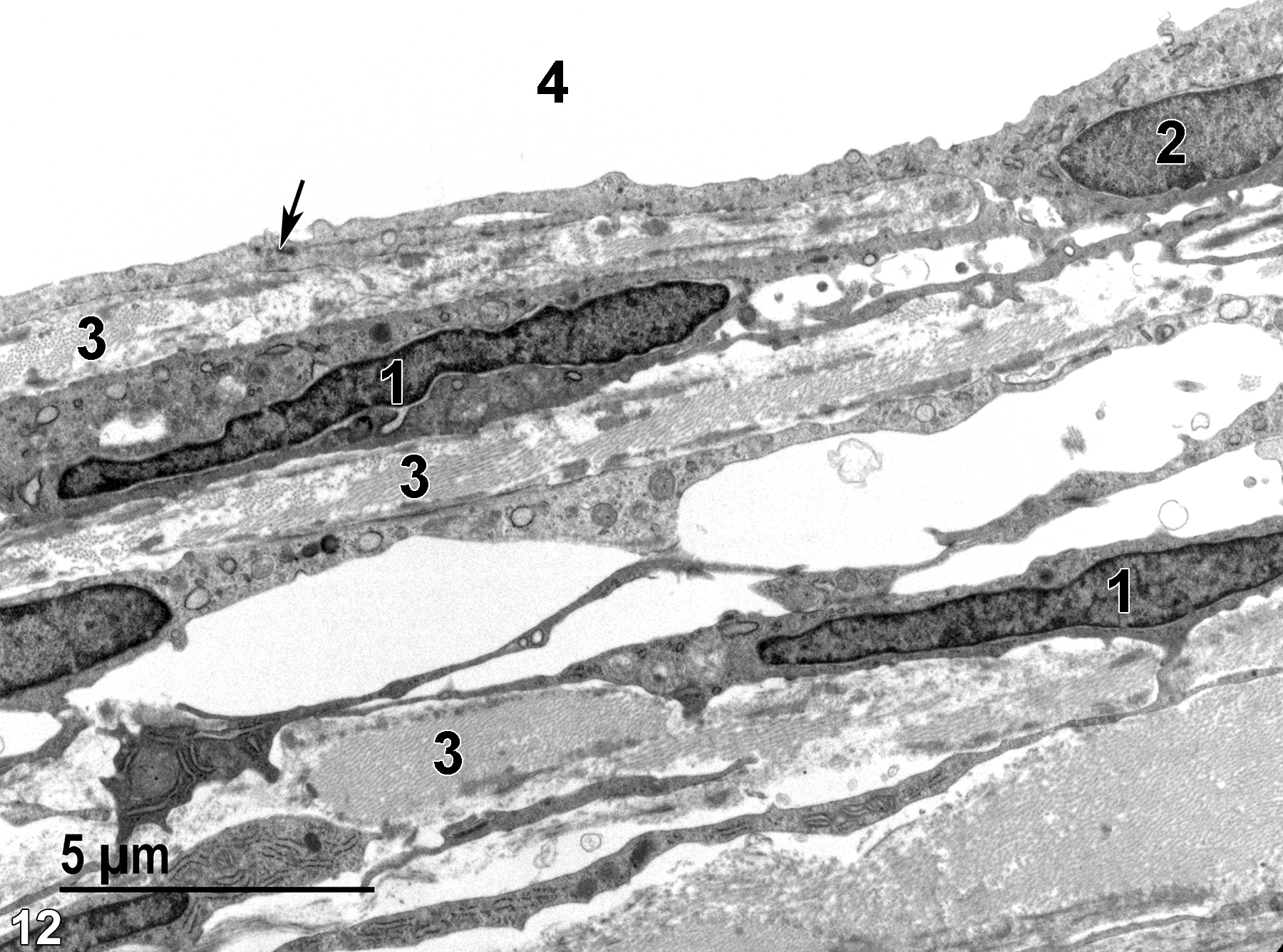
Figure 12. A higher magnification view of the trabecular meshwork showing fibroblasts with elongated nuclei (1) that produce the beams (clusters) of collagen fibrils (3) that regulate the flow of aqueous humor into Schlemm’s canal (4), which is lined with endothelial cells with elongate nuclei (2). The endothelial cells are connected to each other by desmosomes (arrow). 6800x.
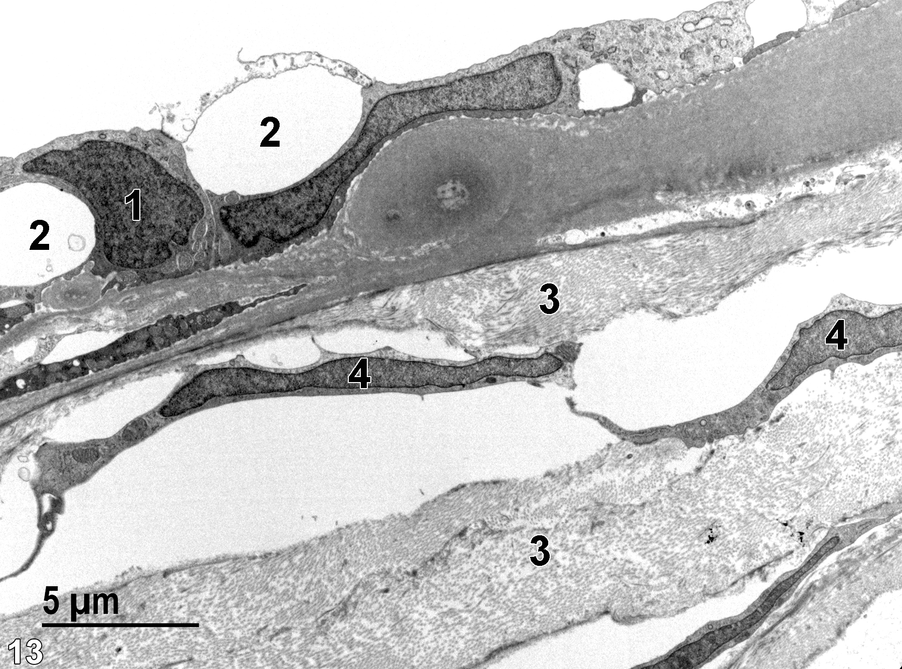
Figure 13. An even higher magnification of the trabecular meshwork. Large areas of the endothelial layer lining Schlemm’s canal often contain large vacuoles (2). The nuclei of the endothelial cells (1) are frequently pleomorphic in the vacuolated area. Note the elongated nuclei (4) in the attenuated fibroblasts and the bundles of collagen fibrils (3) comprising the beams of the trabecular meshwork. 4800x.
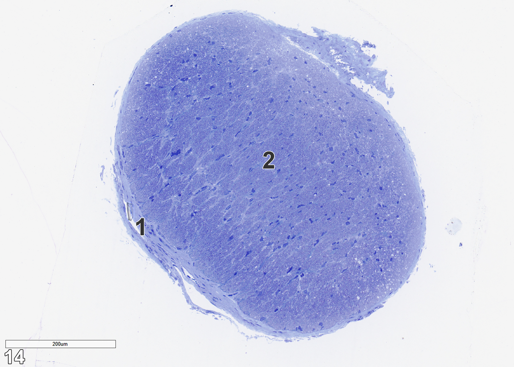
Figure 14. A toluidine blue O-stained semithin section of an optic nerve in transverse section. The outermost layer is a relatively thin epineurium (1) that consist of fibroblasts with flattened and elongated nuclei. The fibroblasts are surrounded by the collagenous matrix they produce. The epineurium encloses the neural tissue (2), which is composed of myelinated and unmyelinated axons and dendrites, oligodendrocytes that produce the myelin, and astrocytes that have long astrocytic processes containing microfilaments. 14x.
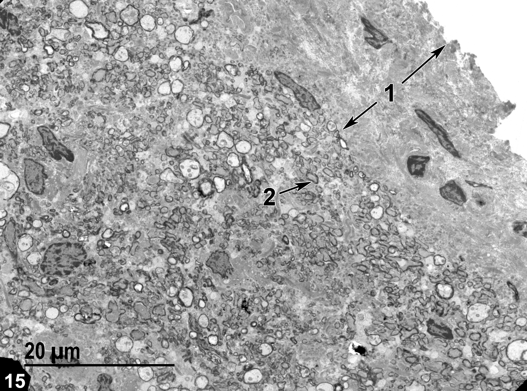
Figure 15. An electron micrograph of a cross section of the optic nerve, showing the epineurium (1, double arrows), which is made up of collagen and fibroblasts. Within the neural portion of the optic nerve, irregular outlines of electron-dense myelin (2, arrow) are seen. 1900x.
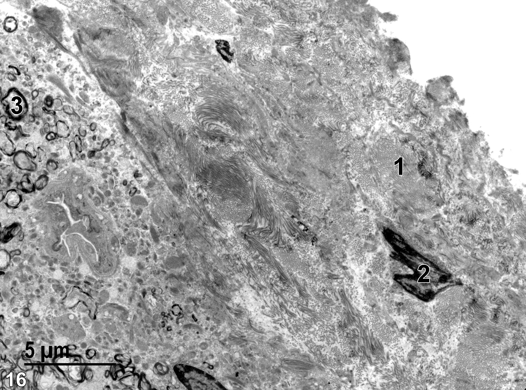
Figure 16. A higher magnification view of the area shown in Figure 15. The collagen fibers (1) that make up the epineurium are clearly visible, as well as a nucleus of a fibroblast (2). A myelinated axon (3) is shown. 4800x.
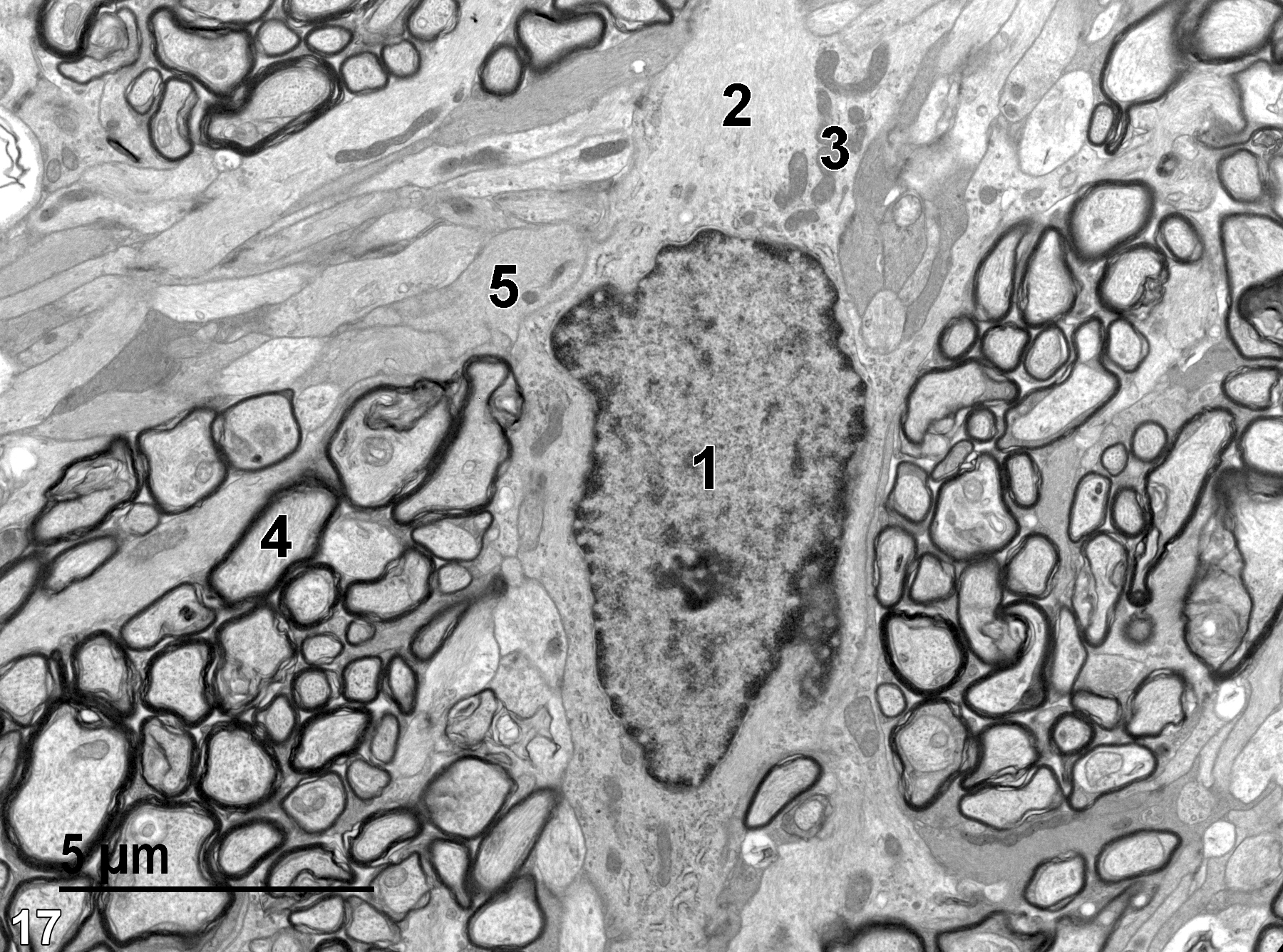
Figure 17. An astrocyte nucleus (1) and an astrocytic process (2) containing numerous microfilaments and a number of mitochondria (3). A myelinated axon (4) with electron-dense layers of membranes is distinct from the unmyelinated axon or dendrite (5) in close proximity. 6800x.
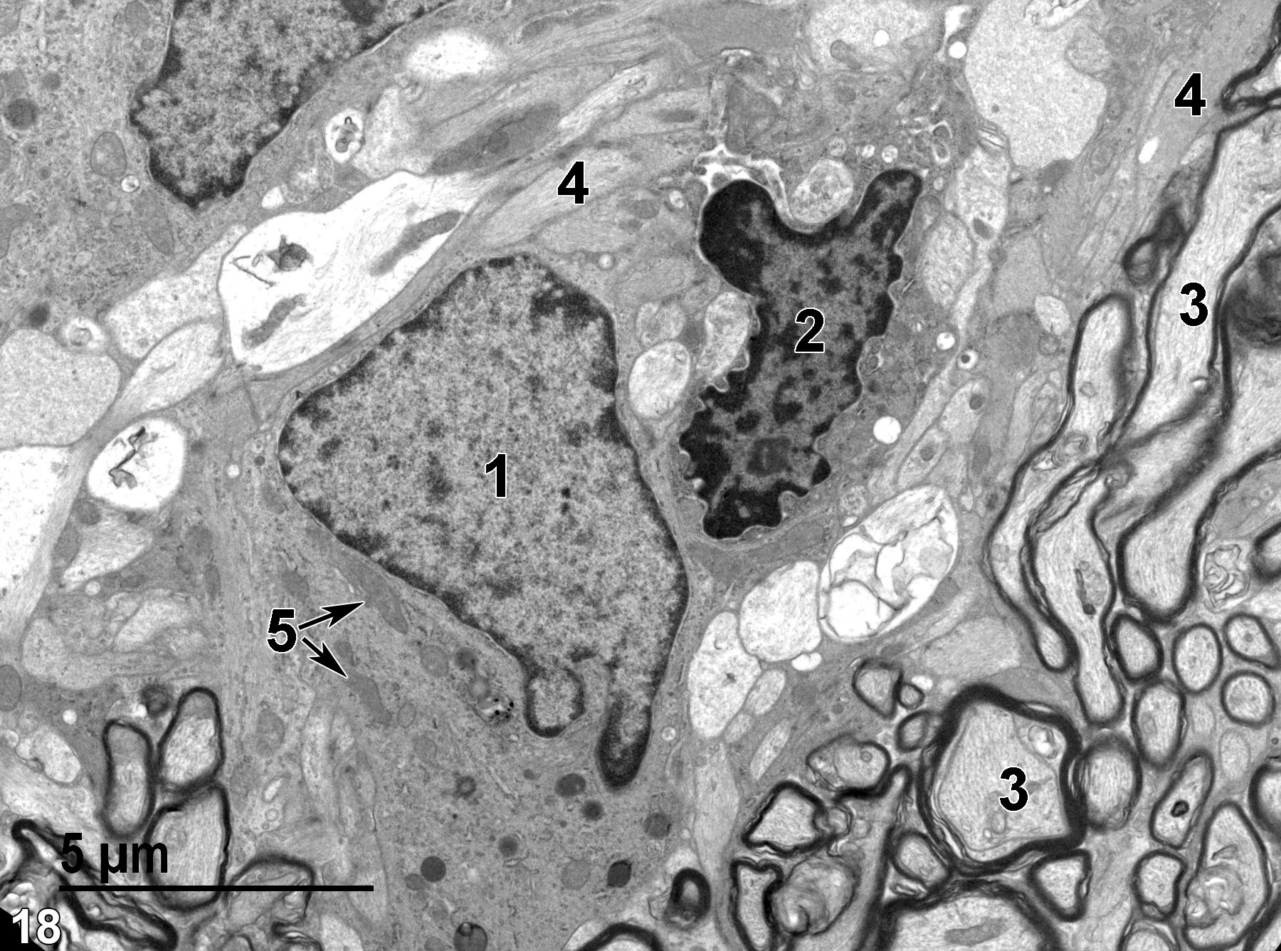
Figure 18. A probable astrocyte with a large nucleus (1) containing mostly euchromatin and a few mitochondria (5, double arrows). A cell with a polymorphic nucleus (2) with much marginated heterochromatin is consistent with a microglial cell. Myelinated axons (3) in a transverse and longitudinal section that contain mostly microtubules are present, as well as unmyelinated neuronal processes (4) that primarily contain microfilaments. 6800x.
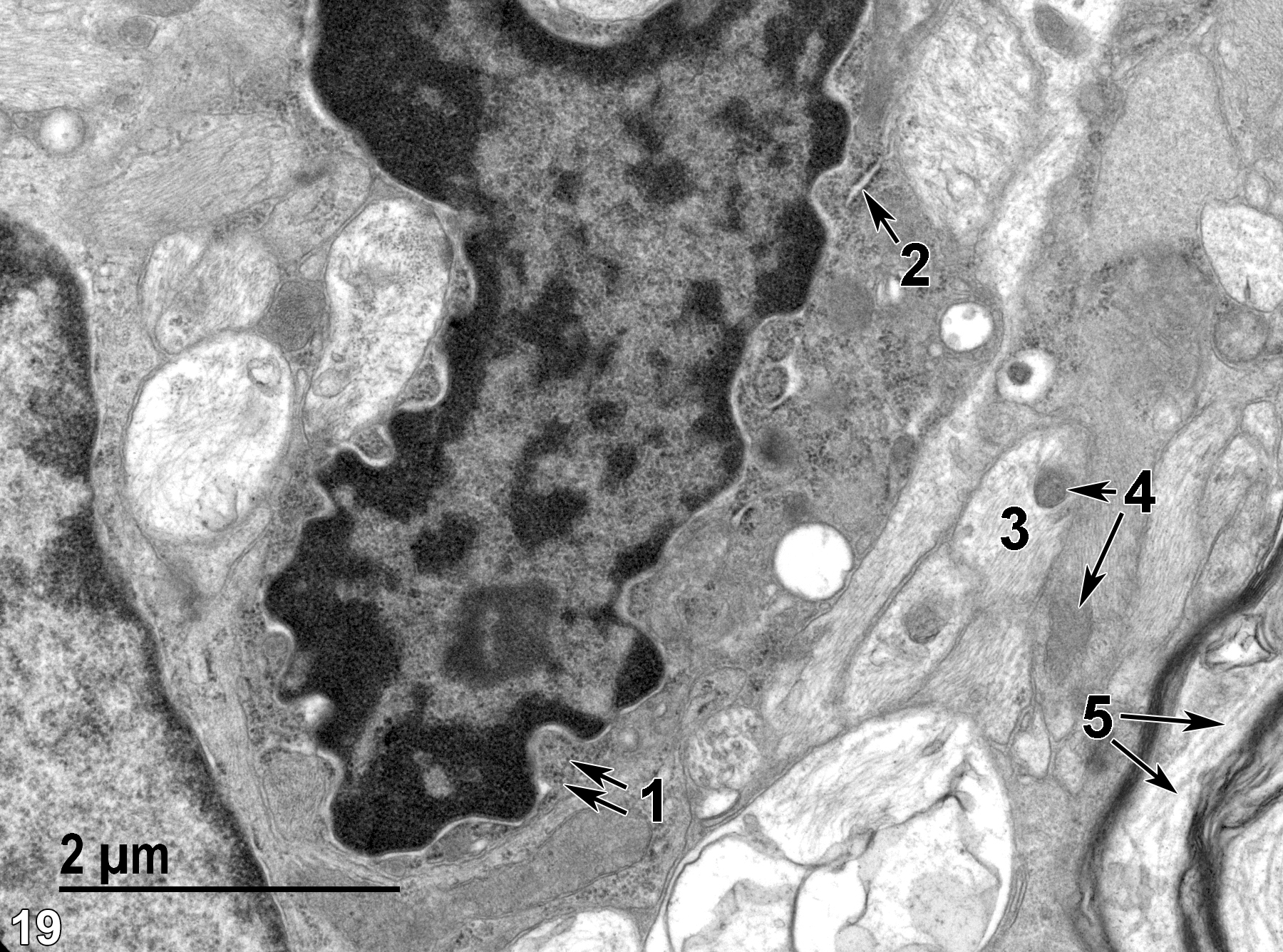
Figure 19. A higher magnification view of the glial cell shown in Figure 18, showing some polysomes (1, double arrows), along with some endoplasmic reticulum (2, arrow). An unmyelinated axon (3) contains microfilaments and microtubules, whereas the myelinated axon (5) contains mostly microtubules (double arrows). Both an unmyelinated axon and an adjacent astrocytic extension containing large amounts of microfilaments contain similar mitochondrial profiles (4, double arrows). 18500x.
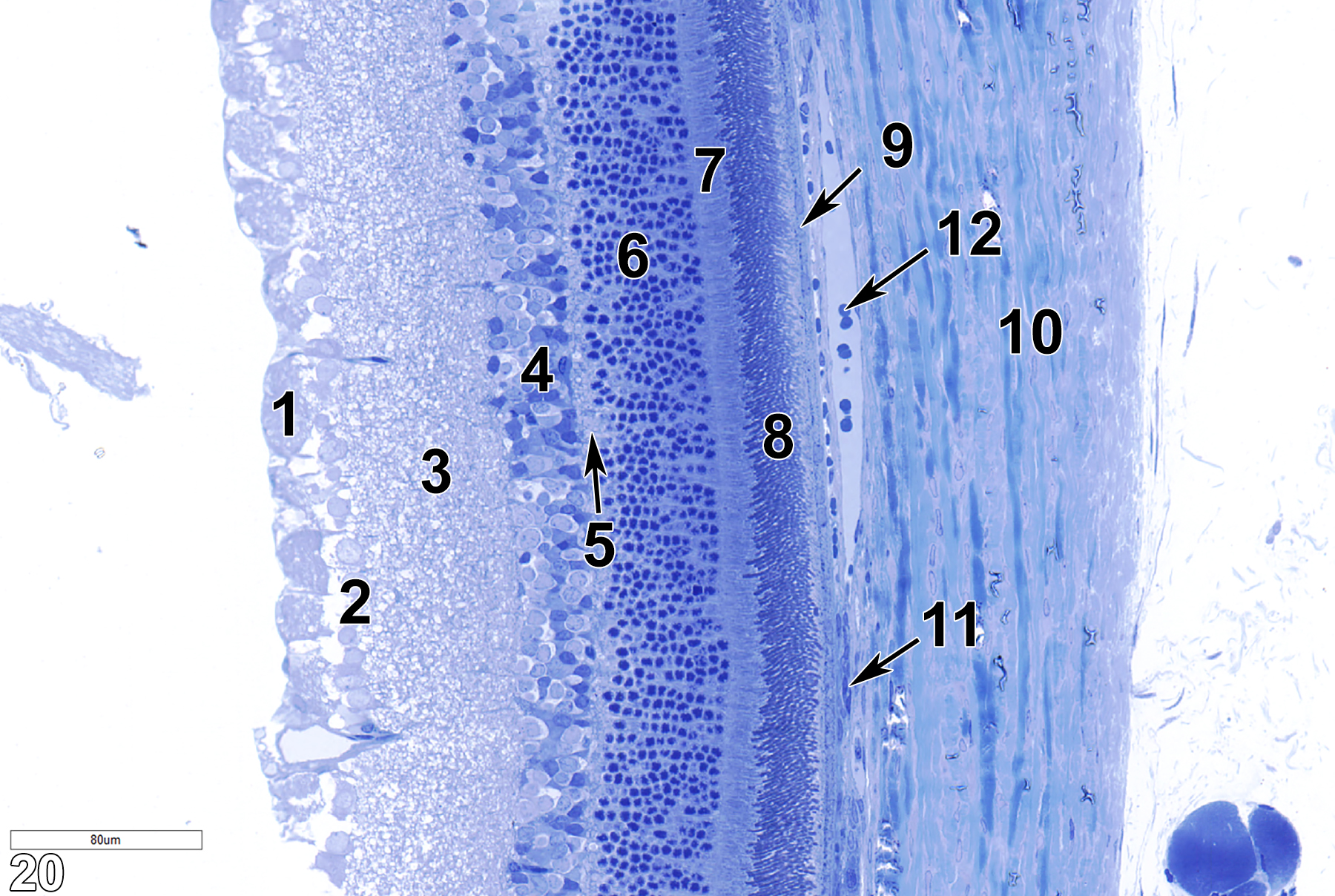
Figure 20. A toluidine blue O-stained semithin section of the retina. The nerve fiber layer (1) is next to the ganglion cell layer (2), which consists of cells with large round nuclei. The next layer is the inner plexiform (synaptic) layer (3), followed by the inner nuclear layer (4). This, in turn, is followed by the outer plexiform layer (5, arrow) and the outer nuclear layer (6). The outer layer of the photoreceptor cells (7) follows and then the inner layer of the photoreceptor cells (8). Next is the pigmented epithelium (9, arrow), followed by Bruch’s membrane (11, arrow). The choroid (10) is a broad collagenous matrix, within which are various blood vessels containing erythrocytes (12, arrow). 25x.
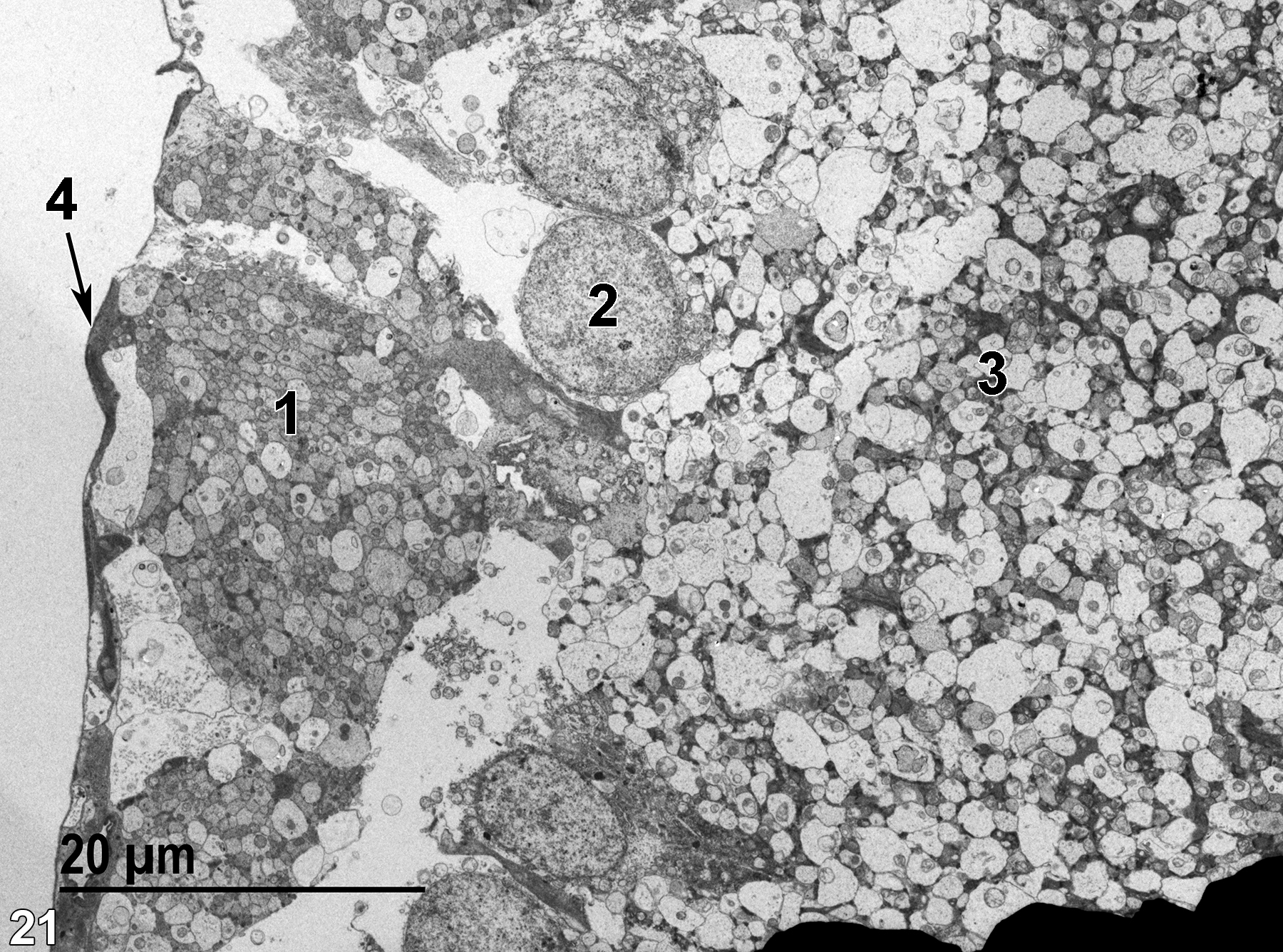
Figure 21. Four of the layers of the retina. The inner limiting membrane (4, arrow) is a thin epithelial cell layer adjacent to the vitreous body. The next layer is the nerve fiber layer (1), which arises from the ganglion cell layer and exits through the optic nerve. The ganglion cell layer cells have axons that form the nerve fiber layer (1) and has large round nuclei (2) and dendrites that connect with the bipolar cells in the outer plexiform layer. The next layer is the inner plexiform (synaptic) layer (3), which is composed of synapses with bipolar cells, amacrine cells, and ganglion cell dendrites (see Weiss 1988, p. 1099). 1900x.
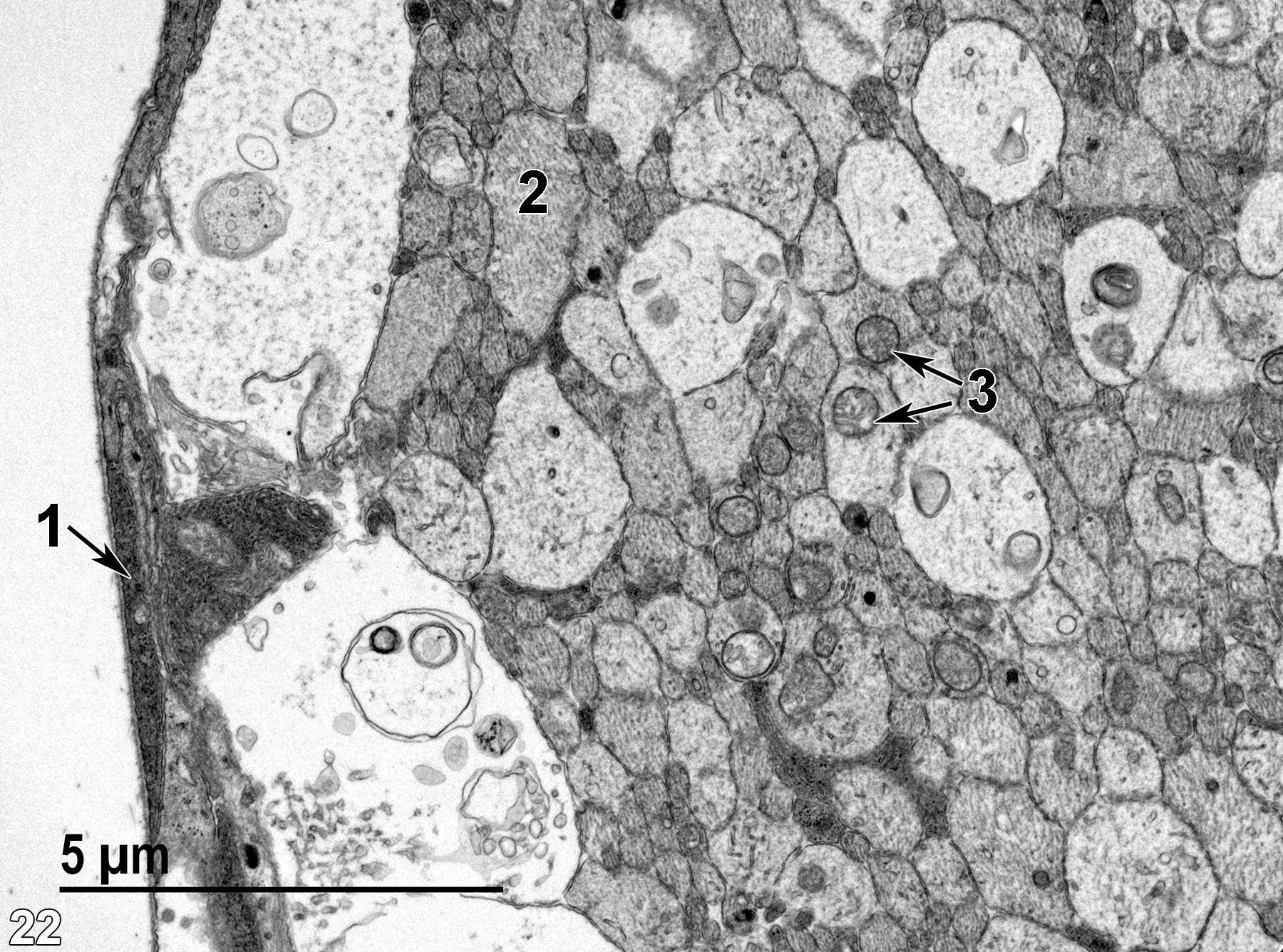
Figure 22. A higher magnification view of a portion of Figure 21, showing the thin epithelial cell layer of the inner limiting membrane (1, arrow) and neural elements of the nerve fiber layer containing microtubules (2) and occasional mitochondria (3, double arrows). 9300x.
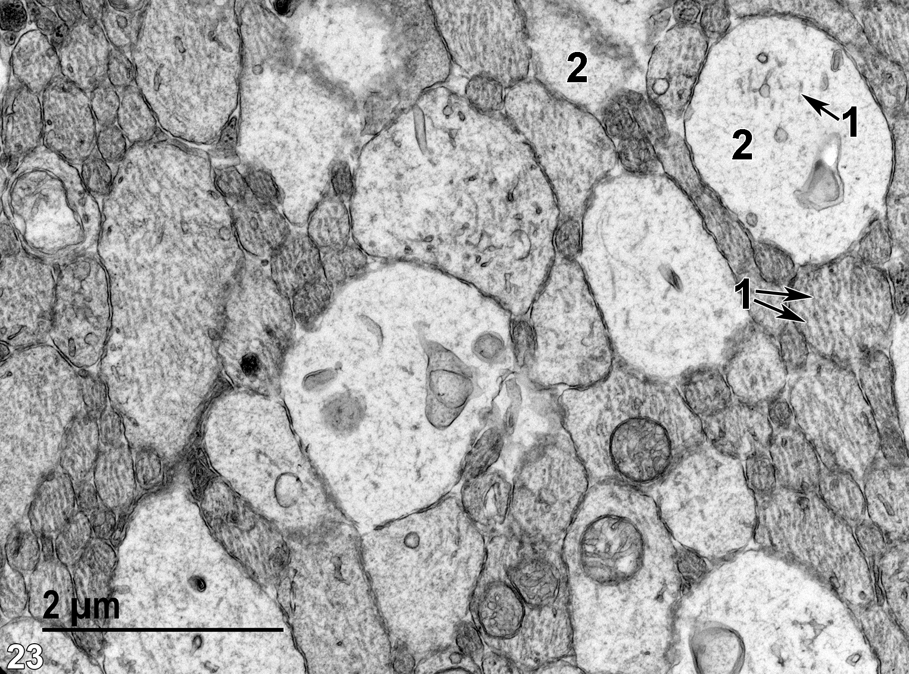
Figure 23. An even higher magnification of the nerve fiber layer showing neural processes containing microtubules (1, arrows) and microfilaments (2). 18500x.
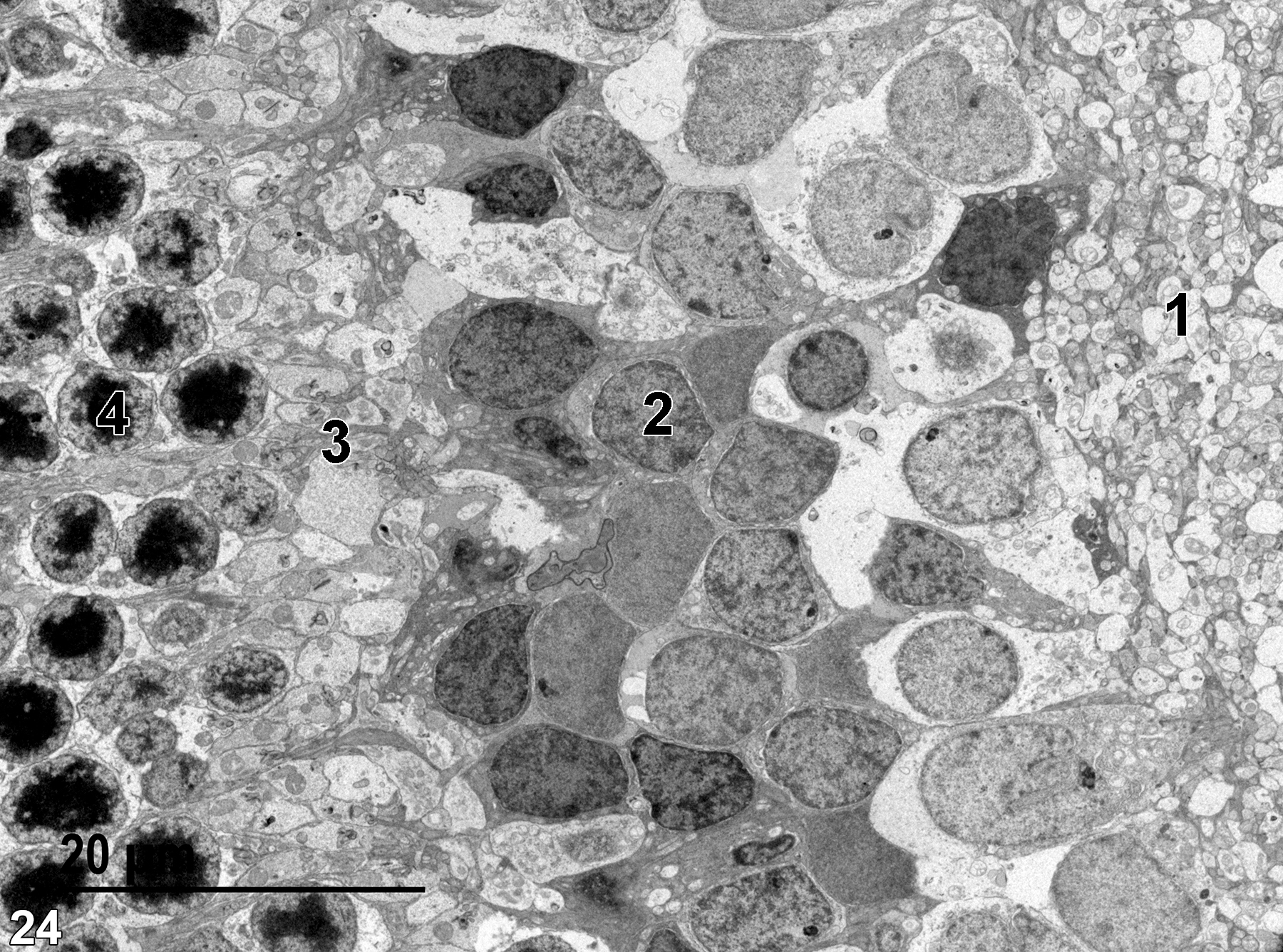
Figure 24. A low magnification view of the inner plexiform layer (1), the inner nuclear layer (2), the outer plexiform layer (3), and the outer nuclear layer (4). 1900x.
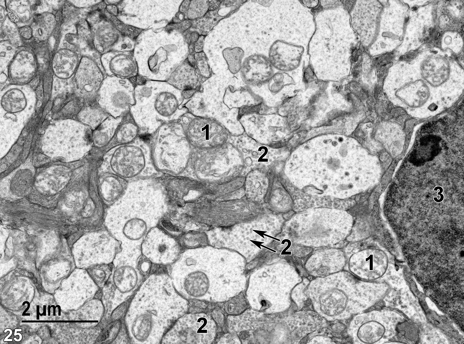
Figure 25. A higher magnification view of the inner plexiform layer, which is composed of neural elements (1) containing mitochondria, and nerve endings (2) with synaptic vesicles (arrows). Part of a nucleus (3) of the inner nuclear layer is shown. 11000x.
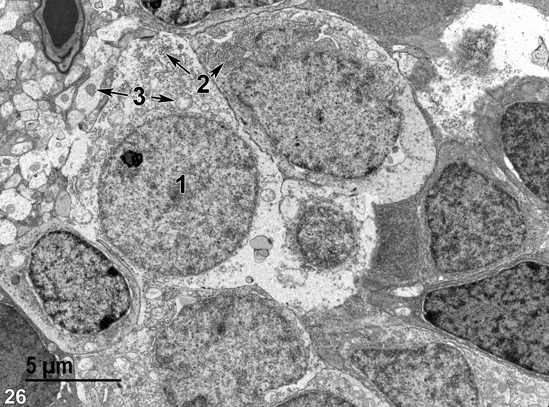
Figure 26. Rounded nuclei (1) of cells in the inner nuclear layer. Elements of rough endoplasmic reticulum (2, double arrows) are also visible within the cells. Note the difference in morphology of the mitochondria of the inner nuclear layer cell and those of the plexiform layer neural processes (3, double arrows). 4800x.
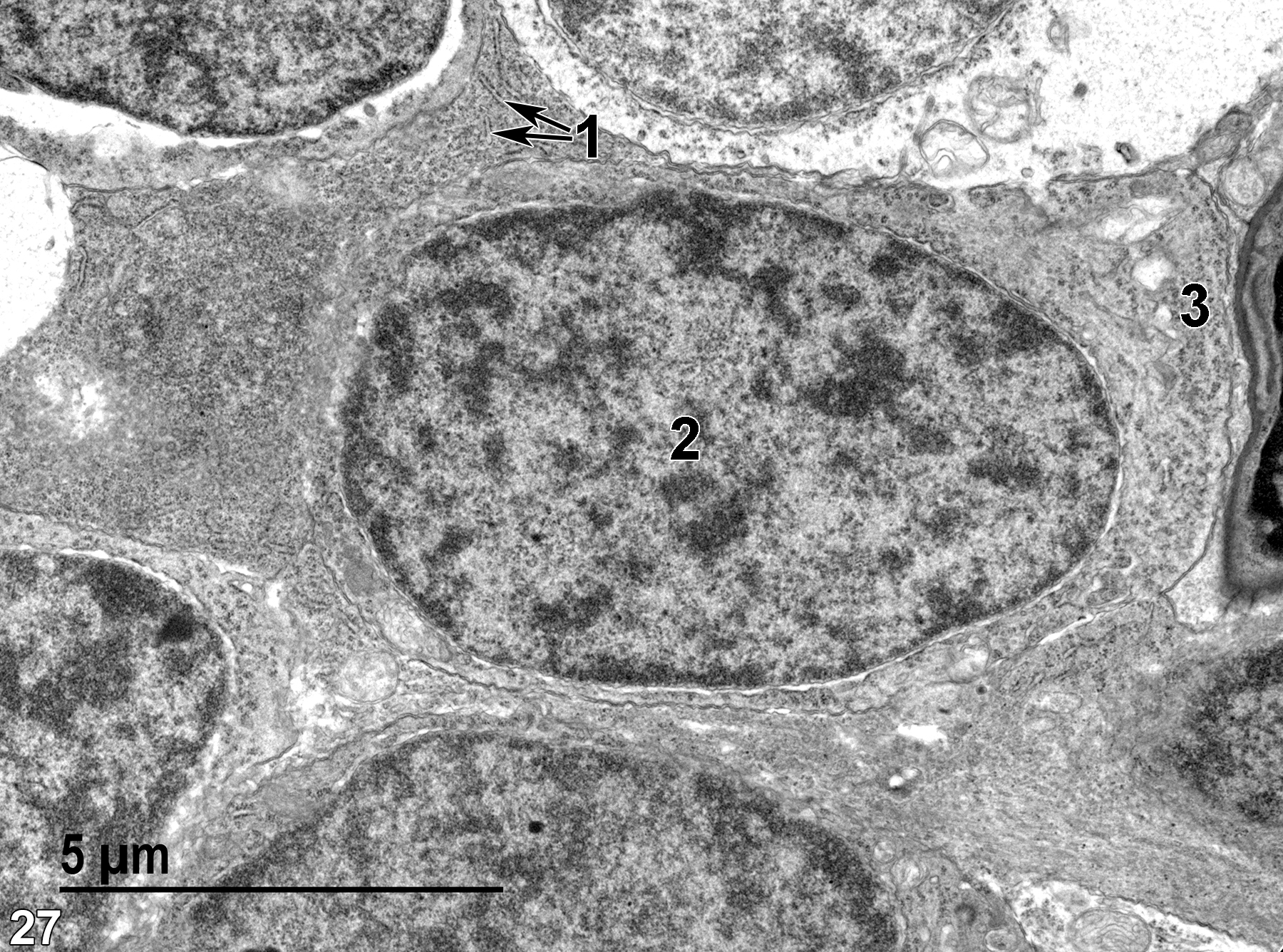
Figure 27. A higher magnification view of a cell in the inner nuclear layer, which is filled with rough endoplasmic reticulum (1, double arrows), a single ovoid nucleus (2) with scattered heterochromatin, and numerous polysomes (3). 9300x.
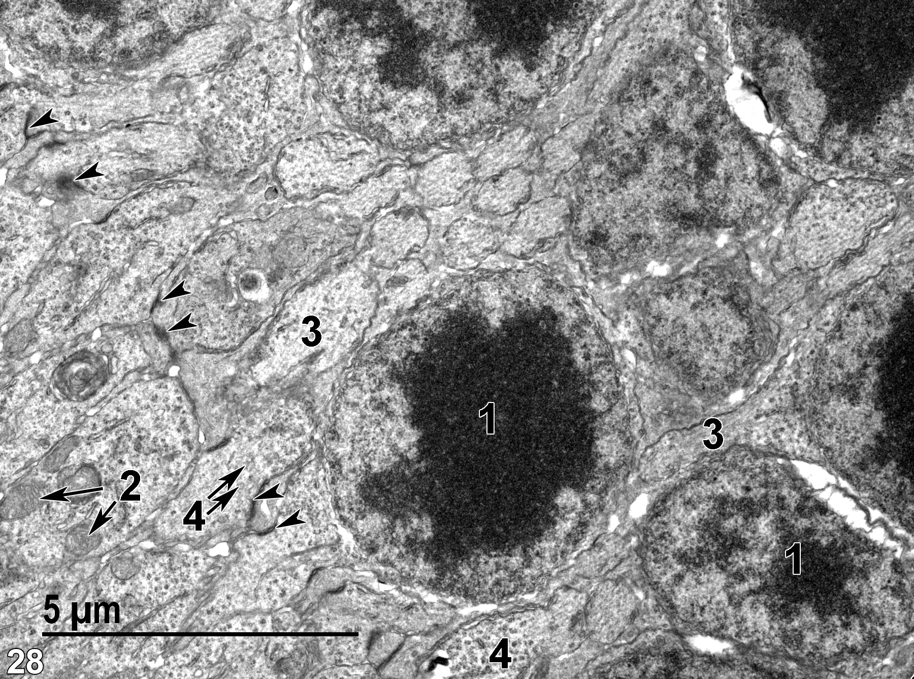
Figure 28. Nuclei (1) of the outer nuclear layer (the perikarya of the rods and cones) and the zonulae adherens (arrowheads) making up the outer limiting membrane of the retina. Mitochondria (2) are scarce (double arrows). Some of the neural processes contain obvious microfilaments (3). Nerve endings containing synaptic vesicles (4) are shown (double arrows). 9300x.
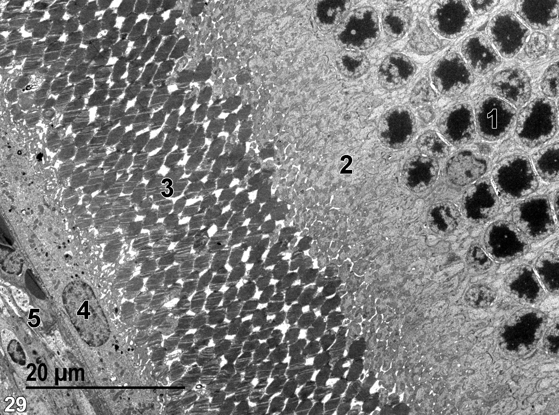
Figure 29. A low-magnification view of the outer nuclear layer (1), the inner segments of rods and cones (2), the outer segments of rods and cones (3), the pigmented epithelium (4), and Bruch’s membrane (5). 1900x.
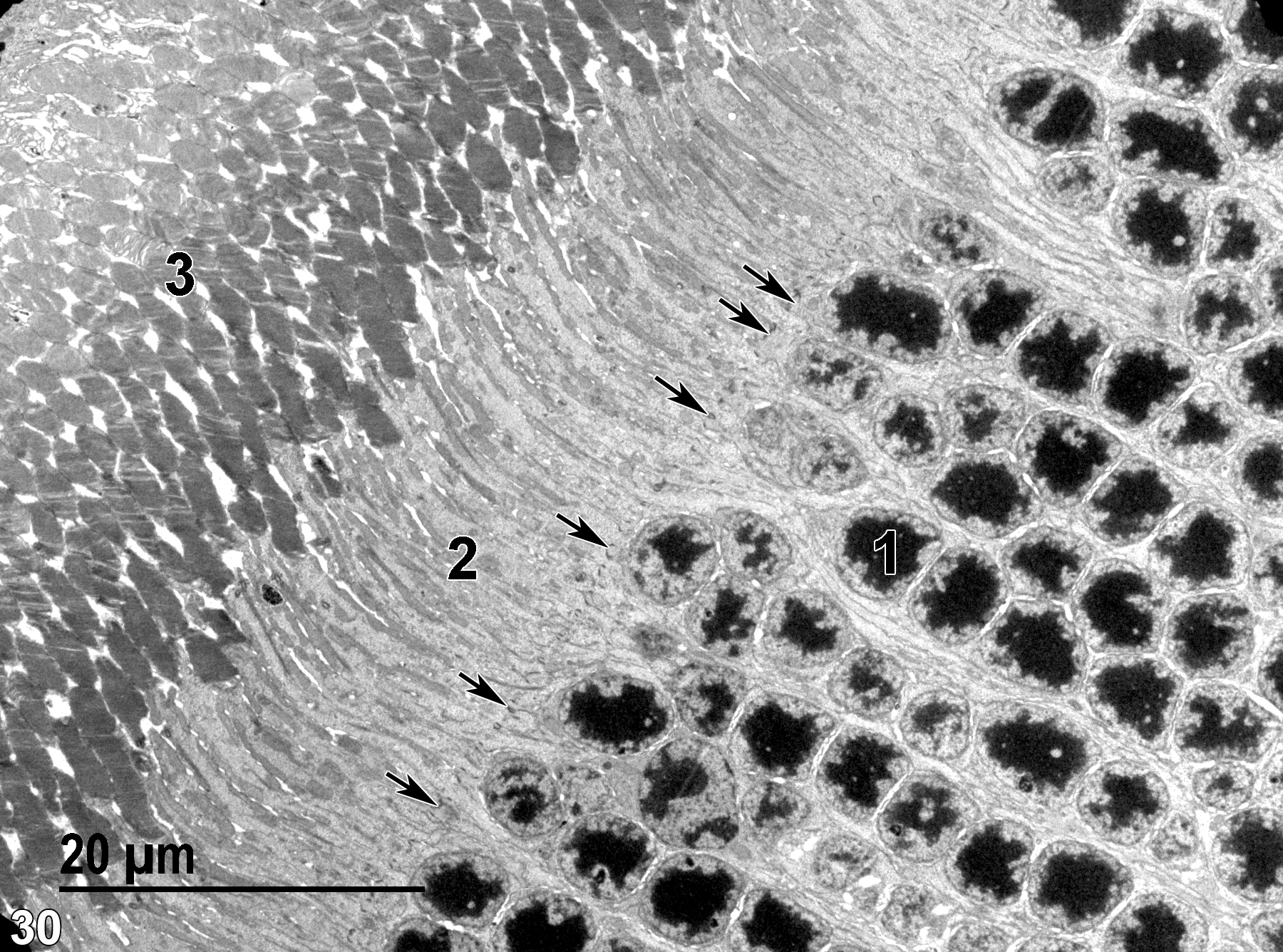
Figure 30. Another low magnification view of the retina, showing the outer nuclear layer (1), the inner segments of rods and cones (2) and the zonulae adherens consisting of the outer limiting membrane (arrows), and the outer segments of the rods and cones (3). 1900x.
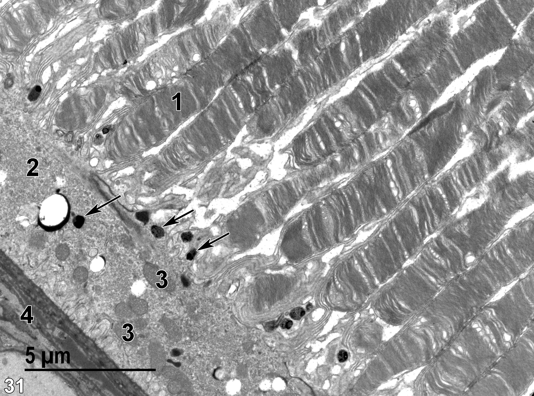
Figure 31. A higher magnification view of the outer segments of the rods and cones (1) overlaid by the pigmented epithelium (2), which contains melanin granules (arrows), and a number of mitochondria (3). The base of Bruch’s membrane (4) is shown. 6800x.
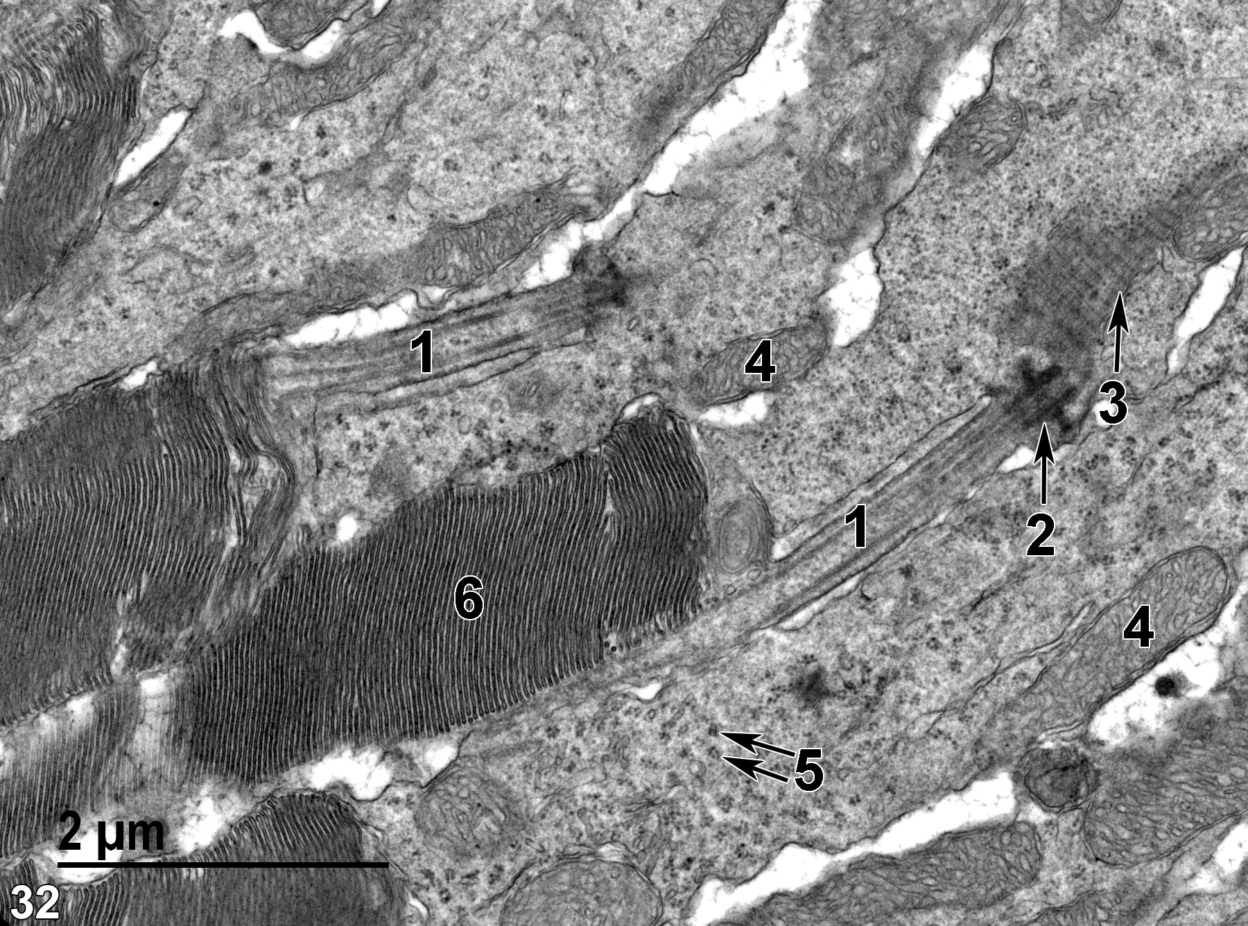
Figure 32. A higher magnification image of the membranous discs of the outer segment of rods (6). Within the rod cell cytoplasm, a single modified cilium (1) is present with a well-defined basal body (2, arrow) and a rootlet (3, arrow). The cytoplasm also contains numerous polysomes (5, double arrows) and mitochondria (4). 18500x.
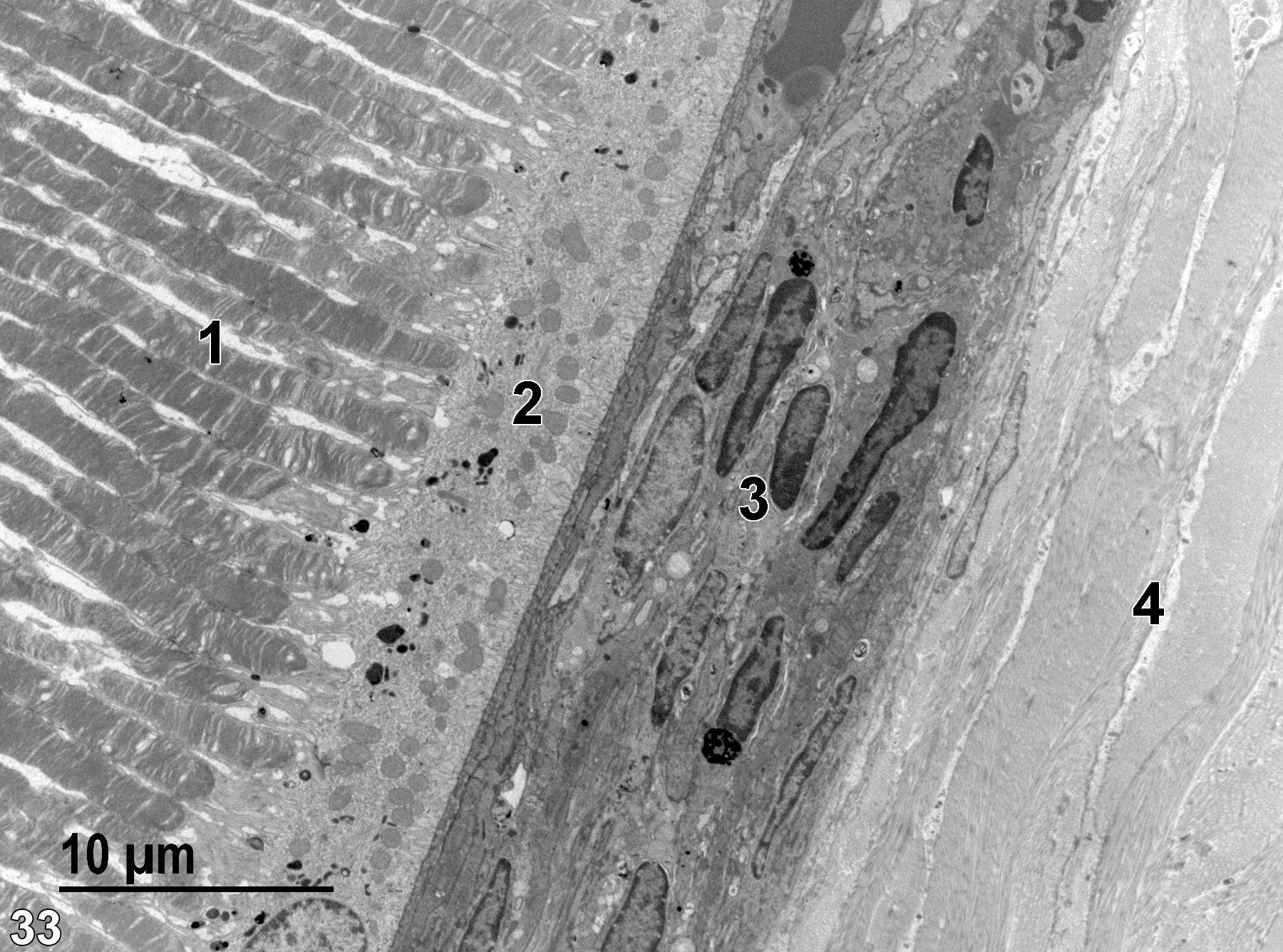
Figure 33. The outer segments of rods and cones (1), the pigmented epithelium (2), Bruch’s membrane (3), and the choroid (4), which is made up of layers of collagen and fibroblasts. 2900x.
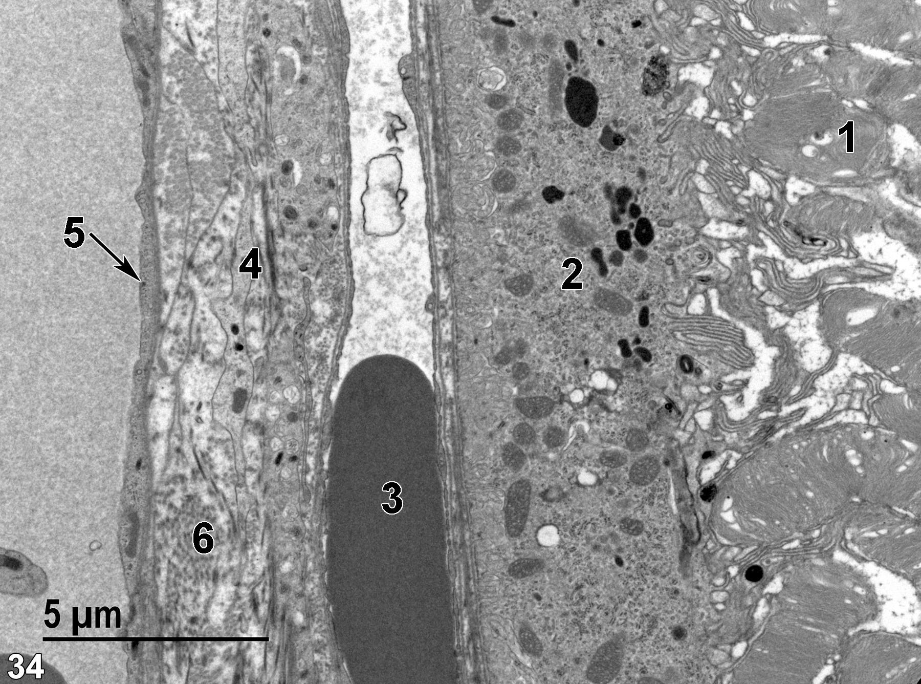
Figure 34. A higher magnification view of the membranous discs of the outer segments of rods and cones (1), the pigmented epithelium (2), which contains melanin granules, a blood vessel containing an erythrocyte (3), Bruch’s membrane (4) with numerous cell processes intermixed with collagen fibrils (6), and another vessel with a thin endothelial lining (5, arrow). 6800x.
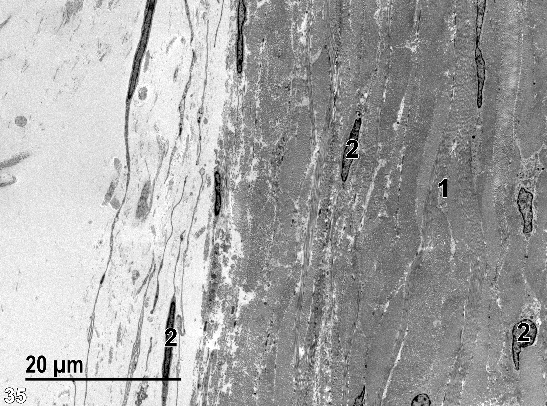
Figure 35. The outer edges of the choroid with large bundles of collagen fibrils (1) and various elongated fibroblasts (2). 1900x.
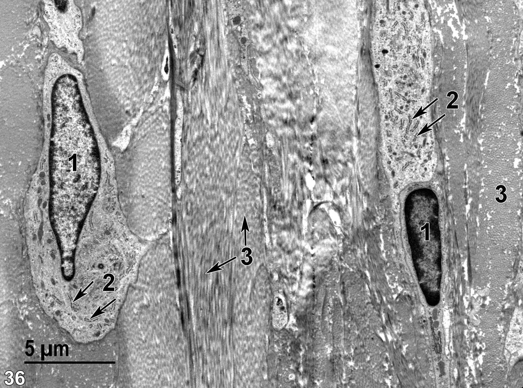
Figure 36. The structure of the elements within the choroid. Bundles of collagen fibrils (3) can be seen in the transverse and longitudinal section (double arrows), associated with the fibroblasts (1) that produce the collagen. The fibroblasts have elongated nuclei with prominent marginal heterochromatin and numerous profiles of rough endoplasmic reticulum (2, arrows). 4800x.
| Banks WJ. 1993. Chapter 28: Eye and ear. In: Applied Veterinary Histology. 3rd ed. St. Louis, MO: Mosby-Yearbook, Inc., 469-497. |
| Dunn DG, Baker JFM, Sorden SD. 2018. Chapter 16: Eye and associated glands. In Boorman’s Pathology of the Rat (Suttie AW, ed.). 2nd ed. London: Academic Press, 251-278. |
| Eurell JA, Frappier BL, eds. 2006. Dellmann’s Textbook of Veterinary Histology. 6th ed. Ames, IA: Blackwell Publishing. |
| Fawcett DW. 1994. Bloom and Fawcett: A Textbook of Histology. 12th ed. New York: Chapman and Hall. |
| Rhodin JAG. 1974. Histology: A Text and Atlas. New York: Oxford University Press. |
| Ross MH, Kaye GI, Pawlina W. 2003. Histology: A Text and Atlas. 4th ed. Philadelphia: Lippincott Williams & Wilkins. |
| Weiss L, ed. 1988. Cell and Tissue Biology: A Textbook of Histology. 6th ed. Baltimore: Urban & Schwarzenberg. |
All Images


