

Immune System
Thymus
Narrative
The thymus consists of lobes subdivided into lobules composed of lymphoid tissue. The entire thymus is covered by a connective tissue capsule that is continuous, with connective tissue septa separating the thymus lobules. Each lobule consists of a cortex that has a sparse stroma composed of epithelial reticular cells and collagenous fibrils, along with large numbers of small lymphocytes. The reticular epithelial cells have desmosomes connecting them to each other, branching cellular processes, and inconspicuous organelles. They typically have a small number of tonofilaments near the nucleus and at the cell periphery, stacks of rough endoplasmic reticulum, free ribosomes, glycogen granules, and lysosomes. The medulla inside the cortical layer contains larger reticular epithelial cells (forming Hassall’s corpuscles) that possess more mitochondria and rough endoplasmic reticulum than that of the cortical reticular epithelial cells. Fewer lymphocytes are present than are seen in the cortical region. Small lymphocytes (T cells) are most prevalent in the cortex, whereas medium and large lymphocytes are more frequent in the medulla.
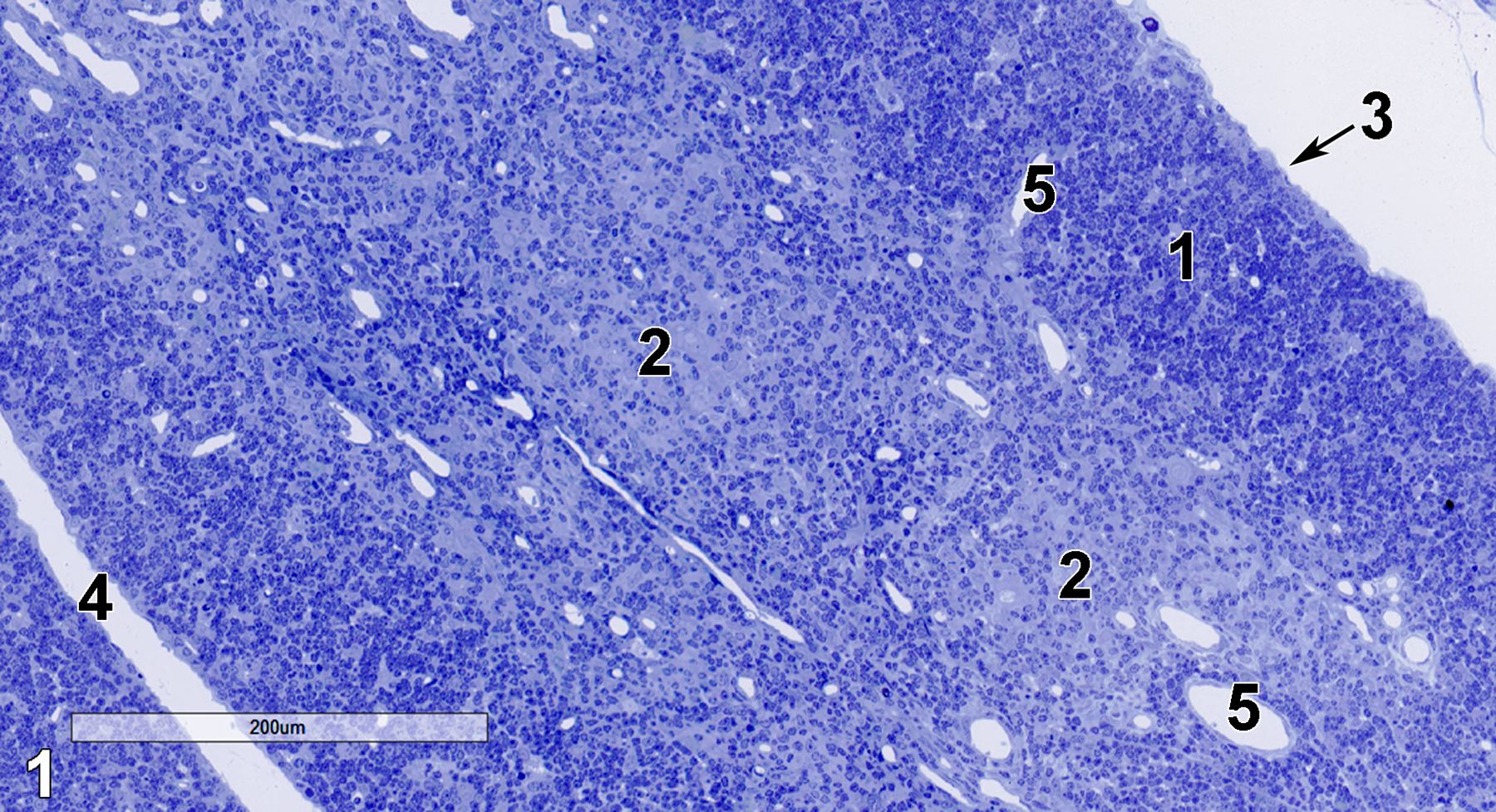
Figure 1. A semithin section (0.5 micrometer thick) of a toluidine blue O-stained portion of the thymus showing the cortical region (1), which is rich in small lymphocytes and the medullary region (2), with more lightly stained reticular epithelial cells, along with a lesser population of lymphocytes. A septum (4) of collagenous tissue separates the two lobules shown. The capsule (3, arrow) is located in the upper right of the image. Numerous blood vessels (5) are present. 14x.
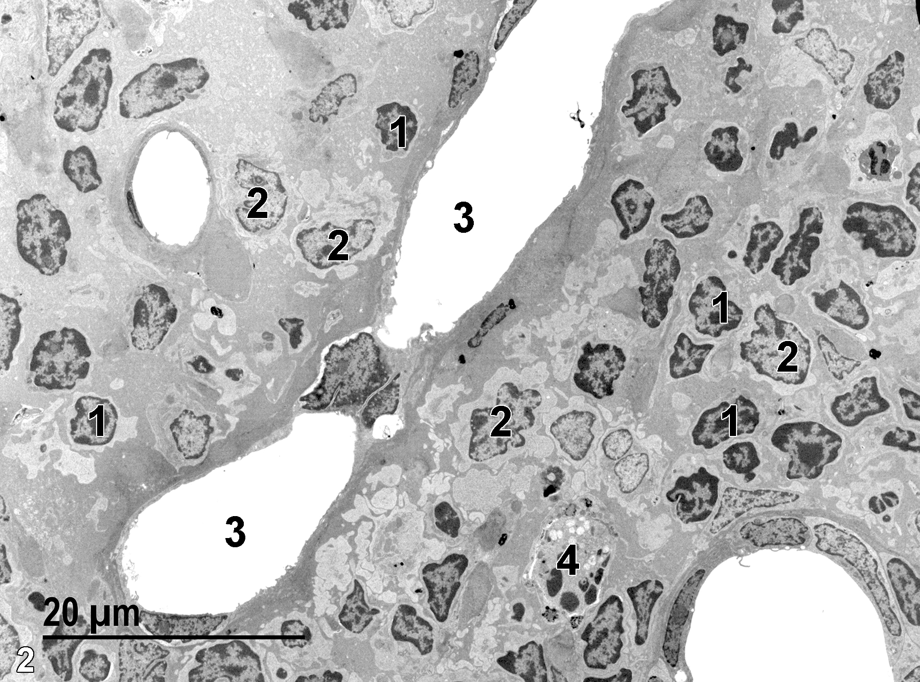
Figure 2. A low magnification electron micrograph showing lymphocytes (1) in the cortical region to the right of a longitudinal section of a blood vessel (3). A portion of the medulla is seen to the left of the long blood vessel along with another lymphocyte (1). Note the larger, somewhat lighter stained nuclei of reticular epithelial cells (2). One macrophage (4) is seen in the cortical region. 1900x.
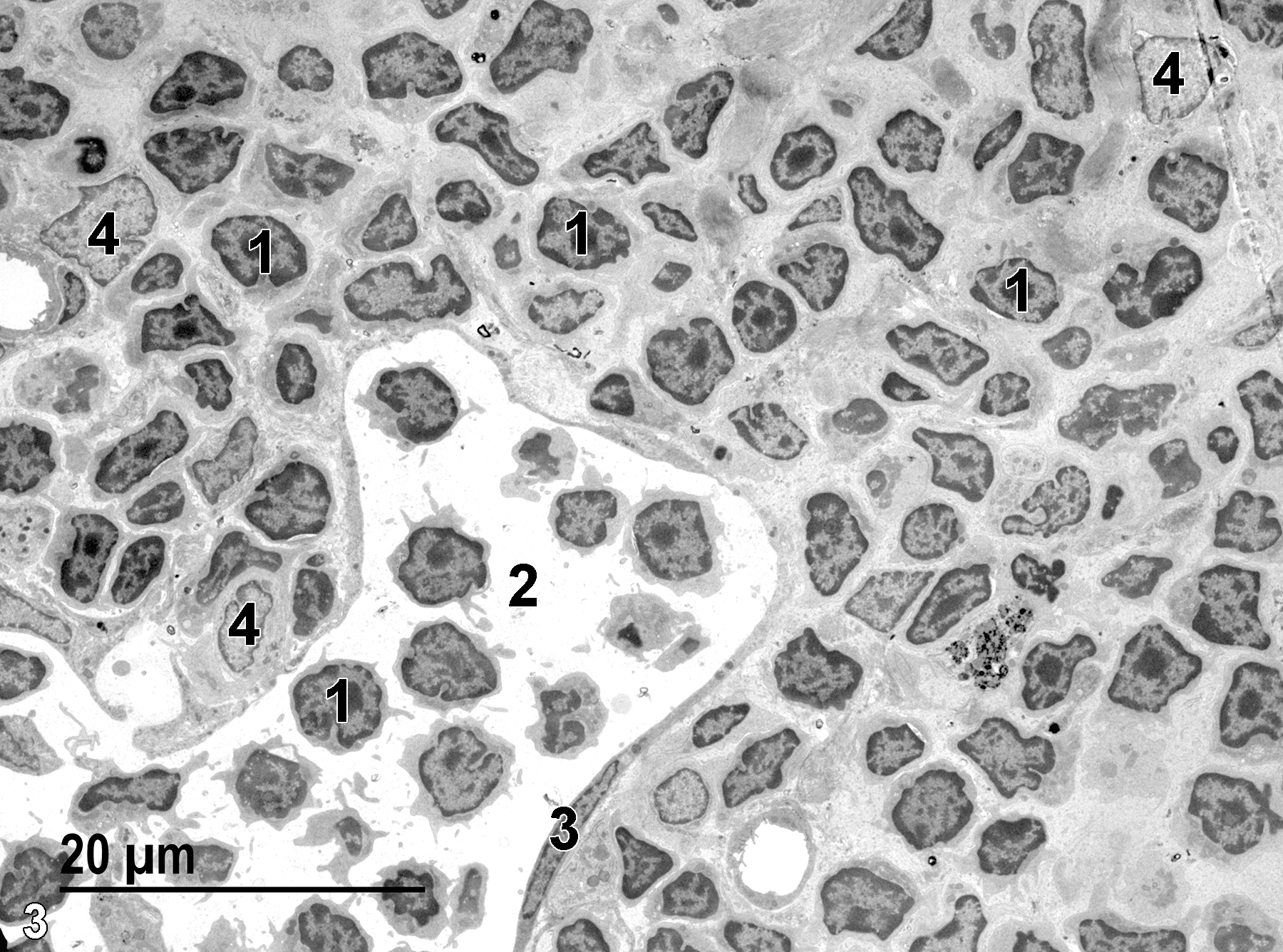
Figure 3. Another low magnification view of the thymus cortex with a dense accumulation of small lymphocytes (1) with prominent darkly stained nuclei and scant cytoplasm, a capillary (2) with an elongated endothelial cell nucleus (3), and reticular epithelial cells (4) with lighter stained nuclei than that seen in the lymphocytes. 1900x.
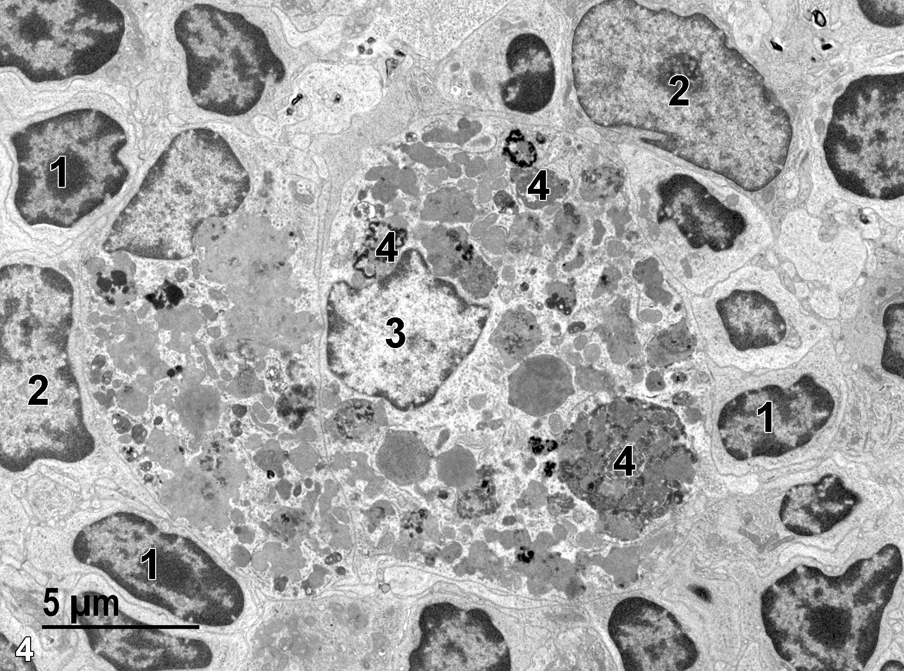
Figure 4. A portion of the cortex containing small lymphocytes with scant cytoplasm and nuclei (1), with prominent nucleoli and marginated heterochromatin. Two nuclei of reticular epithelial cells (2) are shown, which are large and less densely stained than the lymphocyte nuclei. A macrophage is shown with a single nucleus (3) and numerous lysosomes (4). 4800x.

Figure 5. Another view showing a reticular epithelial cell (1) with abundant cytoplasm, an eosinophil (2) with characteristic elliptical granules containing linear inclusions, and lymphocytes (3) with densely stained marginated chromatin and nucleoli. 4800x.
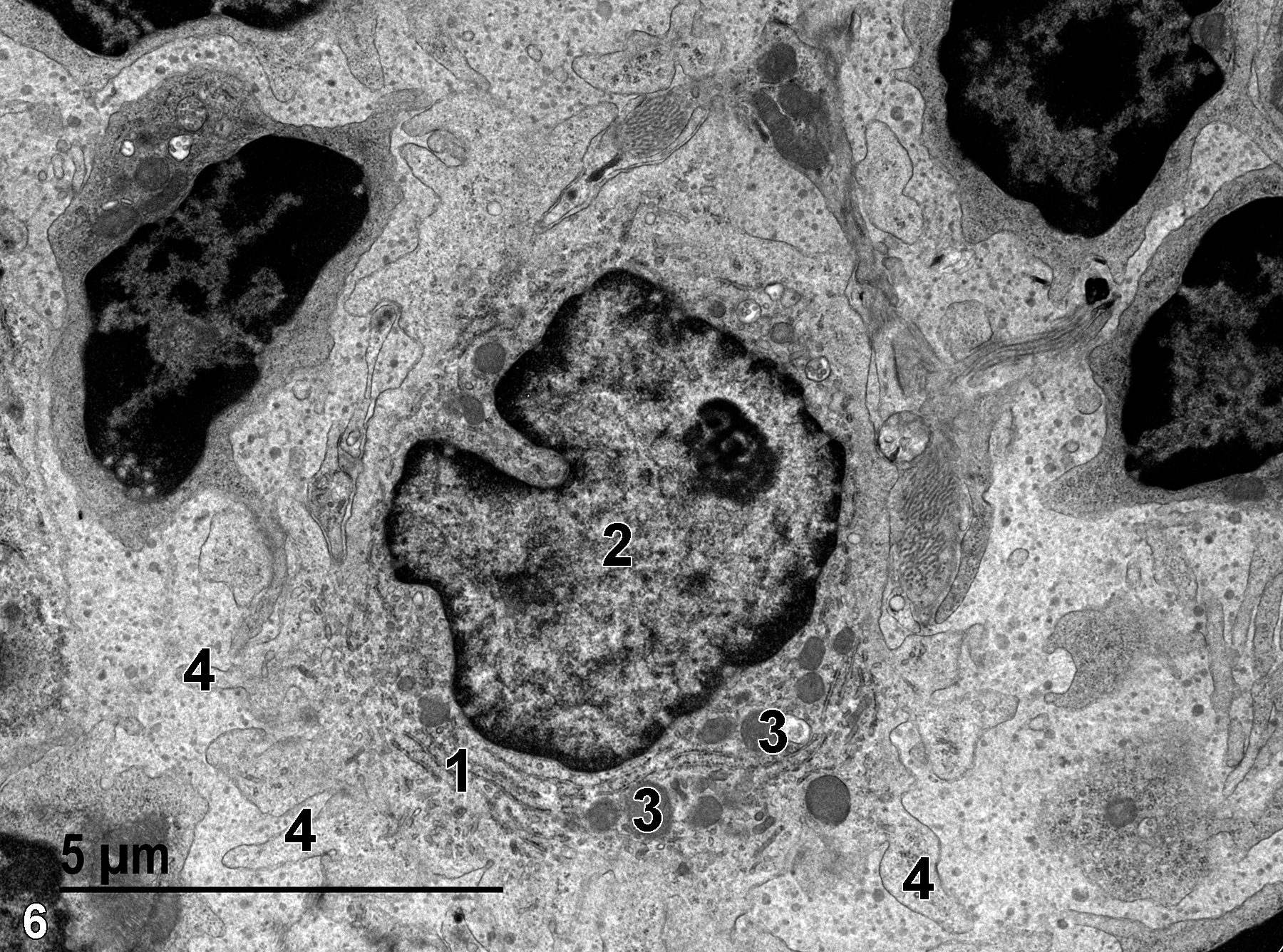
Figure 6. A higher magnification view of a reticular epithelial cell with a single nucleus (2), stacks of rough endoplasmic reticulum (1), several mitochondria (3), and cytoplasmic extensions (4) from the cells’ surface, which are characteristic of this cell type. 9300x.
| Dellmann HD, Eurell J, eds. 1998. Textbook of Veterinary Histology. 5th ed. Philadelphia: Lippincott Williams & Wilkins. |
| Rhodin JAG. 1974. Histology: A Text and Atlas. New York: Oxford University Press. |
| Weiss L, ed. 1988. Cell and Tissue Biology: A Textbook of Histology. 6th ed. Baltimore: Urban & Schwarzenberg. |
All Images


