

Alimentary System
Large Intestine
Narrative
The large intestine consists of the cecum, colon, and rectum. The cecum and colon are included in this Atlas as representative sections of the large intestine. Segments of the large intestine lack villi. The cecum, colon, and rectum have short infoldings of the mucosa with straight tubular intestinal glands (crypts of Lieberkühn). The surface epithelial cells lining the large intestine lumen consist mostly of absorptive cells (enterocytes) with some mucous cells, whereas the epithelial cells of the crypts have less absorptive cells and more mucous cells. A thin smooth muscle layer (muscularis mucosae) separates the lamina propria from the underlying submucosa that contains nerve fibers, loose connective tissue, and blood vessels. Lymphoid nodules are located within the submucosal layer. The submucosa is subtended by the tunica muscularis. The outermost layer of the intestines is the serosal (adventitious) tissue, which is a mesothelium that consists of squamous epithelial cells.
Cecum
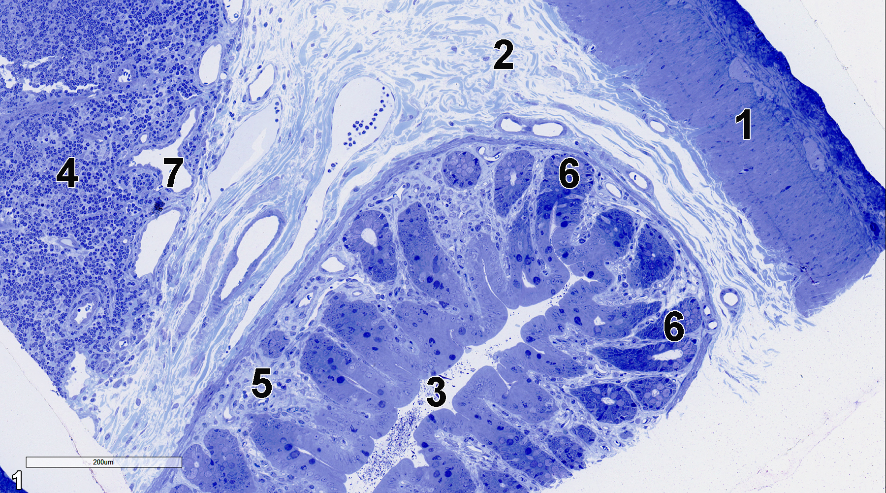
Figure 1. A semithin section (0.5 micrometer thick) of a toluidine blue O-stained portion of the cecum. The outermost layer of the cecum shown is the tunica muscularis (1). The submucosa (2) is composed primarily of connective tissue with vessels and fibroblasts. The lumen (3) of the cecum is shown, surrounded by the mucosa. A crypt of Lieberkühn is lined with absorptive epithelial cells (6) and goblet cells, with the latter in higher numbers near the base of the crypt. The lamina propria (5) consists of connective tissue and vessels. Lymphoid nodules (4) with lymphocytes and lymphatic vessels (7) are found in the submucosa. 15x.
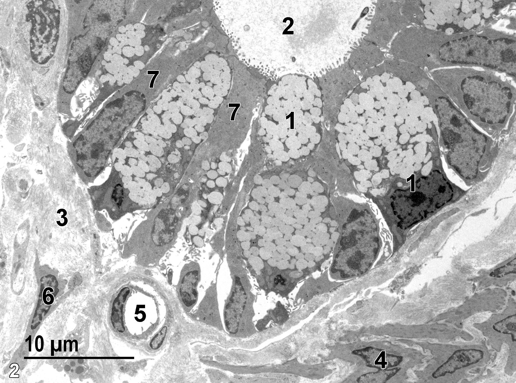
Figure 2. The basal aspect of a crypt of Lieberkühn. The lumen (2) of the crypt is lined with absorptive epithelial cells (7) with microvilli at the luminal surface and mucous cells (1) with no microvilli and containing numerous mucous droplets. The lamina propria (3) has bundles of collagen fibrils, a capillary (5), and occasional fibroblasts (6). A layer of smooth muscle cells, the muscularis mucosae (4), forms a layer below the lamina propria. 2900x.
Colon
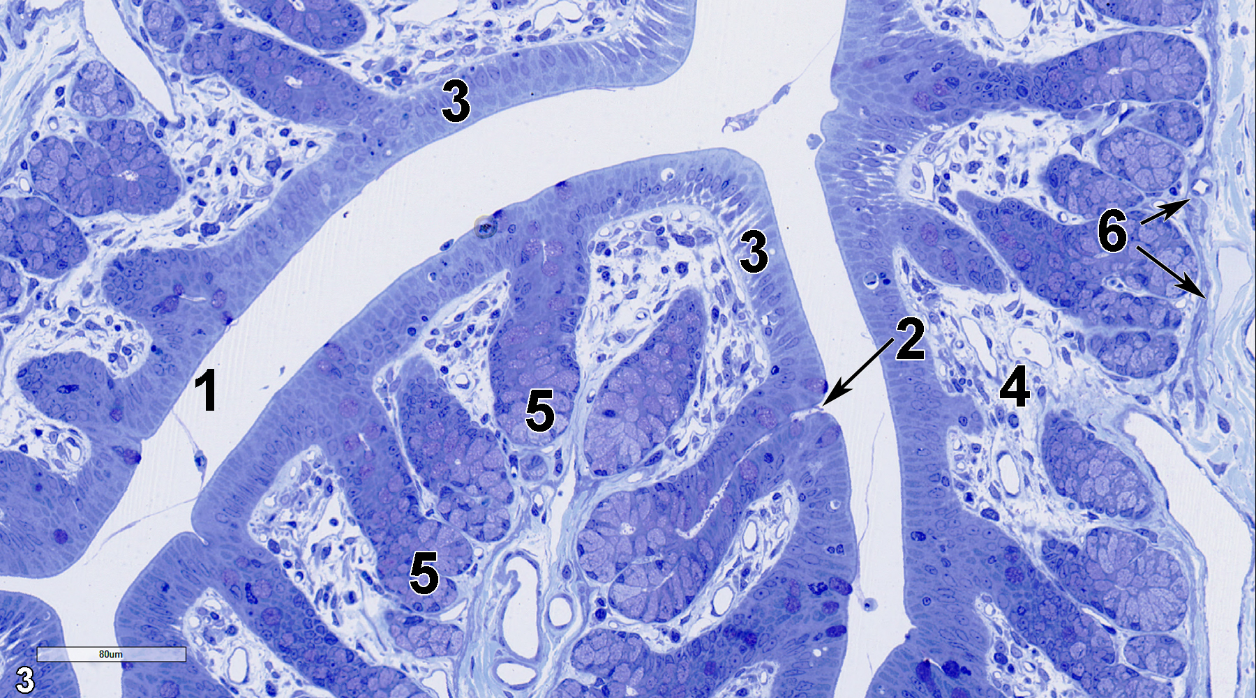
Figure 3. A semithin section (0.5 micron thick) of a toluidine blue O-stained portion of the colon showing the lumen (1). Superficial absorptive epithelial cells line the mucosa and also extend down into the intestinal gland (crypt of Lieberkühn) from the entrance to the gland (2, arrow). The more basal aspects of the intestinal glands have higher numbers of mucous cells (5) that contain numerous mucous droplets. The lamina propria (4) contains fibroblasts, collagen fibrils, and vessels. A thin layer of smooth muscle cells makes up the muscularis mucosae (6, arrows). 25x.
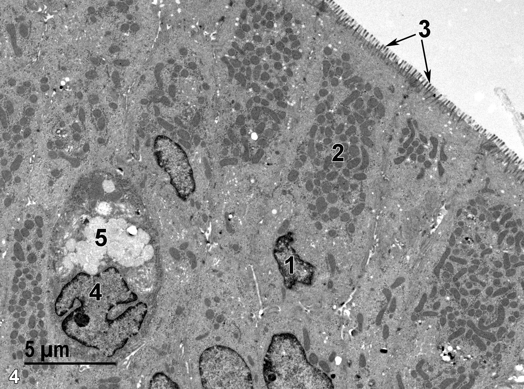
Figure 4. A portion of the colonic mucosal epithelium. An absorptive epithelial cell has a basal nucleus (1), a large number of more apical mitochondria (2) and numerous microvilli (3, arrows) that extend into the colon lumen. A mucous cell is shown with a single nucleus (4) and a cluster of mucous droplets (5). 4800x.
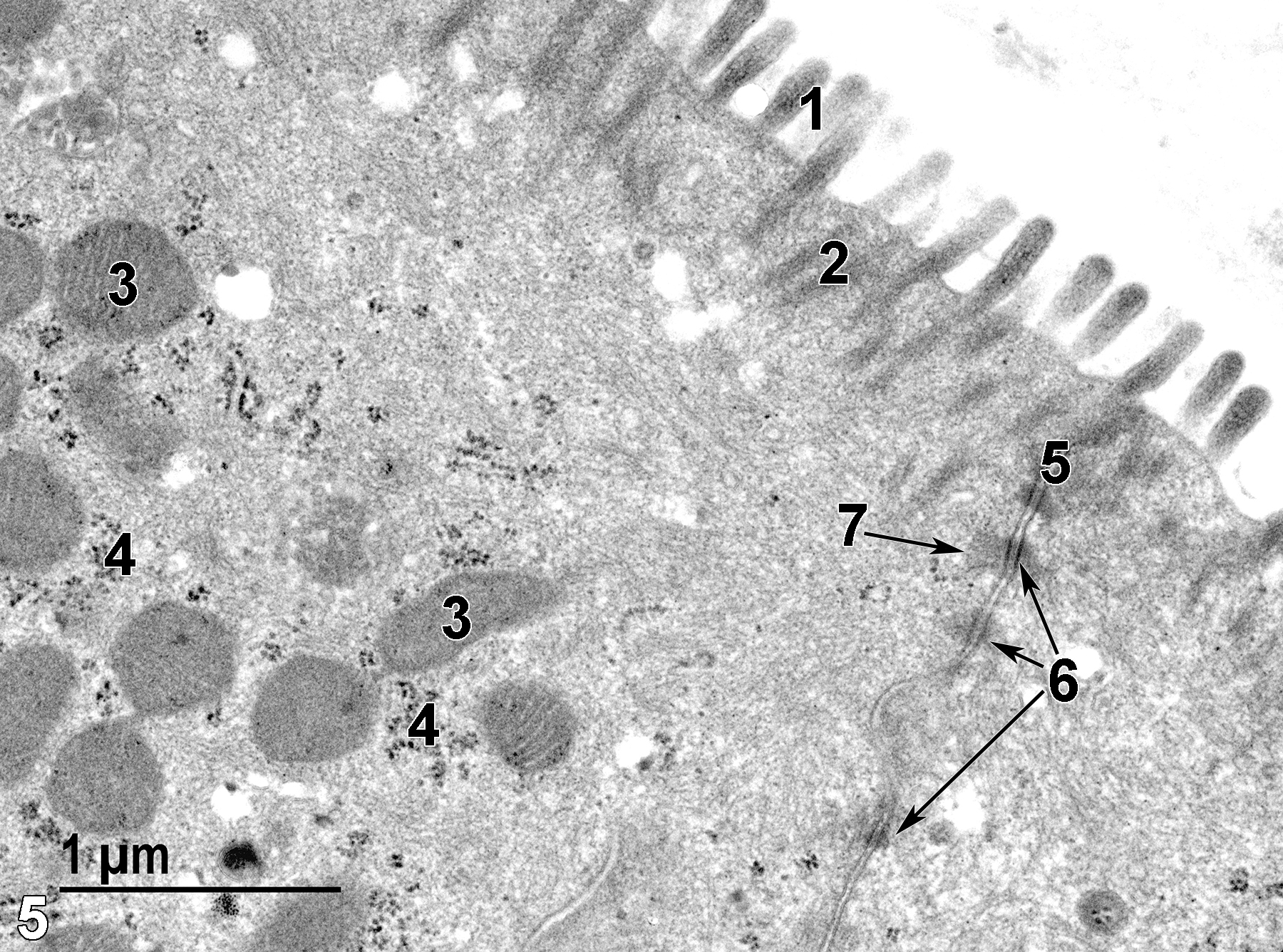
Figure 5. A high magnification view of two absorptive epithelial cells. The cell surface has microvilli (1) that protrude into the colon lumen and has bundles of proteinaceous fibrils that extend into the cytoplasm, forming the rootlets (2) of the microvilli. Mitochondria (3) are present primarily in the apical aspect of these cells. Clusters of free ribosomes (4) are common. A junctional complex (5) binds the apical aspect of the epithelial cells to each other. Desmosomes (6, arrows) with associated tonofilaments (7, arrow) are part of the junctional complex. 30000x.
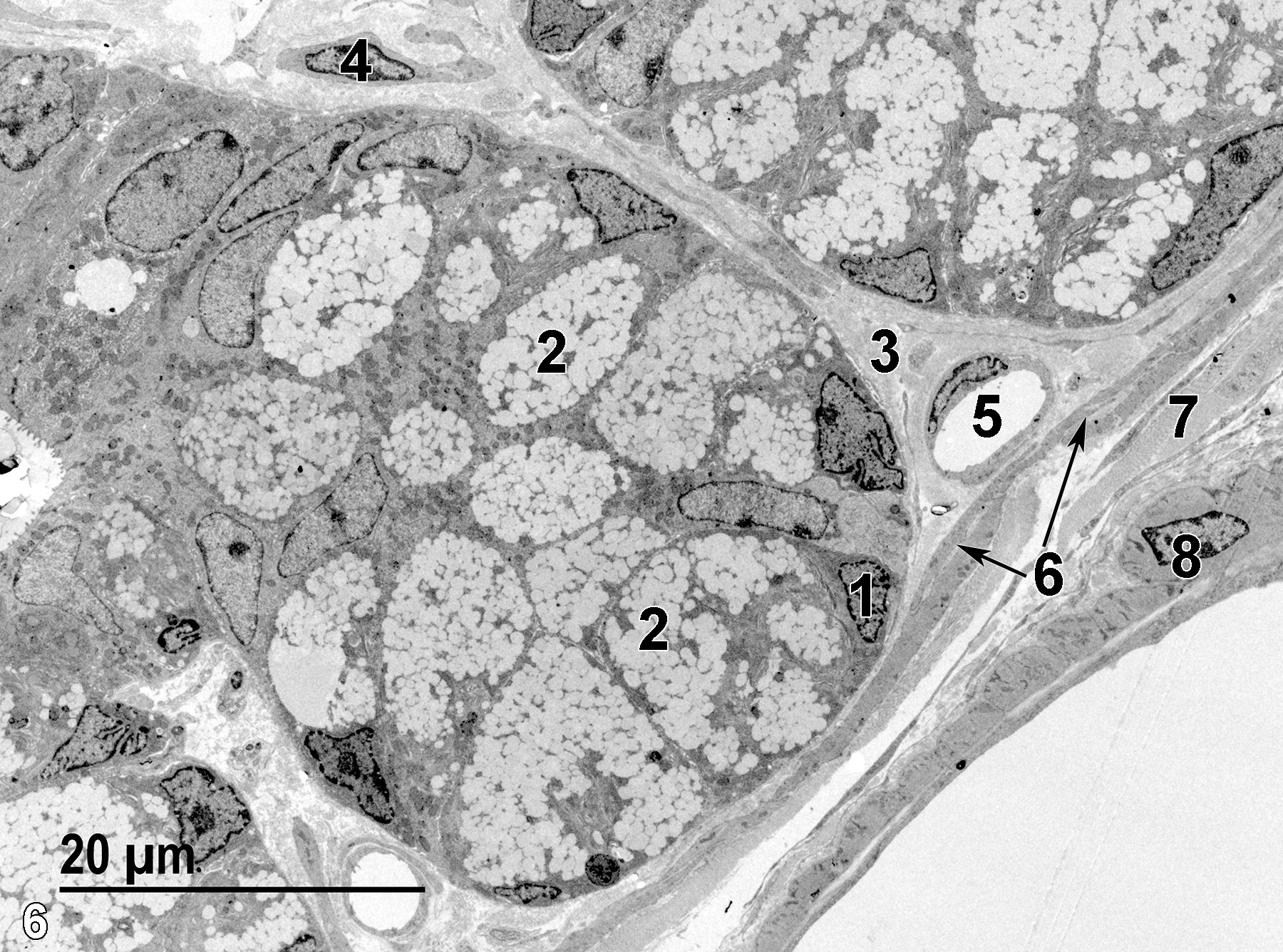
Figure 6. The basal nucleus (1) of a mucous cell. These cells are more numerous in the basal aspect of intestinal glands. Each cell contains large numbers of mucous droplets (2). The lamina propria (3) consists primarily of a collagenous matrix, shown with a fibroblast (4) and a capillary (5). A thin band of smooth muscle cells (6, arrows) form the muscularis mucosae that overlies the submucosa, which contains many bundles of collagen fibrils (7). The outermost muscular layer is the tunica muscularis (8). 1900x.
| Cross PC, Mercer KL. 1993. Cell and Tissue Ultrastructure: A Functional Perspective. New York: W.H. Freeman and Company. |
| Dellmann HD, Eurell J, eds. 1998. Textbook of Veterinary Histology. 5th ed. Philadelphia: Lippincott Williams & Wilkins. |
| Rhodin JAG. 1974. Histology: A Text and Atlas. New York: Oxford University Press. |
| Ross MH, Kaye GI, Pawlina W. 2003. Histology: A Text and Atlas. 4th ed. Philadelphia: Lippincott Williams & Wilkins. |
| Uehara T, Elmore SA, K.A. Szabo KA. 2017. Chapter 6: Esophagus and stomach. In Boorman’s Pathology of the Rat (Suttie AW, ed). 2nd ed. London: Academic Press, 35-50. |
| Weiss L, ed. 1988. Cell and Tissue Biology: A Textbook of Histology. 6th ed. Baltimore: Urban & Schwarzenberg. |
All Images


