

Reproductive System, Male
Prostate
Narrative
The prostate is surrounded by a thin capsule, continuous with trabeculae and septa that penetrates the parenchyma and forms a fibroblastic stroma that contains collagen, smooth muscle cells, and few fibroblasts. The septa divide the prostate into lobes. The paired dorsolateral lobes, which consist of dorsal and lateral portions and the paired ventral lobes, are grouped together around the central urethra. The parenchyma of the prostate consists of tubulo-alveolar serous glands with cuboidal to columnar epithelial cells that rest on a basal lamina. The epithelial cells have small sparse mitochondria, a small number of lysosomes, large quantities of rough endoplasmic reticulum, and a large Golgi zone that produce secretory granules apical to the basal nucleus. Peri-urethral mucosal glands surround the central urethra. Occasional concentric concretions are found in tubules and alveoli, particularly in aged animals.
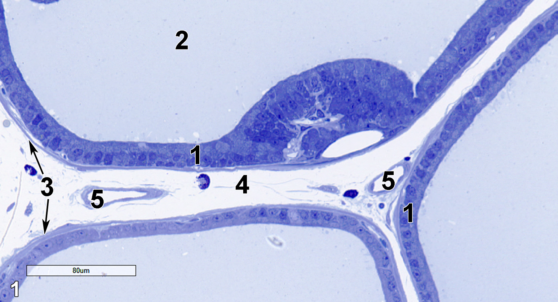
Figure 1. A semithin section (0.5 micrometer thick) of a toluidine blue O-stained portion of the dorsolateral part of the prostate gland. A tall columnar epithelial portion (1) lines the alveolar lumen (2). The stroma consists of a thin layer of smooth muscle cells beneath the epithelial cell layer (3, arrows) and loose connective tissue (4) with several blood vessels (5). 25x.
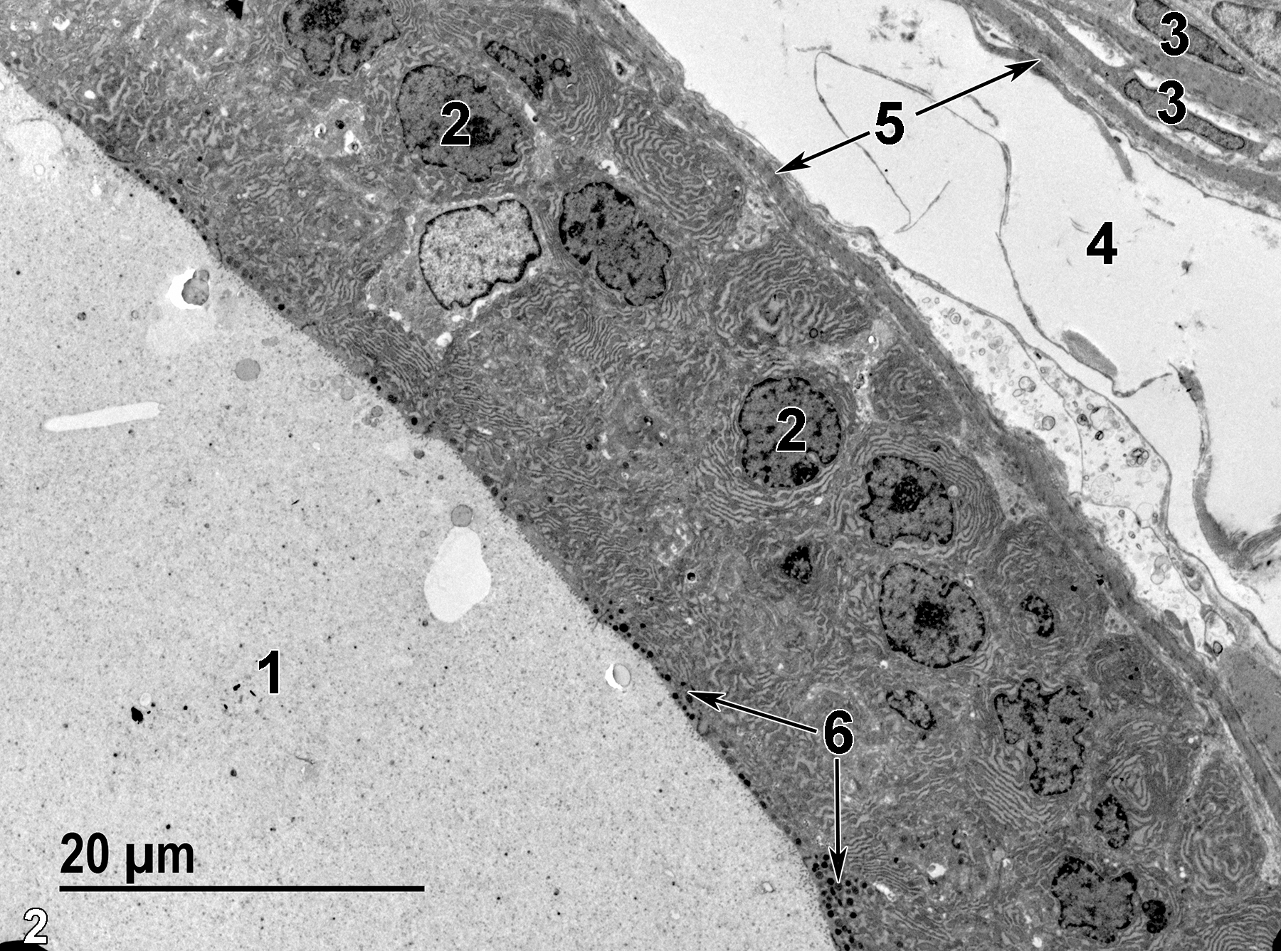
Figure 2. A low magnification ultrastructural view of a dorsolateral prostatic alveolus (1) with finely granular secretory material in the alveolar lumen. The epithelial cells have basal nuclei (2) and apical secretory granules (6, arrows). Smooth muscle cells (5, arrows) surround the basal exterior of the alveolar epithelial cells. Note the elongated and flattened nuclei (3) of the smooth muscle cells in the upper right of the image. Connective tissue (4) lies between two adjacent alveoli. 1900x.
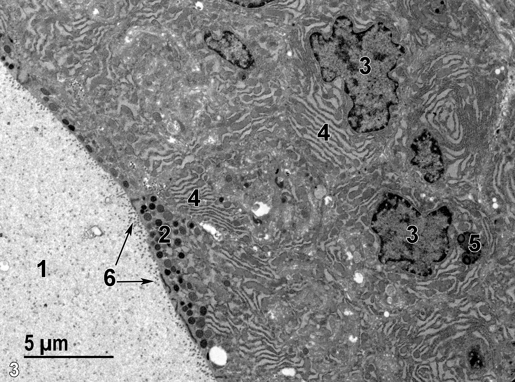
Figure 3. A higher magnification view of dorsolateral lobe alveolar epithelial cells. Note the finely granular secretory material in the alveolar lumen (1), the apical secretory granules (2), the basal nuclei (3), the large stacks of rough endoplasmic reticulum (4), a lysosome (5), and microvilli (6, arrows) on the apical surface of the epithelial cells. 4800x.
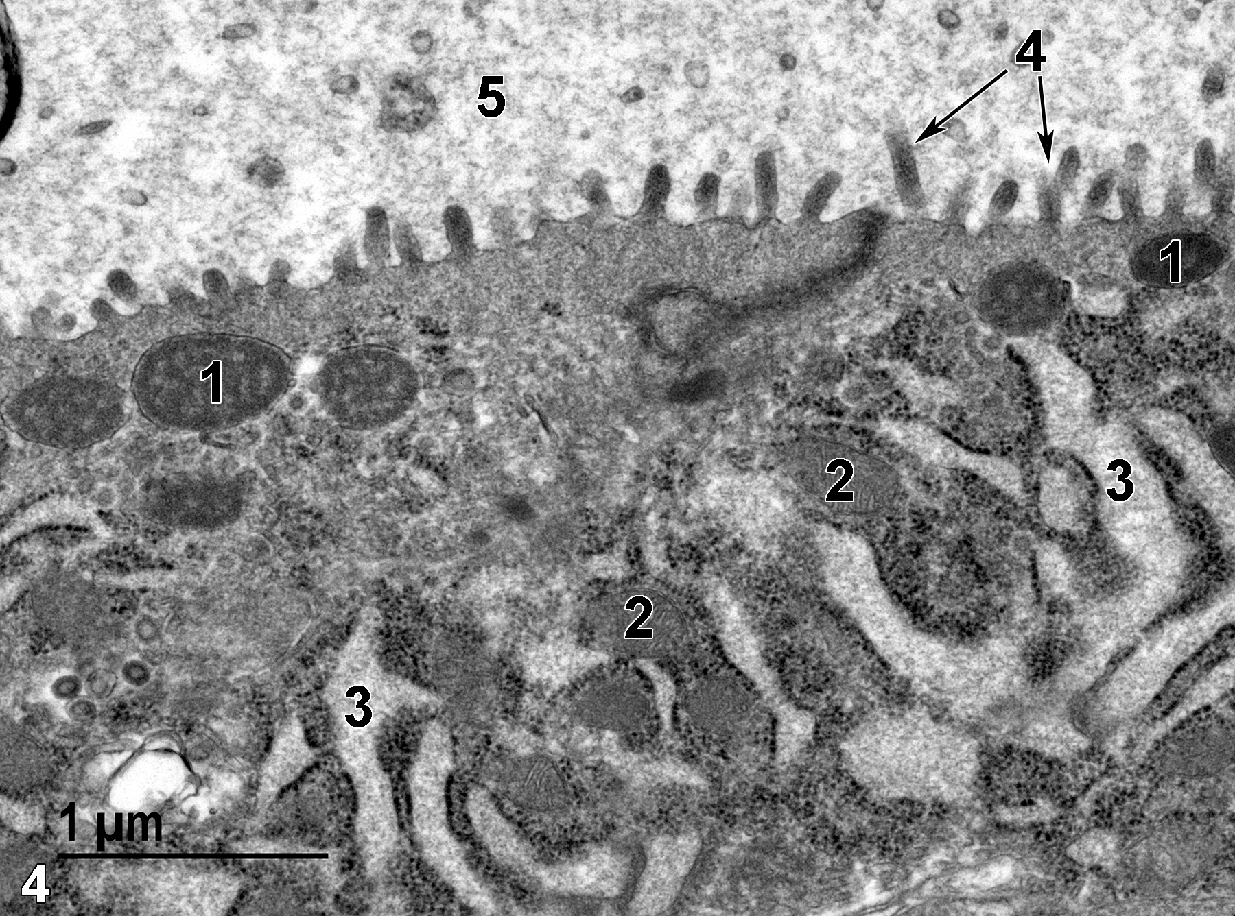
Figure 4. An even higher magnification image of the surface of a dorsolateral lobe alveolar epithelial cell. Several secretory granules (1) are present, along with a number of mitochondria (2) intermixed with the rough endoplasmic reticulum cisternae (3). Microvilli (4, arrows) line the alveolar lumen (5), which contains finely granular secretory material. 30000x.
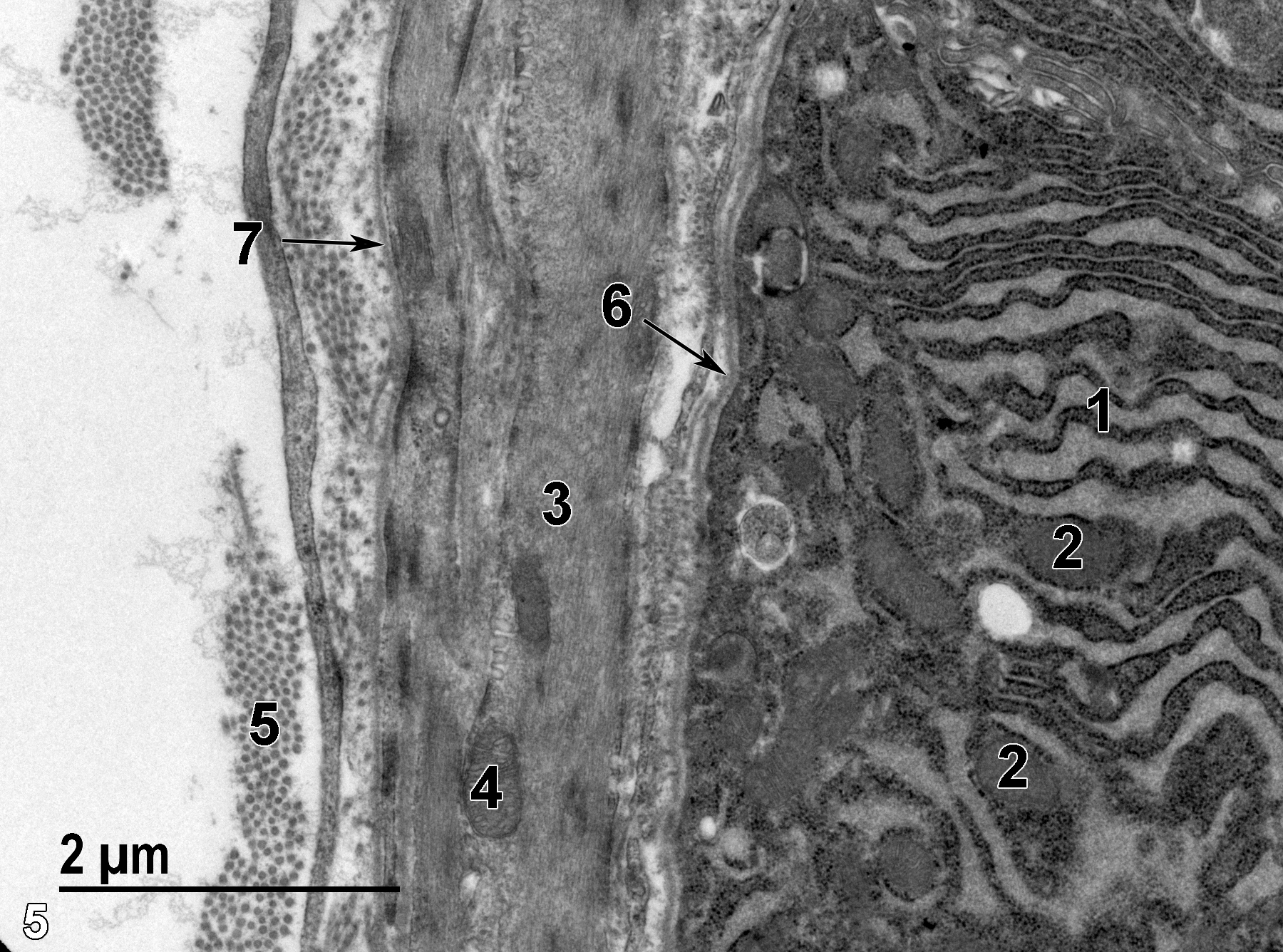
Figure 5. A high magnification of the base of an alveolar epithelial cell with stacks of rough endoplasmic reticulum (1) intermixed with mitochondria (2). The underlying smooth muscle cells of the stroma contain large amounts of actin and myosin filaments (3) and a few mitochondria (4). The collagen in the stromal tissue (5) is seen in dense clusters. A thin basal lamina is located at the base of the epithelial cells of the alveolus (6, arrow). In addition, a thin basal lamina (7, arrow) surrounds the smooth muscle cells. 18500x.
| Creasy D, Bube A, de Riik E, Kandori H, Kuwahara M, Masson R, et al. 2012. Proliferative and nonproliferative lesions of the rat and mouse male reproductive system. Toxicol Pathol 40:40S-121S. |
| Dellmann HD, Eurell J, eds. 1998. Textbook of Veterinary Histology. 5th ed. Philadelphia: Lippincott Williams & Wilkins. |
| Rhodin JAG. 1974. Histology: A Text and Atlas. New York: Oxford University Press. |
| Ross MH, Kaye GI, Pawlina W. 2003. Histology: A Text and Atlas. 4th ed. Philadelphia: Lippincott Williams & Wilkins. |
| Weiss L, ed. 1988. Cell and Tissue Biology: A Textbook of Histology. 6th ed. Baltimore: Urban & Schwarzenberg. |
| Whitney, KM. 2018. Chapter 29: Male accessory sex glands. In Boorman’s Pathology of the Rat (Suttie AW, ed.). 2nd ed. London; Academic Press; 579-587. |
All Images


