

Urinary System
Urinary Bladder
Narrative
The urinary bladder has four basic layers — the mucosa, the submucosa (lamina propria), the muscularis, and the serosa. The mucosa is a layer that consists of urothelium (formerly transitional epithelium) with stratified cuboidal cells with many interdigitations of the surface layer when the bladder is empty, which. These cells decrease in number as the bladder fills. In addition, an empty bladder has more layers of epithelial cells, with less layers when the bladder is distended. The cuboidal epithelial cells also become more flattened as the bladder becomes distended. Relatively scarce desmosomes (part of the junctional complex of the umbrella cells (Jokinen and Seely 2018) are present, binding the epithelial cells to each other. Fusiform vesicles of unknown function can be found within some of the epithelial cells.
The basal side of the mucosa is bounded by a thin basal lamina that is beneath the submucosal layer. This layer consists of capillaries, collagen, fibroblasts, and elastic fibers. The submucosa, in turn, is underlain by the muscularis layer, which has three layers of smooth muscle cells and a number of blood vessels.
Finally, the bladder is surrounded by the outermost serosa layer located on the upper lateral and superior surfaces of the bladder.
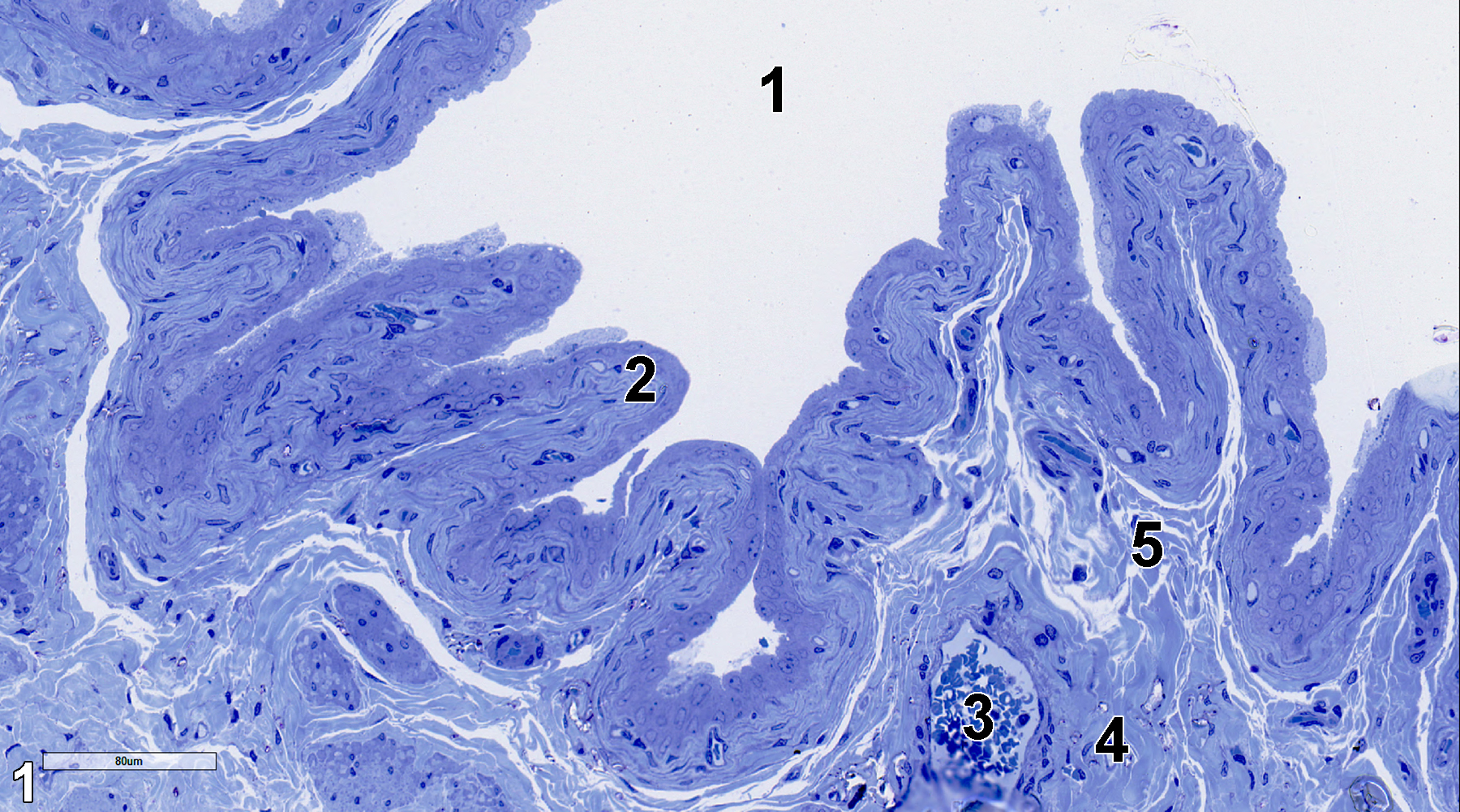
Figure 1. A toluidine blue O-stained semithin section (0.5 micrometer thick). It shows the lumen (1) of the urinary bladder, the folded mucosal layer (2), a blood vessel (3) in the muscularis layer (4) directly below the looser submucosal layer (5). 25x.
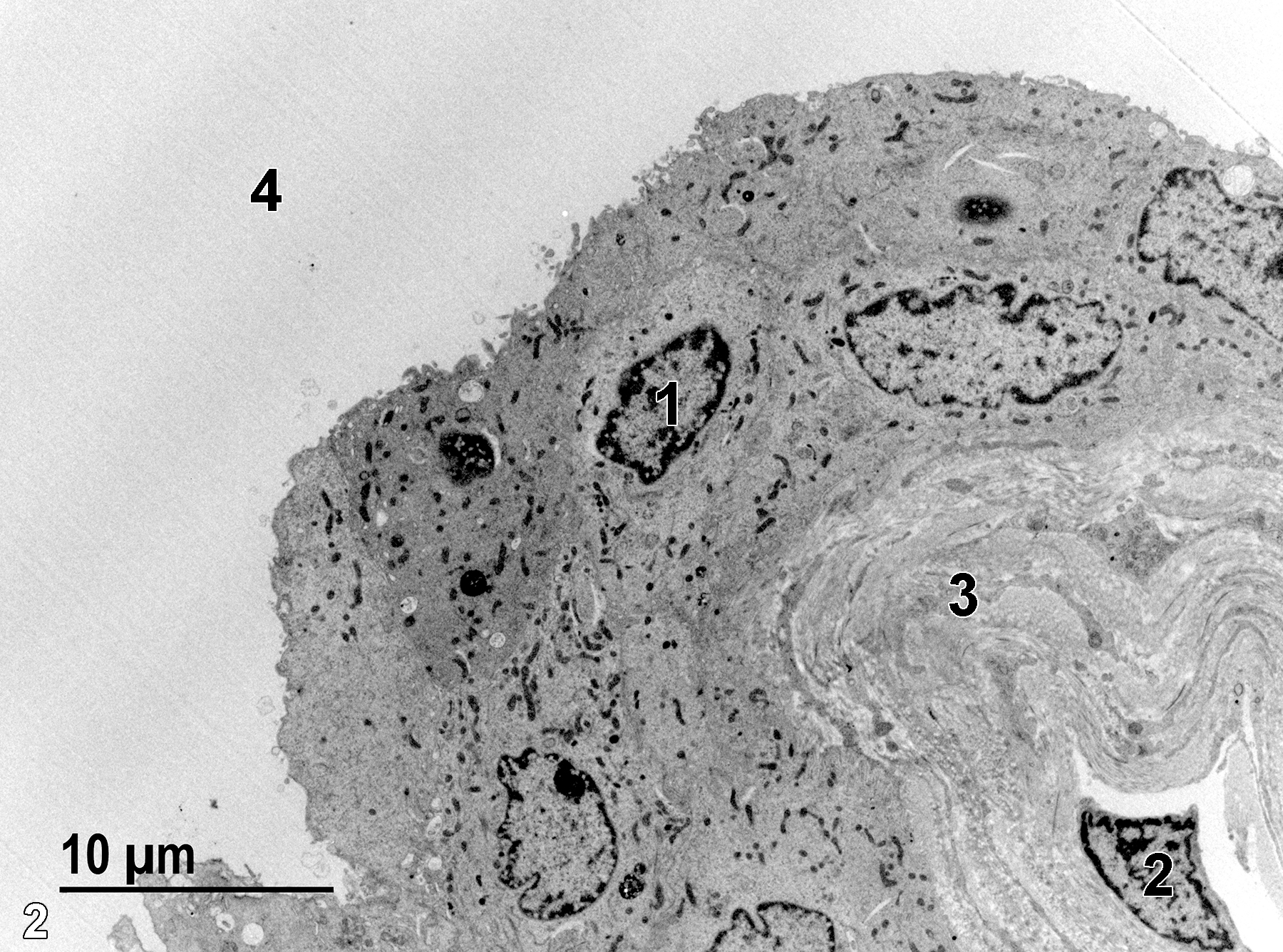
Figure 2. An ultrathin section of the bladder epithelium and underlying submucosal layer. An epithelial cell nucleus (1) has marginated heterochromatin, and some of the nuclei in the epithelial cell layer have crenelated borders. In the submucosa, a single fibroblast cell nucleus (2) is present within the mass of collagen (3), consisting of the bulk of the submucosa. The surface of the mucosa extends into the bladder lumen (4). 2900x.
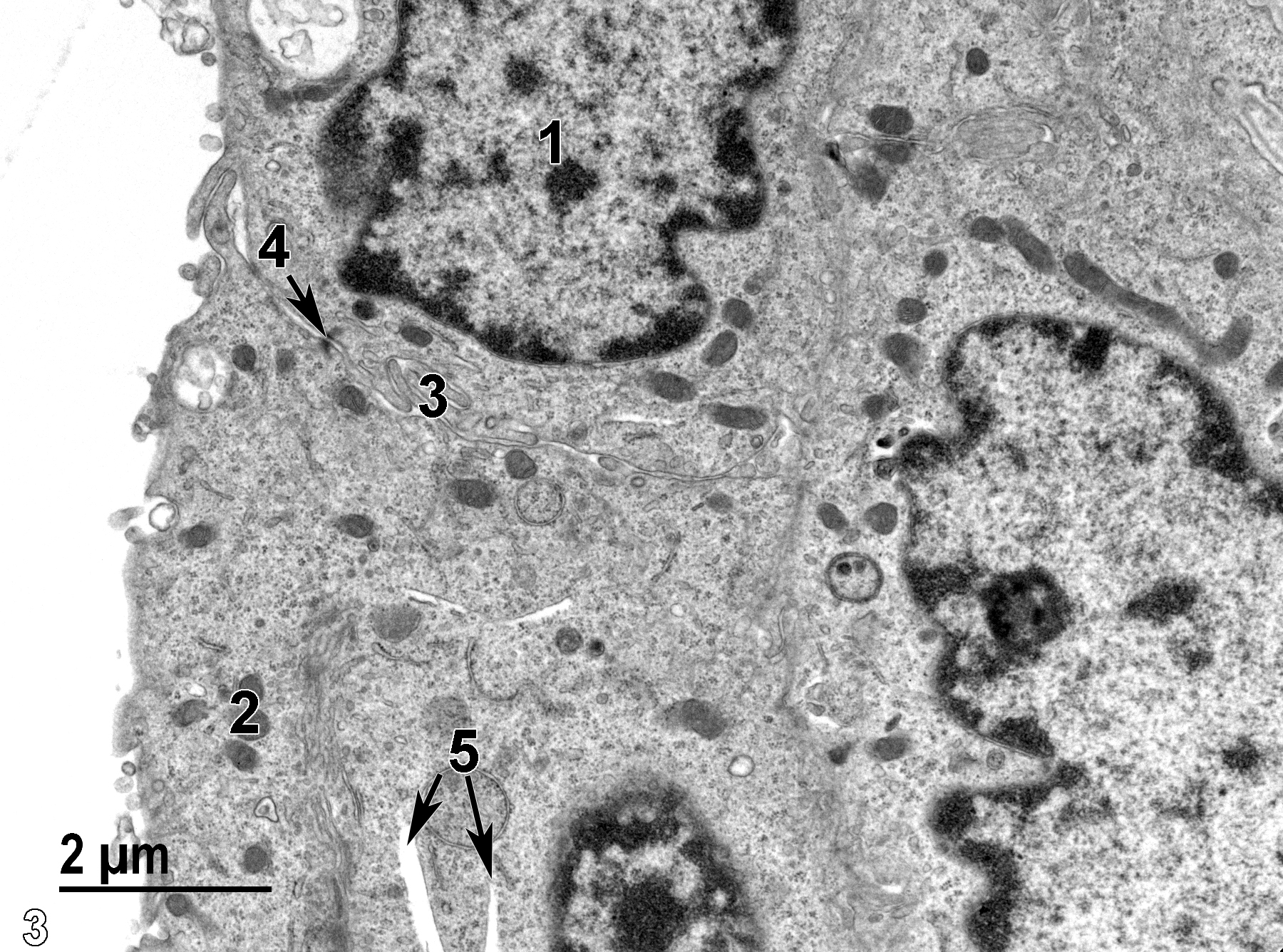
Figure 3. A higher magnification view of cells of the mucosal layer showing an epithelial cell nucleus (1), mitochondria (2), apical infoldings (3) of the epithelial cells, a desmosome (4, arrow), and two fusiform vesicles (5, arrows). 11000x.
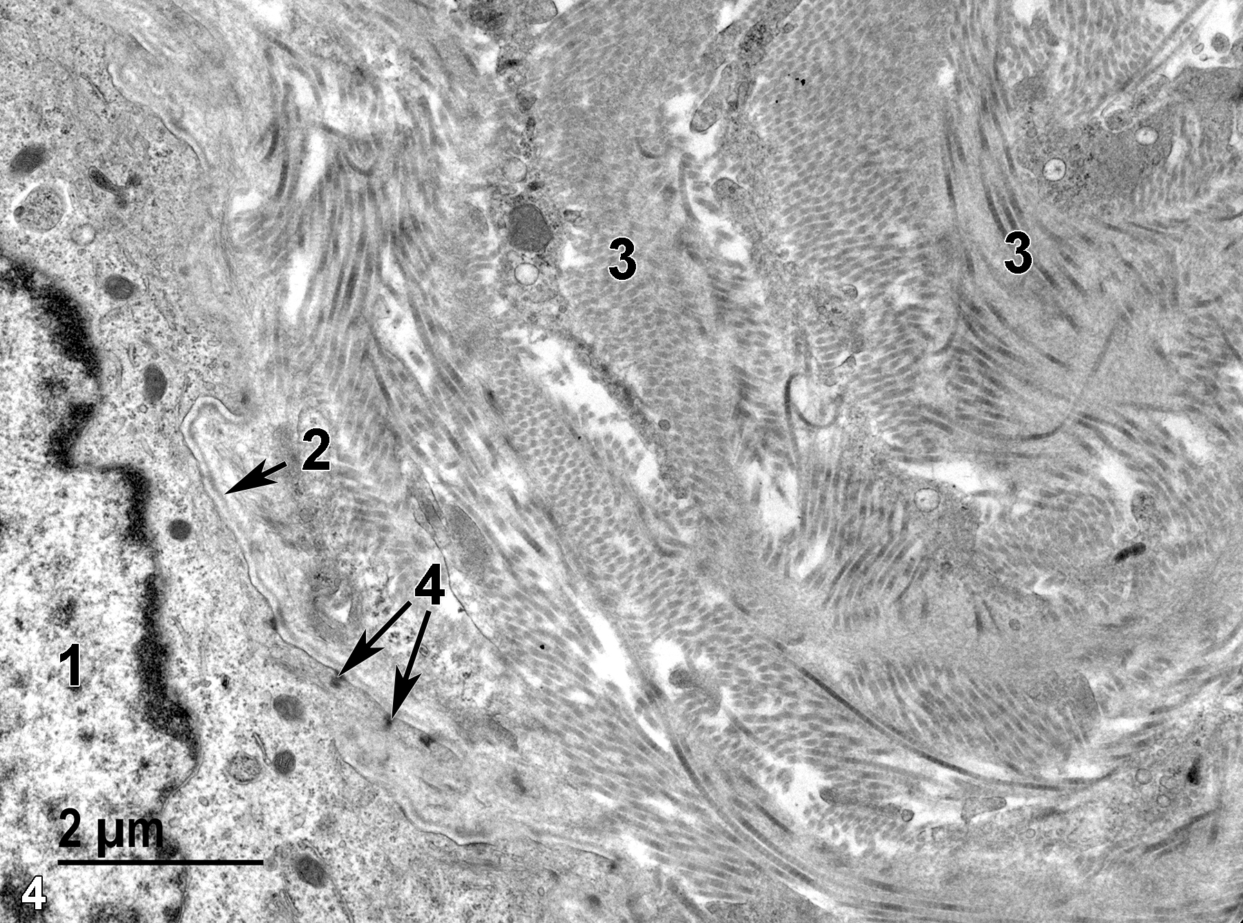
Figure 4. The nucleus (1) of one of the basal epithelial cells of the mucosa. Separating the epithelial cells from the submucosal layer that contain characteristic collagen fibrils (3) is a thin basal lamina (2, arrow). Several hemi-desmosomes (4, arrows) in the basal aspect of the epithelial cell are oriented toward the basal lamina. 11000x.
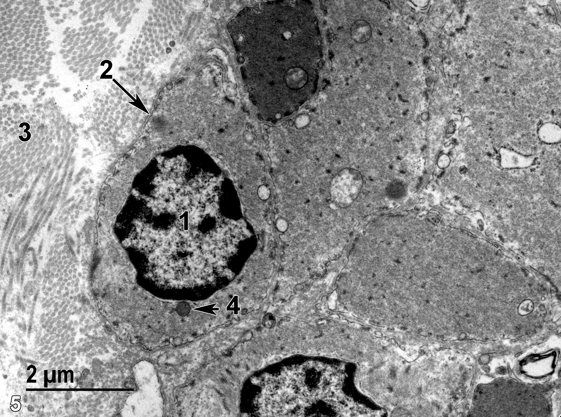
Figure 5. A view of the collagen fibrils (3) of the submucosal layer next to smooth muscle cells of the muscularis layer. A mitochondrion (4, arrow) of a smooth muscle cell is adjacent to the nucleus (1), and the smooth muscle cell has a thin but distinct basal lamina (2, arrow). 13000x.
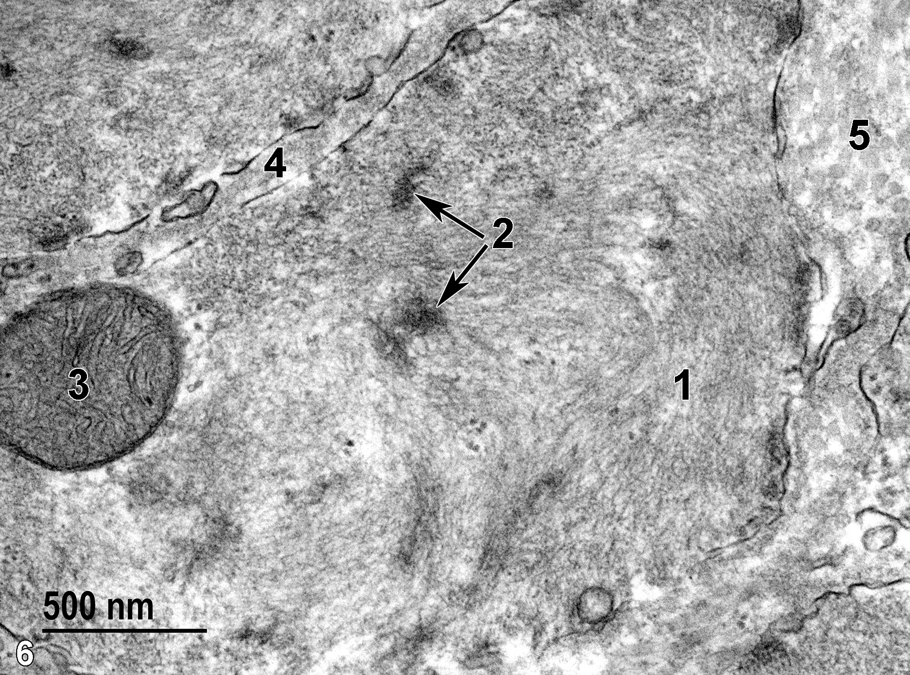
Figure 6. A high magnification view of a smooth muscle cell of the muscularis layer. Numerous contractile filaments of actin and myosin (1) fill the cytoplasm, along with dense bodies (2, arrows).A single mitochondrion (3) with prominent cristae and fairly electron-dense matrical content is present. Smooth muscle cell basal laminae (4) are present between the cells. Collagen fibrils (5) are located between smooth muscle cells in the muscularis layer. 49000x.
| Cross PC, Mercer KL. 1993. Cell and Tissue Ultrastructure: A Functional Perspective. New York: W.H. Freeman and Company. |
| Jokinen MP, Seely JC. 2018. Chapter 12: Urinary bladder. In Boorman’s Pathlogy of the Rat (Suttie AW, ed.). 2nd ed. London: Academic Press;167-188. |
| Rhodin JAG. 1974. Histology: A Text and Atlas. New York: Oxford University Press. |
| Weiss L, ed. 1988. Cell and Tissue Biology: A Textbook of Histology. 6th ed. Baltimore: Urban & Schwarzenberg. |
All Images


