

Reproductive System, Female
Ovary
Narrative
The two ovaries are each connected to the abdominal wall via the hilus and are surrounded by a layer of cuboidal or squamous cells known as the ovarian surface epithelium, which sits on a thin basal lamina. Inside of the surface epithelium is the tunica albuginea, which forms a connective tissue capsule that contains collagen and fibroblasts. The ovary tissue within the capsule is differentiated into cortical and medullary zones. The medulla contains neural tissue, lymphatics, blood vessels, connective tissue, and interstitial cells. The cortex has ovarian follicles, corpora lutea, fibroblast-like cells, and collagen. Some of the fibroblast-like cells of the cortical region in rats contain accumulations of lipid and are referred to as the “interstitial gland” by Rhodin (1974). The ovaries undergo changes depending on the stage of the estrous cycle observed and the age of the animal (Vidal and Dixon 2018).
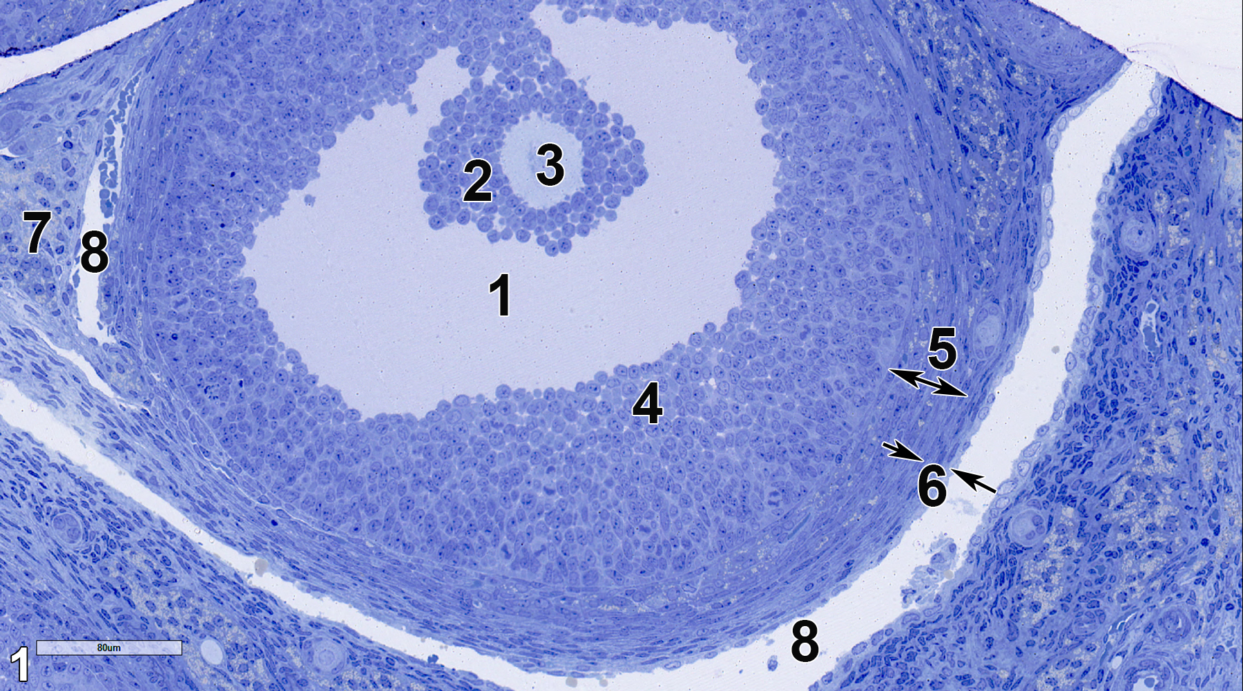
Figure 1. A semithin section (0.5 micrometer thick) of a toluidine blue O-stained section of a secondary follicle of an ovary. The antrum (1) is a space formed by follicular fluid that accumulates between follicular cells. The corona radiata (2) consists of follicular cells surrounding the secondary oocyte (3). The follicular cells at the periphery of the follicle are called the membrana granulosa (4), which is enclosed by the theca interna (5, double-headed arrow), which consists of squamous cells and collagen. Outside of this layer is the theca externa (6, arrows), which consists of squamous cells that are more flattened than those in the theca interna and includes collagen. The theca folliculi (7), which consists of stromal cells near the follicle, contains vascular elements (8). 25x.
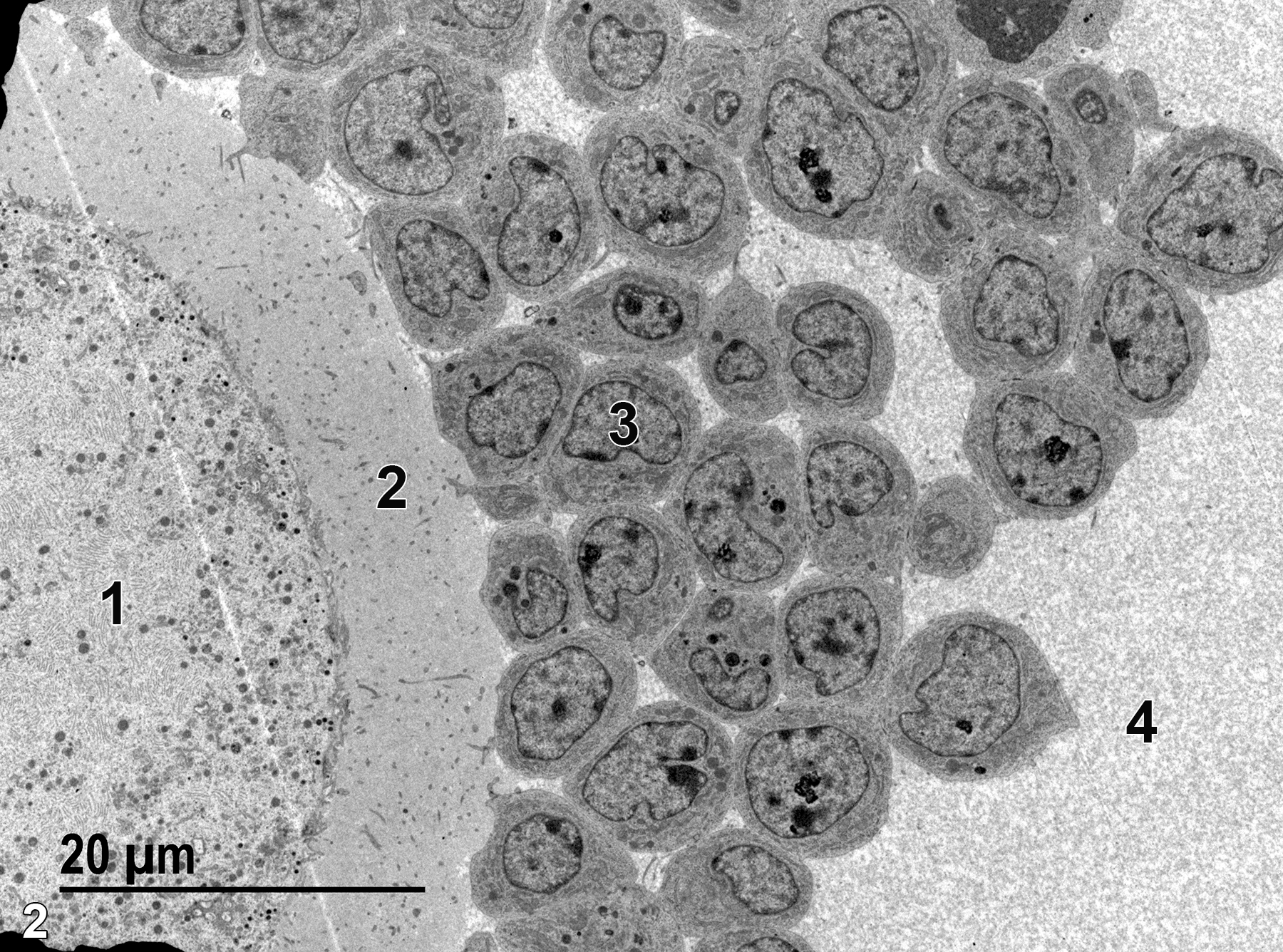
Figure 2. A low magnification electron micrograph of a secondary oocyte. Surrounding the secondary oocyte (1) is the zona pellucida (2), which consists of mucopolysaccharides with numerous secondary oocyte microvilli ramifying through the matrix. A nucleus of a follicular cell (3) in the corona radiata is shown. The spaces between follicular cells is filled with follicular fluid (4), known as liquor folliculi. 1900x.
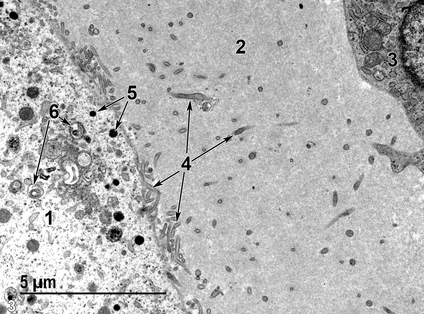
Figure 3. An enlarged view of part of the oocyte (1), zona pellucida (2), and part of a follicular cell (3). The zona pellucida consists of a finely fibrillar material (mucopolysaccharide) penetrated by numerous microvilli (4, arrows) that originate from the surface of the secondary oocyte. The secondary oocyte contains cortical granules (5, arrows) and lysosomes (6, arrows). 9300x.
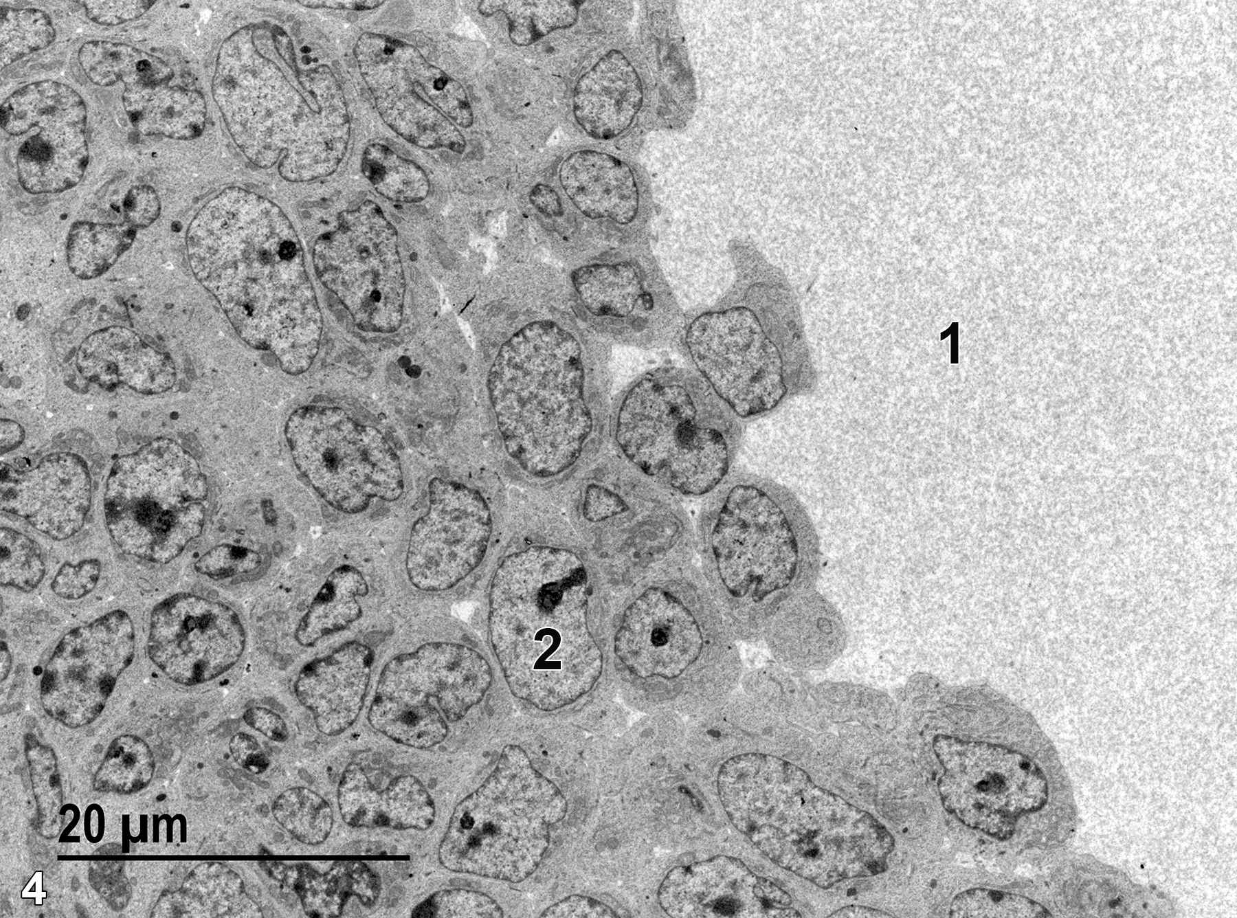
Figure 4. Some of the follicular cells that make up the membrana granulosa surrounding the secondary oocyte. The fibrillar matrix of the liquor folliculi in the antrum (1) is at the surface of and penetrates between the follicular cells (2) of the membrana granulosa. 1900x.
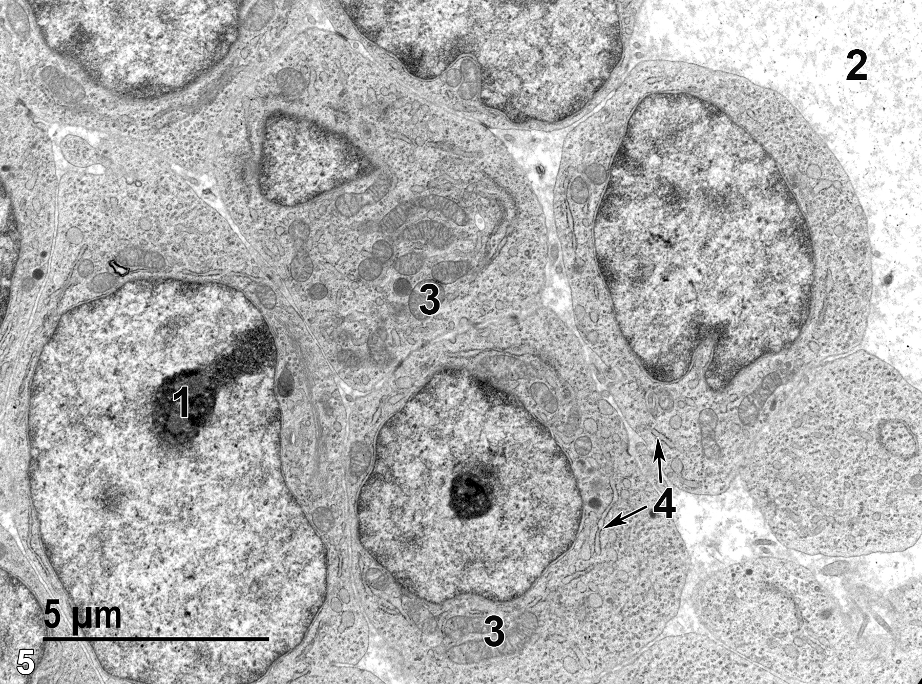
Figure 5. A further enlargement of Figure 4. The nucleolus (1) of a follicular cell nucleus is shown. The fibrillar nature of the liquor folliculi (2) is evident. The follicular cells contain mitochondria (3) and rough endoplasmic reticulum (4, arrows). 6800x.
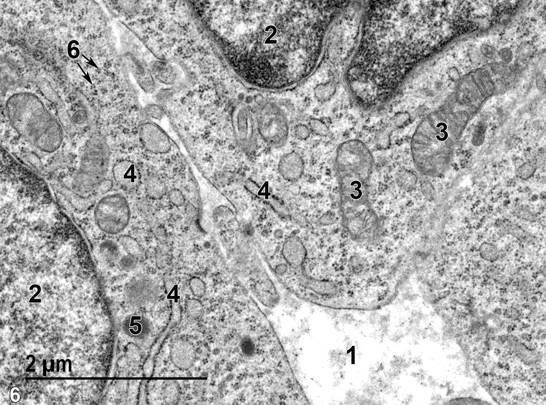
Figure 6. An even higher magnification of follicular cells with interstitial liquor folliculi (1), nuclei (2) with evident nuclear envelopes, mitochondria (3) with a rough endoplasmic reticulum with ribosomes (4) on the membrane surfaces that is both tubular and vesicular in profile, a lipid droplet (5), and clusters of free ribosomes in the cytoplasm (6, arrows). 23000x.
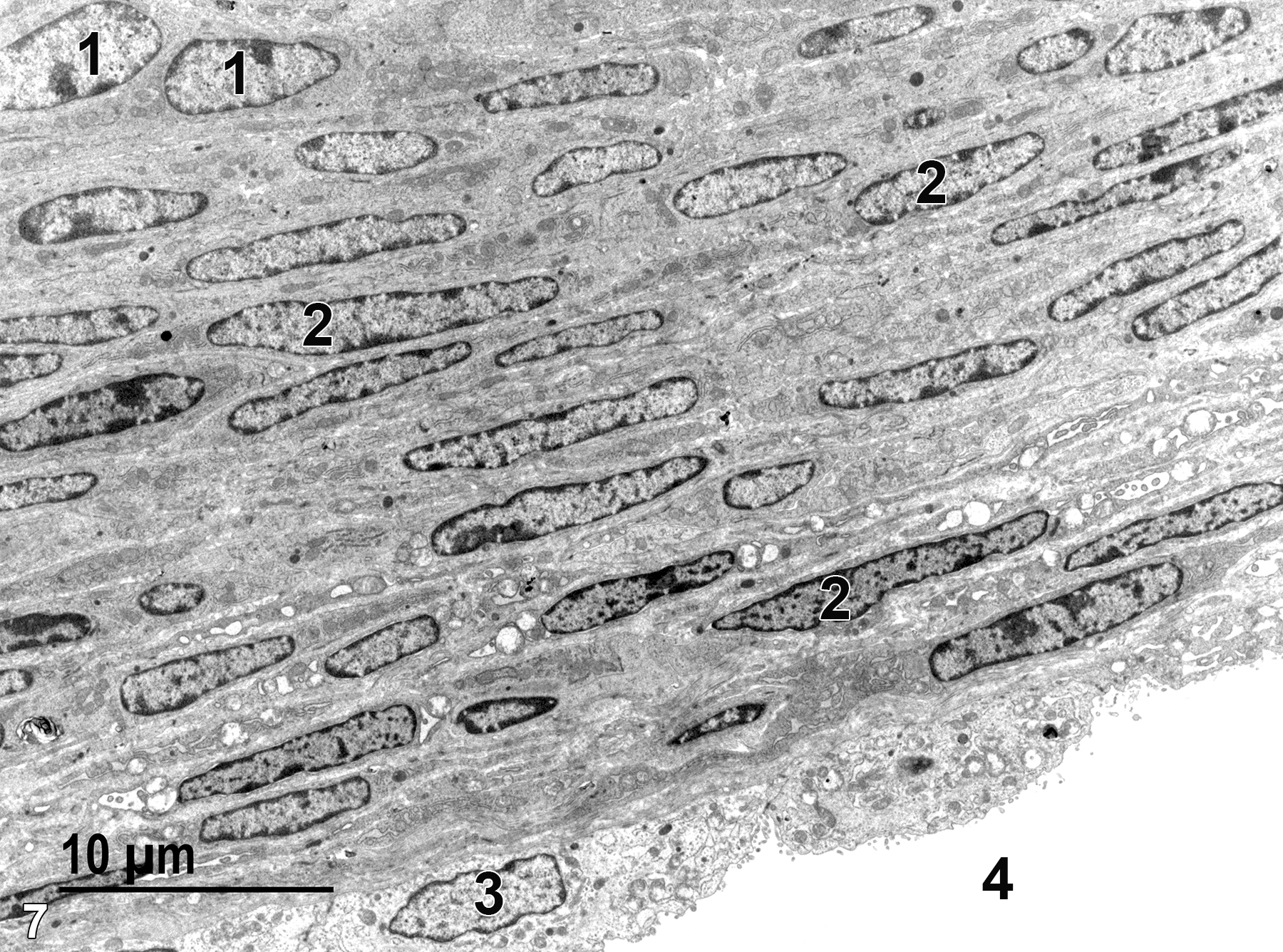
Figure 7. A low magnification view of the edge of the theca interna underlain by the theca externa and a layer of endothelial cells of a vessel. Both the theca interna and theca externa consist of squamous cells and collagen bundles. Theca interna cells (1) are thicker and have somewhat rounded nuclei. The more flattened cells (2) of the theca externa have more elongated and flattened nuclei. The thin basal lamina of the theca externa lying between the most basal cells and the endothelial cells (3) lining a blood vessel (4) is not evident in this micrograph. 2900x.
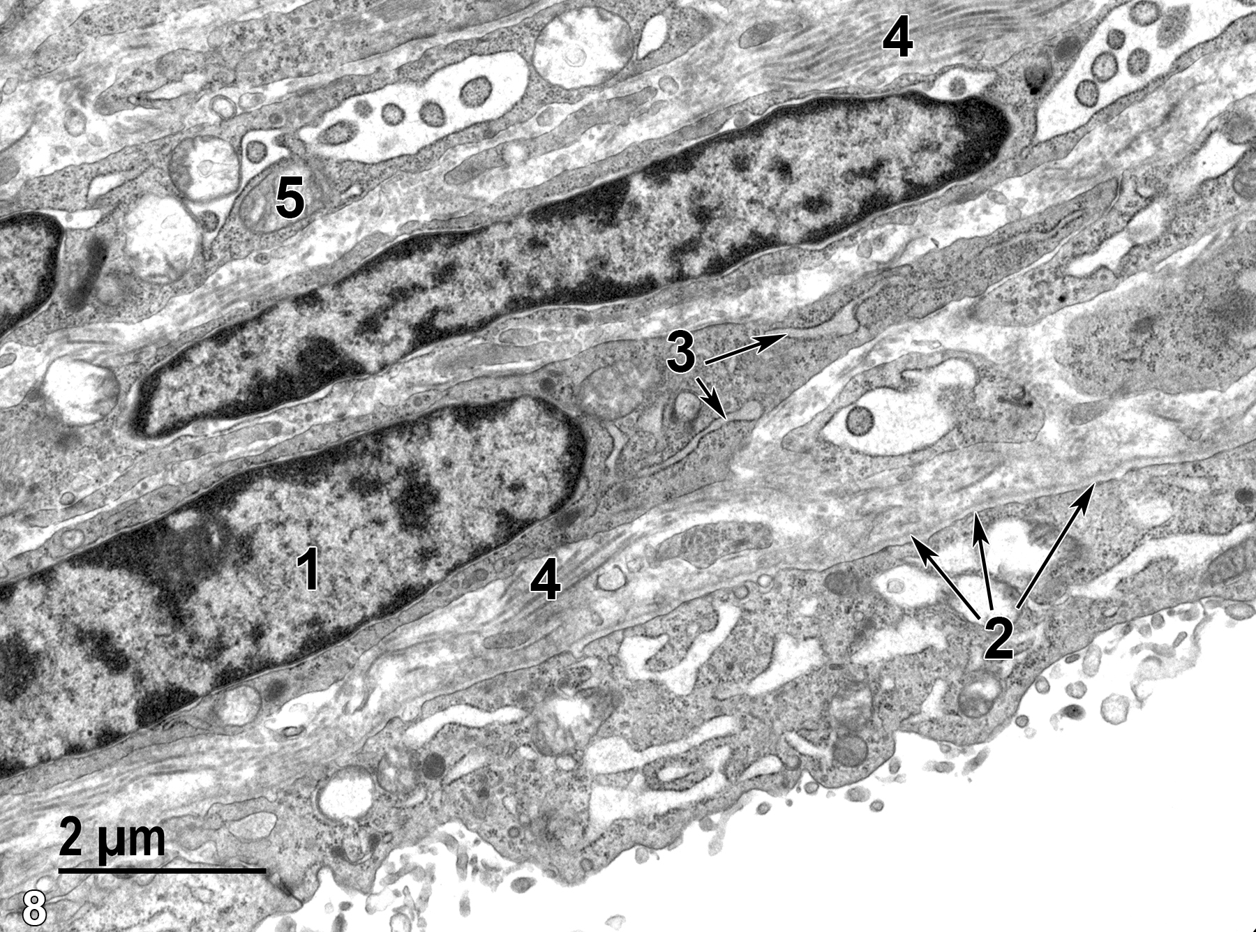
Figure 8. An enlarged view of part of the theca externa layer of Figure 7. The elongated nuclei (1) of the squamous cells have marginated electron-dense heterochromatin. The cells also contain prominent rough endoplasmic reticulum (3, arrows). The thin basal lamina (2, arrows) of the follicle is obvious at this magnification. Collagen bundles (4) are visible in the intercellular spaces. A mitochondrion (5) is shown. 11000x.
| Boorman GA, Eustis SL, Elwell MR, Montgomery CA, Jr., MacKenzie WF, eds. 1990. Pathology of the Fischer Rat: Reference and Atlas. New York: Academic Press. |
| Dellmann HD, Eurell J, eds. 1998. Textbook of Veterinary Histology. 5th ed. Philadelphia: Lippincott Williams & Wilkins. |
| Rhodin JAG. 1974. Histology: A Text and Atlas. New York: Oxford University Press. |
| Vidal JD, Dixon D. 2018. Chapter 26: Ovary. In Boorman’s Pathology of the Rat (Suttie AW, ed.). 2nd ed. London: Academic Press, 523-536. |
| Weiss L, ed. 1988. Cell and Tissue Biology: A Textbook of Histology. 6th ed. Baltimore: Urban & Schwarzenberg. |
All Images


