

Endocrine System
Adrenal Gland
Narrative
The adrenal gland has a capsule made up of fibroblasts, smooth muscle cells, and collagen. Beneath the capsule is an outer cortex that produces steroid hormones that control electrolyte balance (mineralocorticoids), protein and carbohydrate metabolism (glucocorticoids), and sex hormones (Weiss 1988; Suttie and Sutcliffe 2018). The cortex is further subdivided into three zones: a relatively thin outer zona glomerulosa; a thicker intermediate zona fasciculata; and an innermost zona reticularis abutting the medullary layer. The innermost layer, the medulla, secretes catecholamines that regulate smooth muscle function and heart rate.
As described by Rhodin (1974), the zona glomerulosa consists of epithelial cells containing 0.5 micrometer wide lipid droplets, relatively few mitochondria with tubular cristae, scarce rough endoplasmic reticulum, and a limited number of vesicular profiles that make up the smooth endoplasmic reticulum, along with a small component of Golgi vesicles.
The zona fasciculata epithelial cells are larger than those found in the zona glomerulosa and are filled with lipid droplets of variable size. Mitochondria are numerous, spherical, and larger than those found in the zona glomerulosa. Smooth endoplasmic reticulum is vesicular or formed into branching tubules, and rough endoplasmic reticulum is not present (Rhodin 1974). Glycogen and free ribosomes are abundant. Small lysosomes are present.
The zona reticularis cells contain limited numbers of large lipid droplets, large mitochondria with irregular outlines, many lysosomes, and abundant vesicular smooth endoplasmic reticulum.
The medulla has two types of epithelial cells containing secretory granules— epinephrine cells and norepinephrine cells.
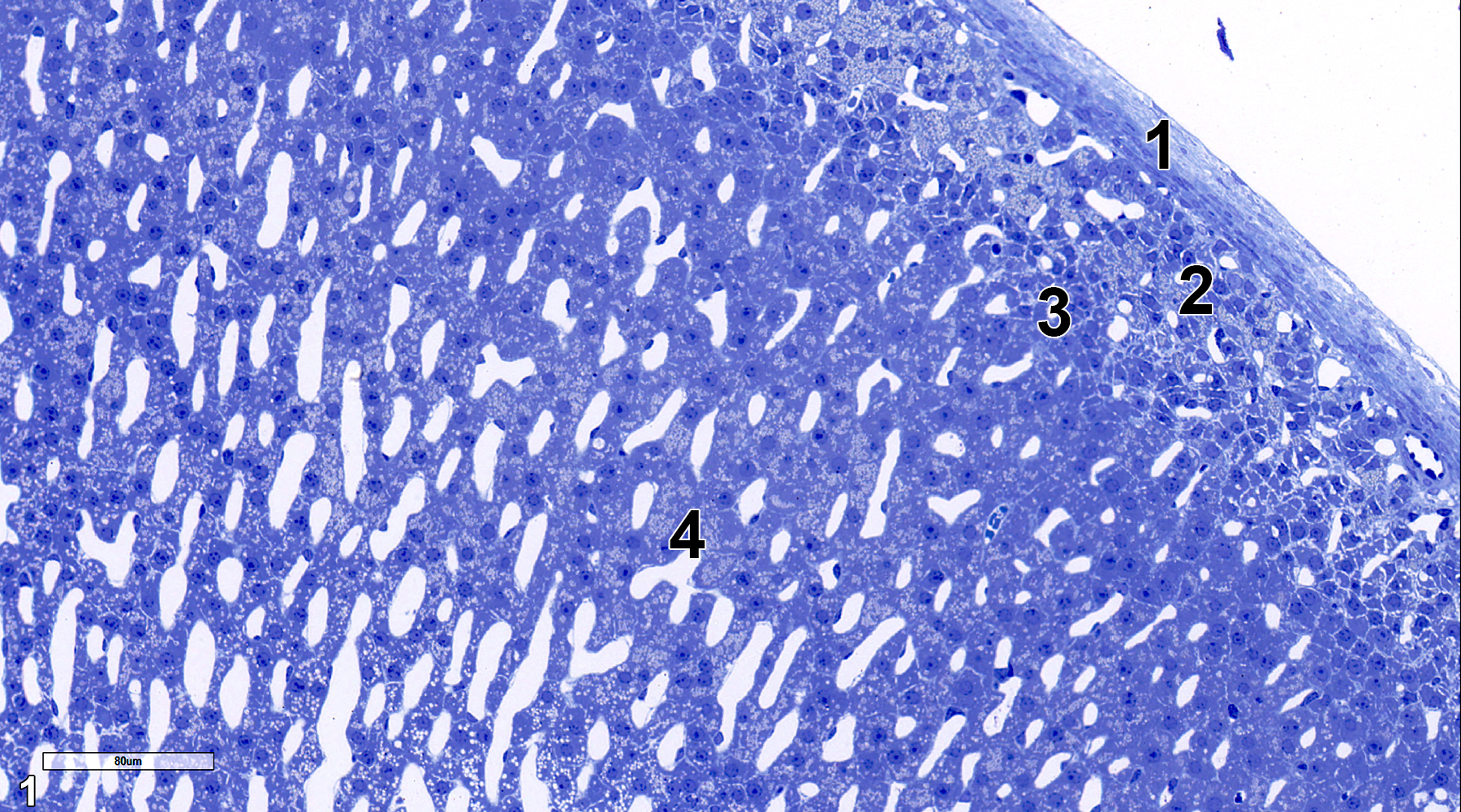
Figure 1. A 0.5 micrometer thick section stained with toluidine blue O, showing the adrenal capsule (1), zona glomerulosa (2), zona fasciculata (3), and the zona reticularis (4). 25x.
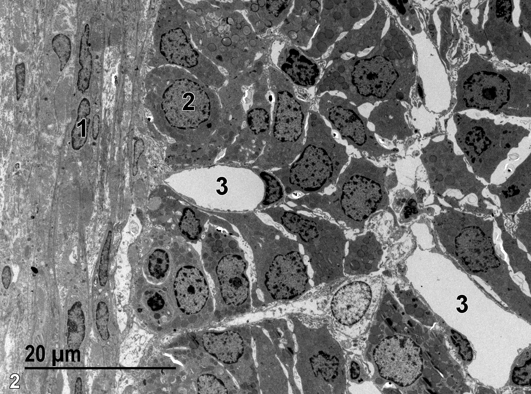
Figure 2. A nucleus of a capsular smooth muscle cell (1) and a nucleus of a zona glomerulosa epithelial cell (2), as well as several capillaries (3). 1900x.
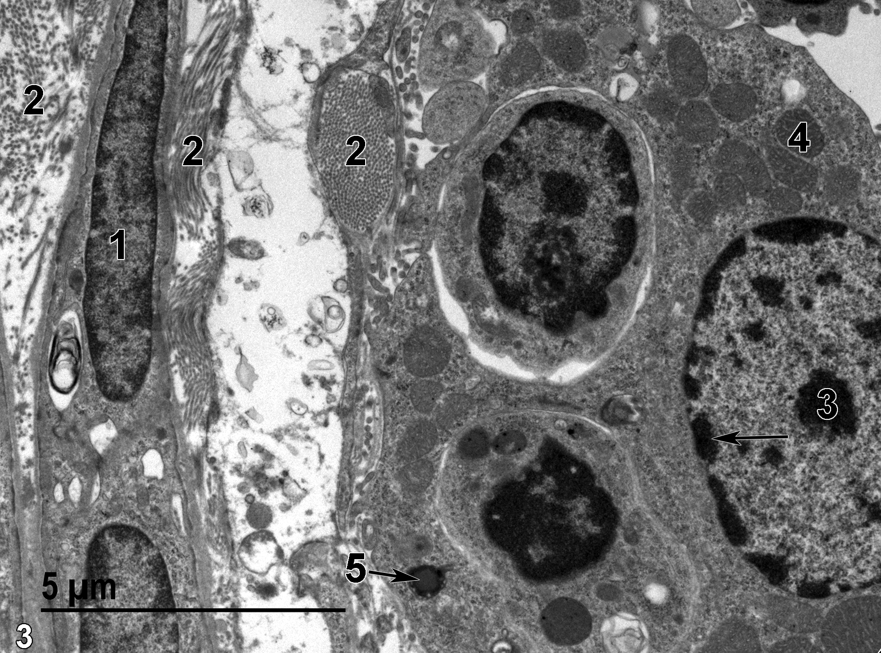
Figure 3. A higher magnification view of the region shown in Figure 2. The smooth muscle cell within the capsular layer (1) is associated with clusters of collagen (2). The nucleus of an epithelial cell has a single nucleolus (3) and marginated heterochromatin (arrow). The cytoplasm has a number of mitochondria (4). A relatively rare lipid droplet (5, arrow) is shown. 9300x.
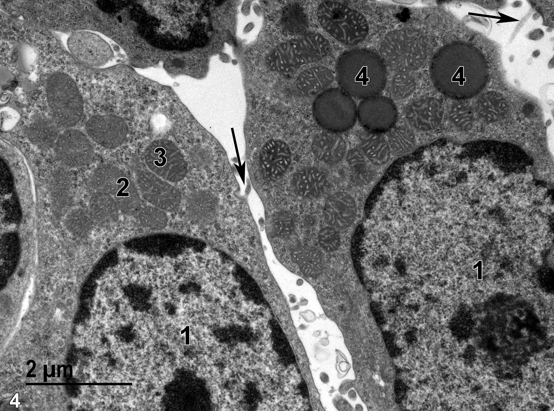
Figure 4. A still higher magnification view of zona glomerulosa epithelial cells showing nuclei (1) with prominent nucleoli and marginated heterochromatin, mitochondria with vesicular cristae (2) when cut in cross section, and tubular cristae (3) when cut in a more longitudinal section. Lipid droplets (4) are relatively electron dense. The surface of the epithelial cells have microvilli (arrows) protruding into the intercellular spaces. Clusters of dark particles are found throughout the cytoplasm, which are particulate glycogen and free ribosomes. 13000x.
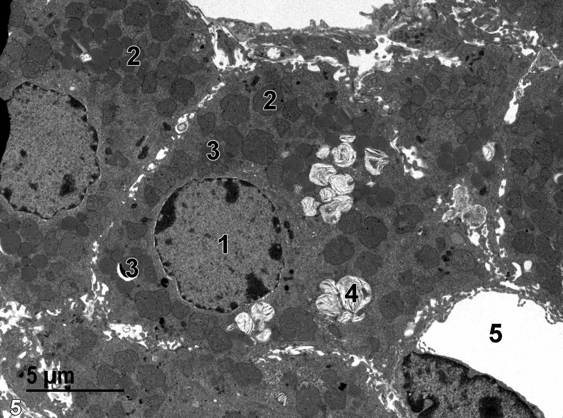
Figure 5. Cells from the zona fasciculata. The epithelial cells have microvilli at the surface, single nuclei (1) with marginated heterochromatin, numerous mitochondria (2), lipid droplets with mostly homogeneous content (3), and lipid droplets that contain large number of membranous profiles (4). A capillary (5) with a prominent endothelial nucleus is shown. 4800x.
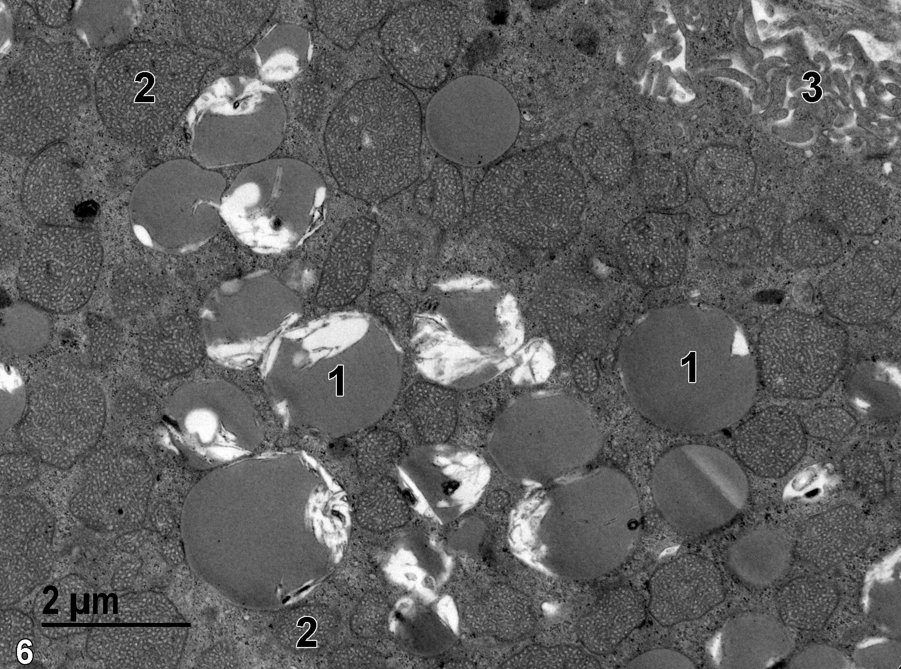
Figure 6. A higher magnification view of the content of a zona fasciculata cell. The cell is densely packed with mitochondria (2) and lipid droplets (1), some of which have homogeneous electron-dense contents, and others with variable amounts of membranous profiles. The microvilli (3) at the surface of the cell are numerous in this view. 11000x.
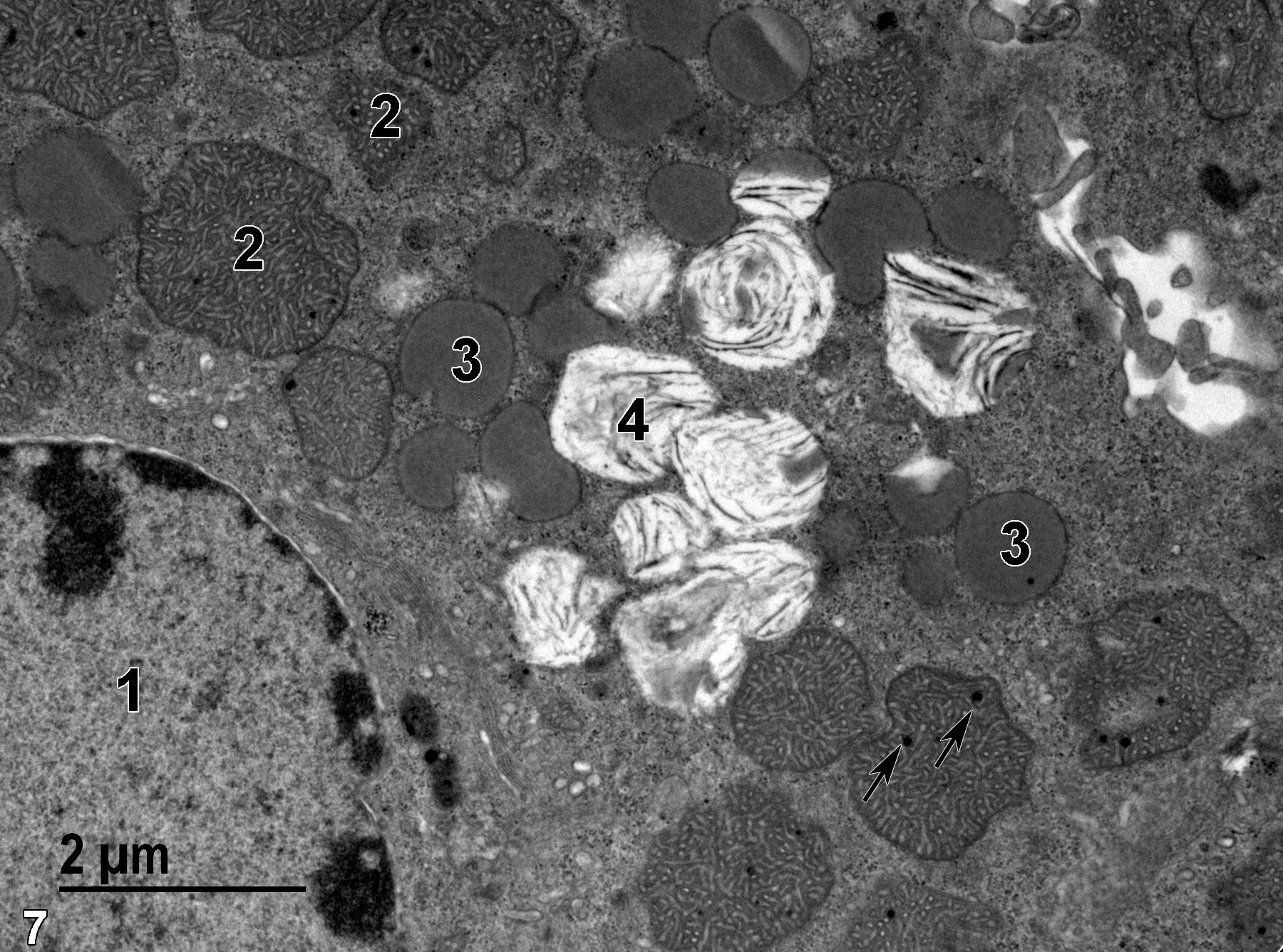
Figure 7. Another zona fasciculata epithelial cell with a nucleus (1) with marginated heterochromatin, numerous mitochondria (2), some of which contain electron-dense granular deposits of calcium (arrows), and the two types of lipid droplet presentation (3 and 4) described in Figure 6. 13000x.
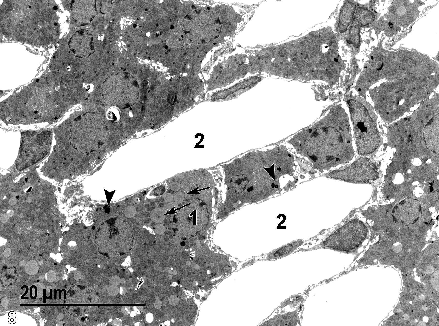
Figure 8. A low magnification view of the zona reticularis, showing a nucleus (1) with marginated heterochromatin, large lipid droplets (arrows), numerous capillaries (2), and prominent electron-dense lysosomes (arrowheads). 1900x.

Figure 9. A binucleate (1) epithelial cell of the zona reticularis, lipid droplets, some containing electron-dense inclusions (2, arrows), many irregular and electron-dense lysosomes (3, arrows), and abundant smooth endoplasmic reticulum, some quite visible at this magnification as vesicular profiles located near the irregular and electron-dense lysosomes (3). Mitochondria (4) are quite numerous. Note the microvillous surface of the epithelial cells. 4800x.
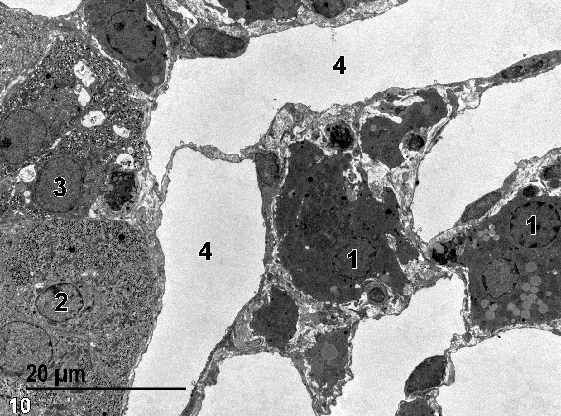
Figure 10. Two zona reticularis cells (1) are shown to the right, as well as an epinephrine medullary cell (2) and a norepinephrine medullary cell (3). The zona reticularis cells are surrounded by numerous capillaries (4). 1900x.
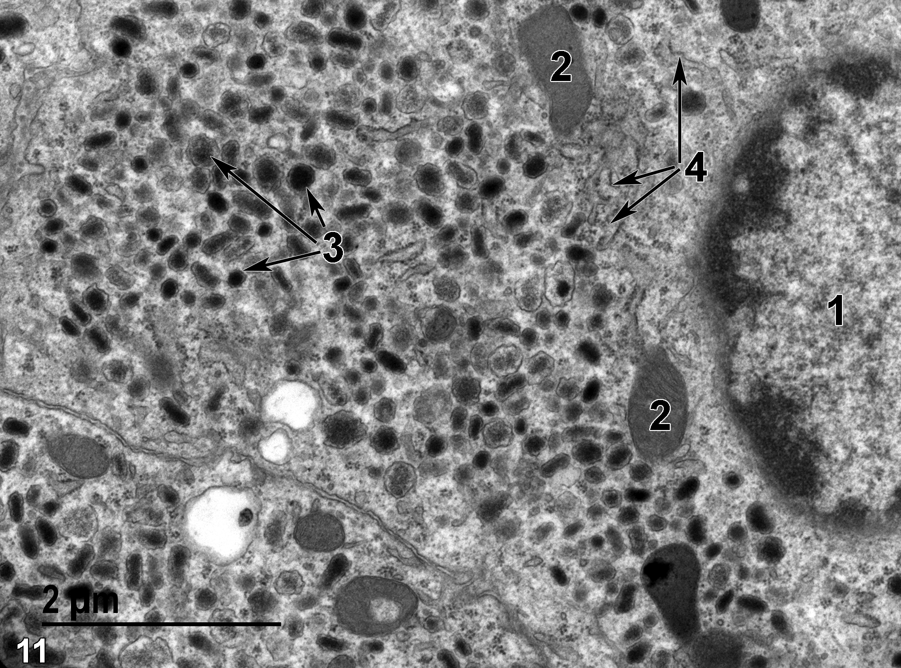
Figure 11. A high magnification view of an epinephrine cell of the medulla with a single nucleus (1), some mitochondria (2), a large number of variably sized secretory granules with variable electron-dense contents (3, arrows), and some profiles of rough endoplasmic reticulum (4, arrows). Aggregates of particulate glycogen and free ribosomes are scattered throughout the cytoplasm. 18500x.
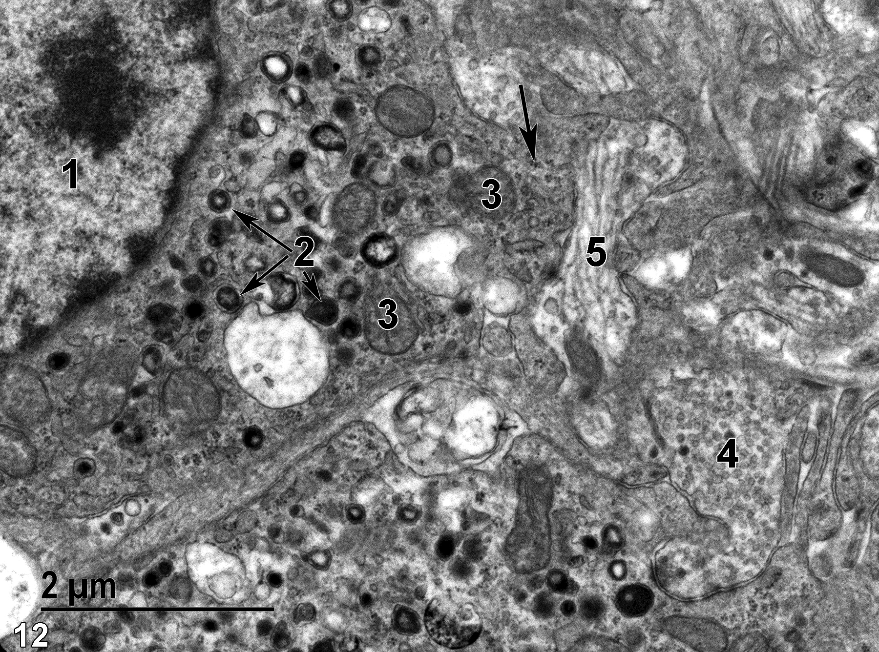
Figure 12. Two norepinephrine cells of the medullary layer. One contains a nucleus (1) with a prominent nucleolus and marginated heterochromatin, as well as a variable population of secretory granules, many with electron-lucent central cores (2, arrows). Mitochondria (3) have elongated tubular cristae. An area of neural cell processes is to the right of the upper norepinephrine cell. Some contain synaptic vesicles (4), whereas others are filled with microtubules and a few vesicles (5). Particulate glycogen clusters are found throughout the cytoplasm (arrow). 18500x.
| Rhodin JAG. 1974. Histology: A Text and Atlas. New York: Oxford University Press. |
| Suttie AW, Sutcliffe C. 2018. Chapter 32: Adrenal gland. In Boorman’s Pathology of the Rat (Suttie AW, ed.). 2nd ed. London: Academic Press, 649−667. |
| Weiss L, ed. 1988. Cell and Tissue Biology: A Textbook of Histology. 6th ed. Baltimore: Urban & Schwarzenberg. |
All Images


