

Hepatobiliary System
Liver
Narrative
The liver produces circulating proteins (lipoproteins, albumins, prothrombin, and fibronectin), secretes bile, stores metabolic reserves (glycogen and lipid), detoxifies drugs and toxins, excretes waste products, and metabolizes various materials. The rat does not have a gallbladder.
The liver lobes are surrounded by a capsule, and the parenchyma of the liver is organized into approximately hexagonal lobules. The lobules consist of sheets of hepatocytes separated by sinusoids that carry blood to the hepatocytes. The lobule contains a central vein and portal triads at the periphery of the lobule that consist of hepatic arteries, bile ducts, and portal veins. The space of Disse is located between the villous surface of hepatocytes and the basal lamina of the sinusoidal endothelial cells. Within this space of Disse are Ito cells (hepatic stellate cells) filled with lipid droplets. The sinusoids contain various blood cells (leukocytes and erythrocytes), as well as Kupffer cells (sinusoidal macrophages). Small bile canaliculi containing microvilli are located between hepatocytes.
Hepatocytes contain one to several round nuclei with prominent nucleoli and marginated heterochromatin, numerous mitochondria, peri-mitochondrial rough endoplasmic reticulum, other stacks of rough endoplasmic reticulum, lysosomes, peroxisomes, Golgi vesicles, and smooth endoplasmic reticulum. A well-fed rat hepatocyte may contain large lakes of glycogen (mostly composed of clusters [rosettes] of alpha particles, which are called beta particles) and a number of lipid droplets of varying size.
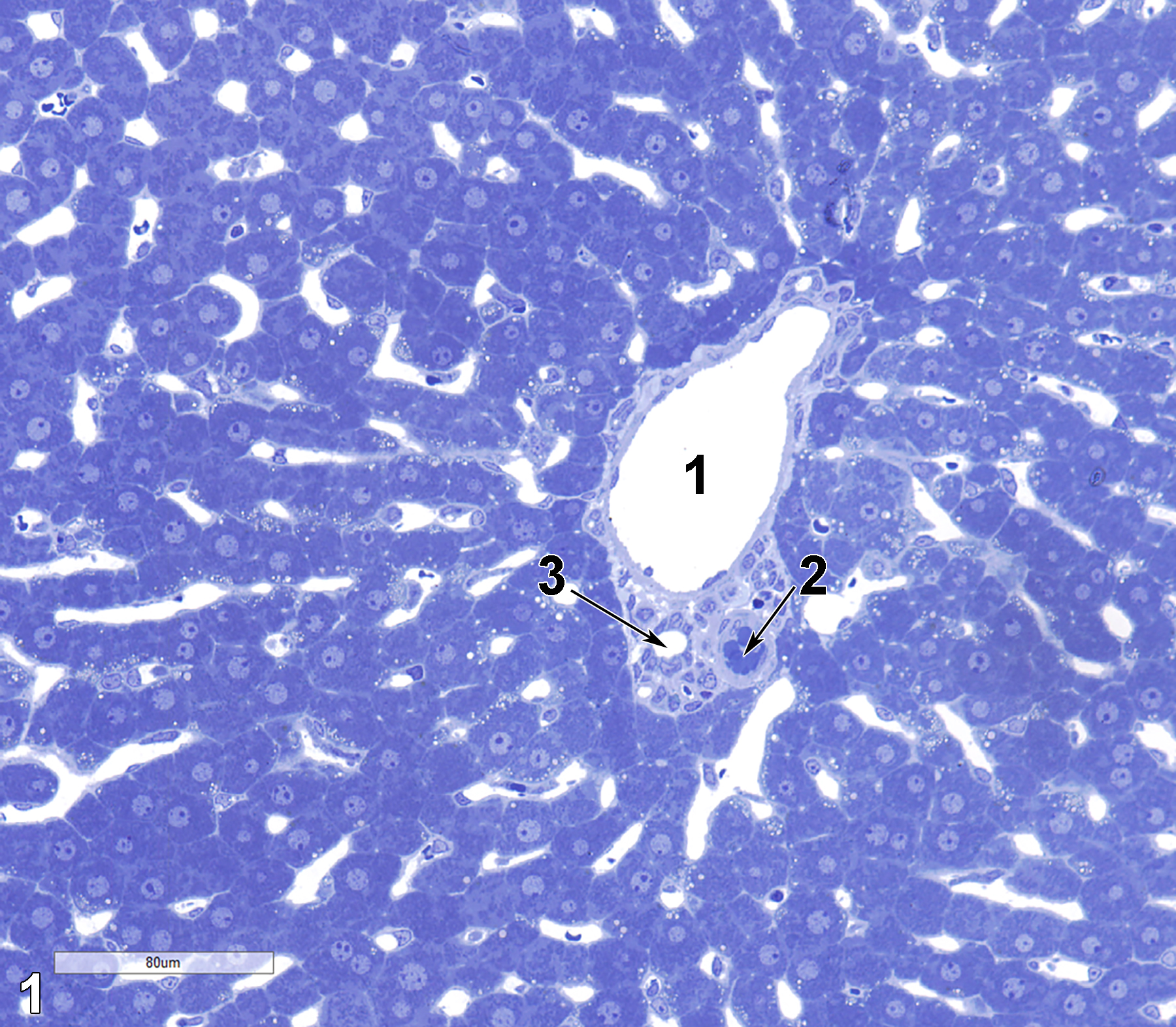
Figure 1. A toluidine blue O-stained semithin section (0.5 micrometer thick) of a portal triad, showing the central vein (1), portal artery (2, arrow), and bile duct (3, arrow). 25x.
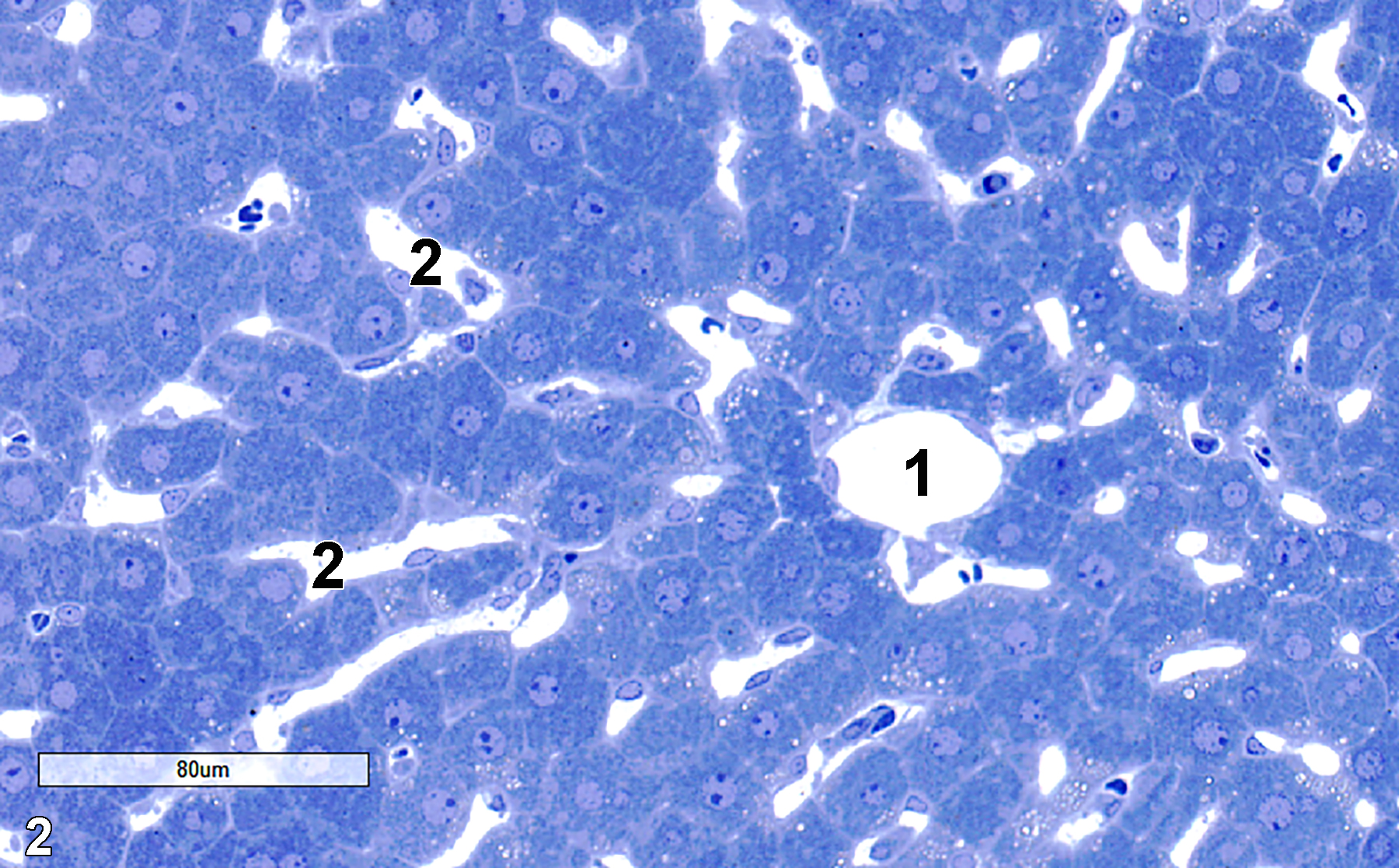
Figure 2. A toluidine blue O-stained semithin section showing the central vein (1) of a liver lobule, with sinusoids (2) generally oriented toward the central vein. The sinusoids contain erythrocytes, leukocytes, Kupffer cells, and have Ito cells just outside the sinusoids in the space of Disse. 25x.
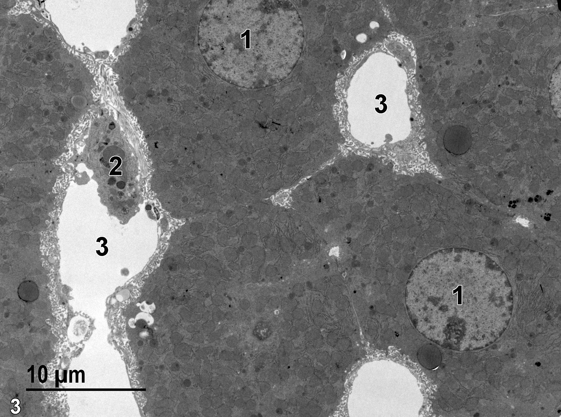
Figure 3. A low magnification image showing two typical round hepatocyte nuclei (1), one of which has a prominent eccentric nucleolus. Three sinusoids (3) are visible, one of which contains a Kupffer cell (2). 2900x.
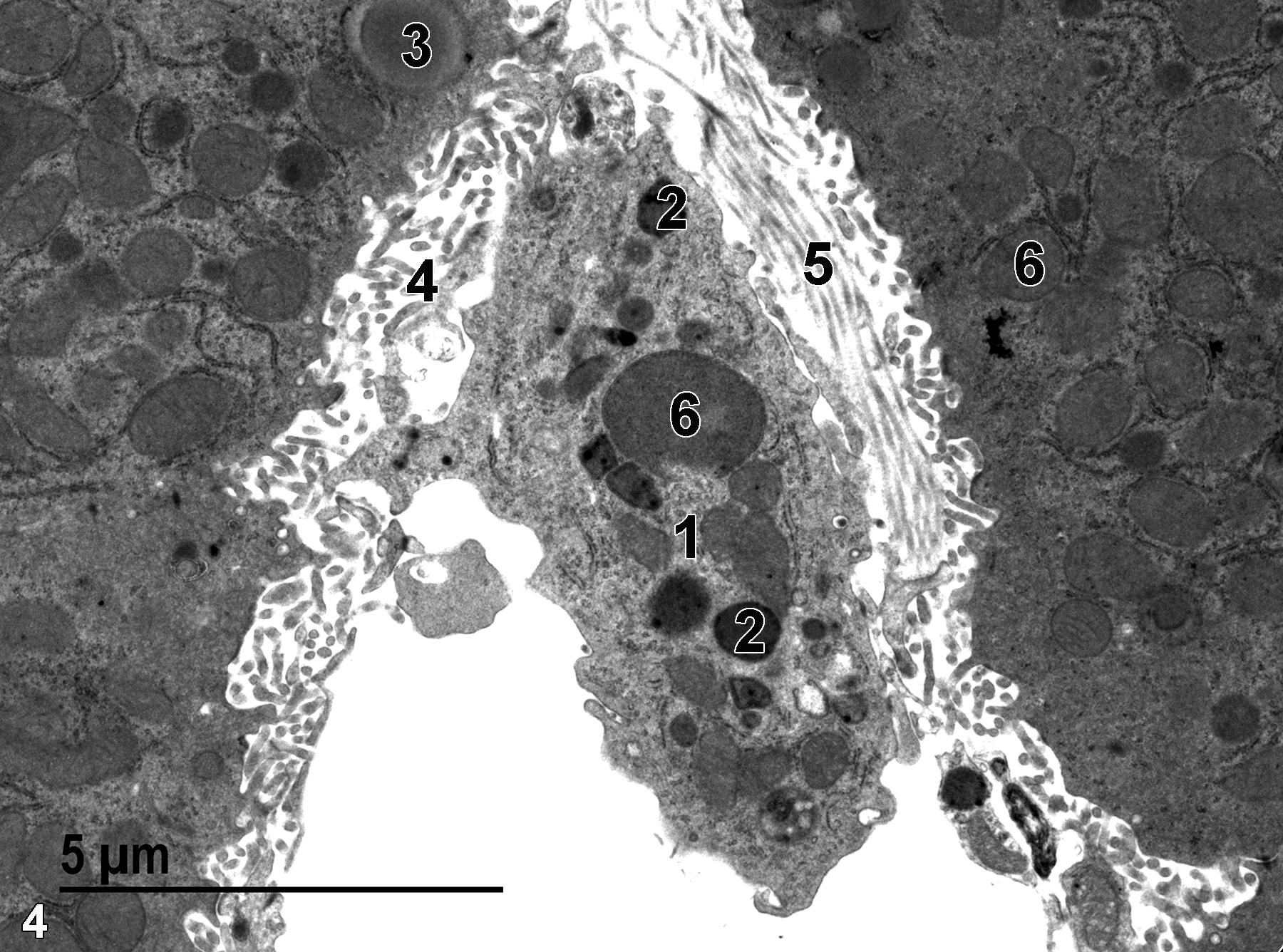
Figure 4. A higher magnification view of the Kupffer cell (1) seen in Figure 3 shows mitochondria (6) in the Kupffer cell and an adjacent hepatocyte. Lysosomes (2) in the Kupffer cell are round and more electron dense than the mitochondria. A lipid droplet (3) is located in one of the hepatocytes. The microvillous surfaces (4) of hepatocytes adjacent to the space of Disse are evident. Bundles of collagenous fibrils (5) that show diagnostic banding patterns in some areas are present. 9300x.
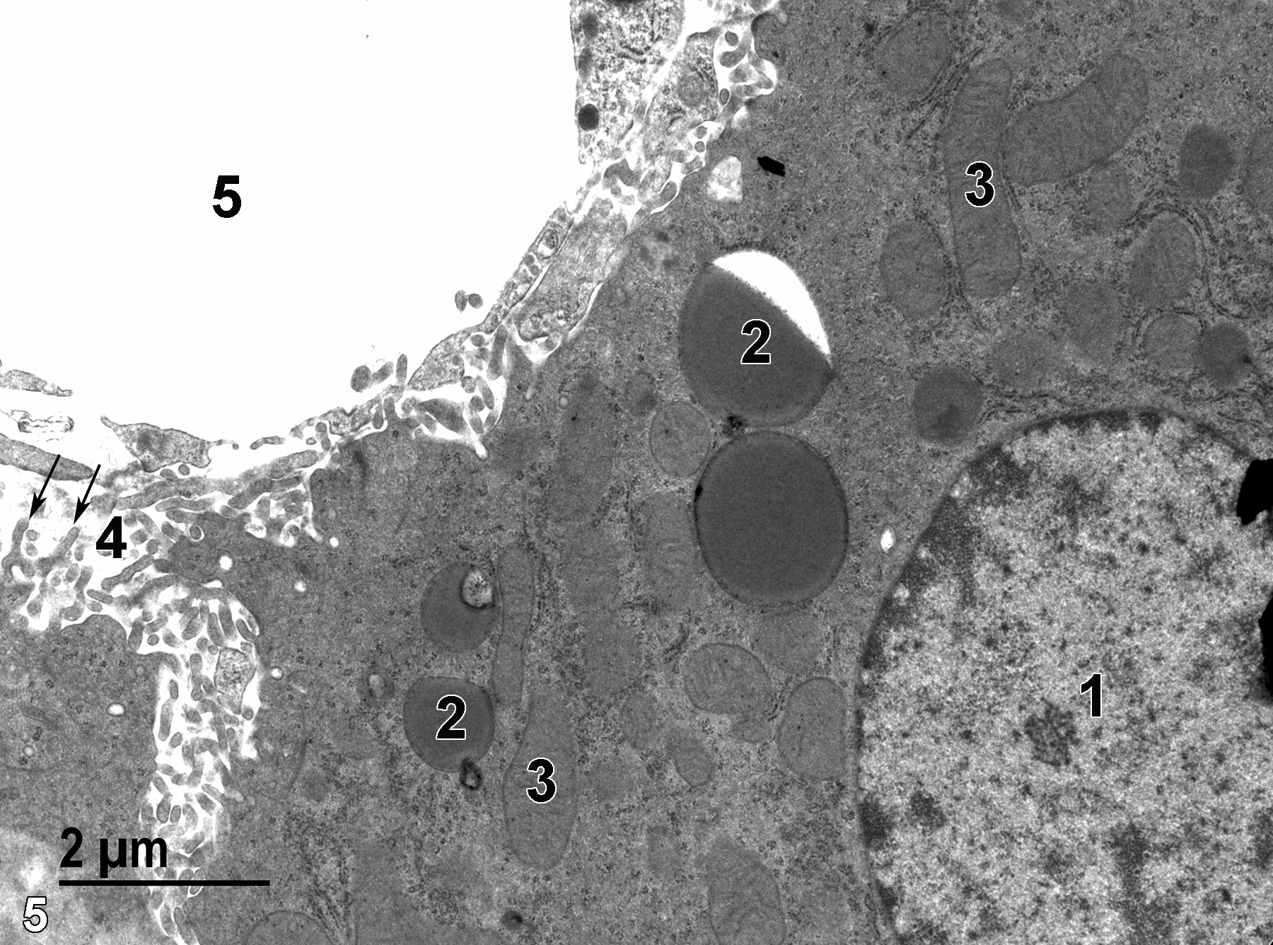
Figure 5. A sinusoidal lumen (5) with the adjacent space of Disse occupied by hepatocyte microvilli (4, arrows) shows how some of the microvilli are branched. The hepatocyte contains prominent lipid droplets (2) and mitochondria (3), in addition to the round nucleus (1) with electron-dense marginated heterochromatin. 11000x.
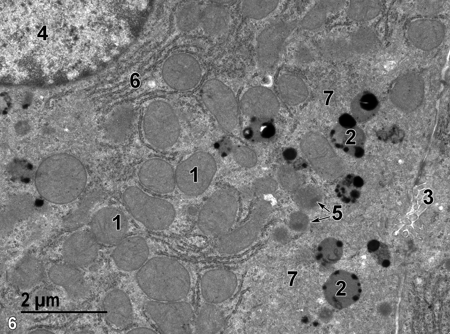
Figure 6. Some hepatocytes containing prominent lysosomes (2) with extremely electron-dense contents, along with mitochondria (1) that are often associated with peri-mitochondrial rough endoplasmic reticulum elements. A bile canaliculus (3) with microvilli projecting from adjacent hepatocytes is shown on the far right in the image. The round nucleus (4) with marginated heterochromatin has an associated stack of rough endoplasmic reticulum (6). Two peroxisomes with electron-dense paracrystalline inclusions (5, arrows) are shown. Areas with vesicular profiles (7) are consistent with smooth endoplasmic reticulum. 13000x.
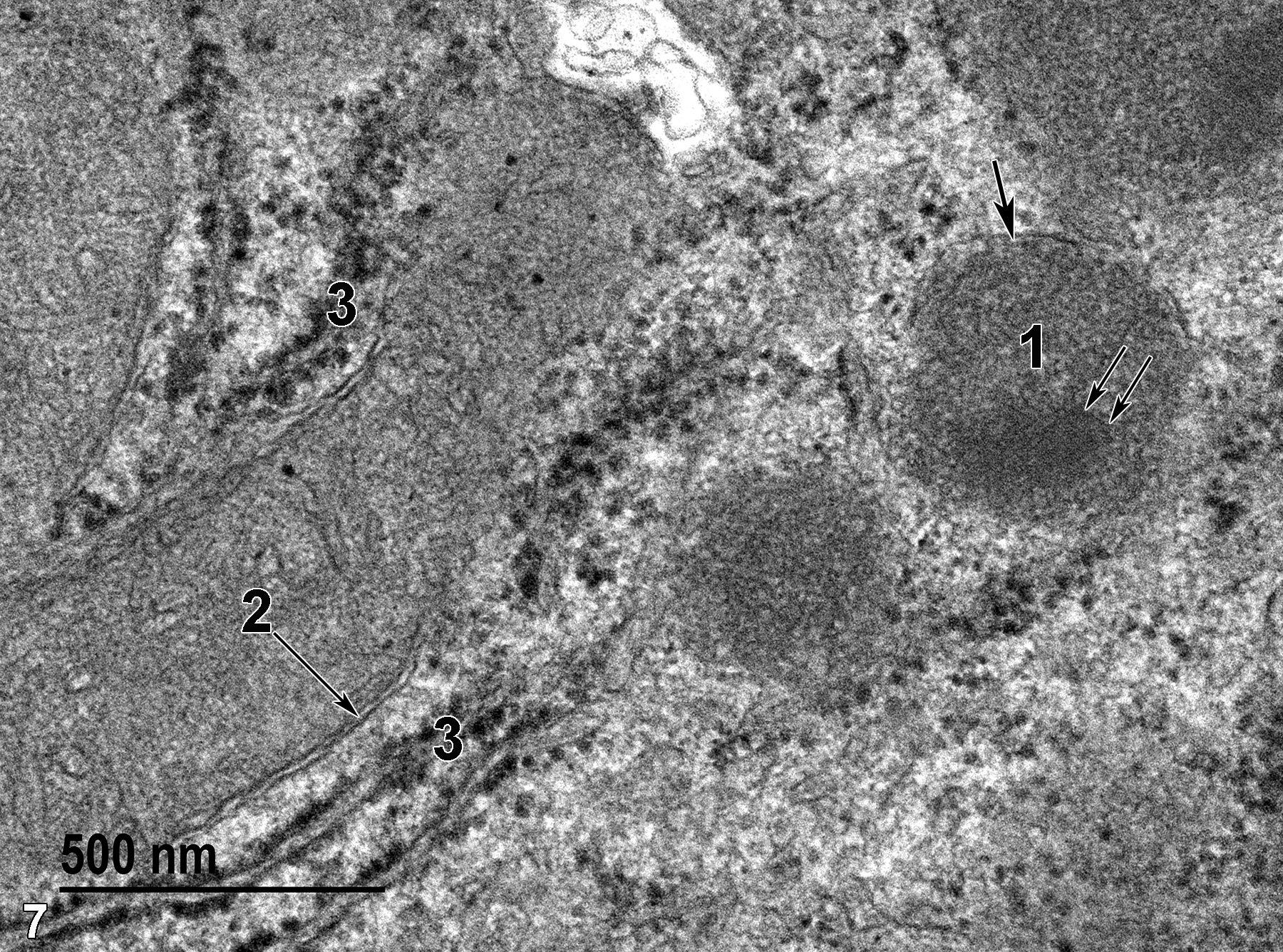
Figure 7. A high magnification view of structures within a hepatocyte. A mitochondrion with a double membrane-bound border (2, arrow) is easily distinguished from the somewhat more electron-dense peroxisome that has a single membrane-bound border (1, thick arrow) and a paracrystalline array (1, thin arrows). Rough endoplasmic reticulum elements (3) with their ribosomal covering are seen adjacent to the mitochondria. 68000x.
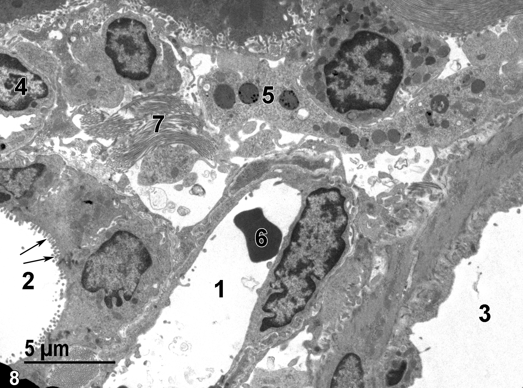
Figure 8. A low magnification image showing parts of the portal triad of a liver lobule. The portal vein (1) contains a single erythrocyte (6). The bile duct is lined with short microvilli (2, arrows). The portal artery (3) is lined with an endothelium underlain by smooth muscle cells. An endothelial cell (4) lines a capillary. A sinusoid is shown which contains a Kupffer cell (5) with lysosomes. A bundle of collagenous fibrils (7) found in the space of Disse is shown. 4800x.
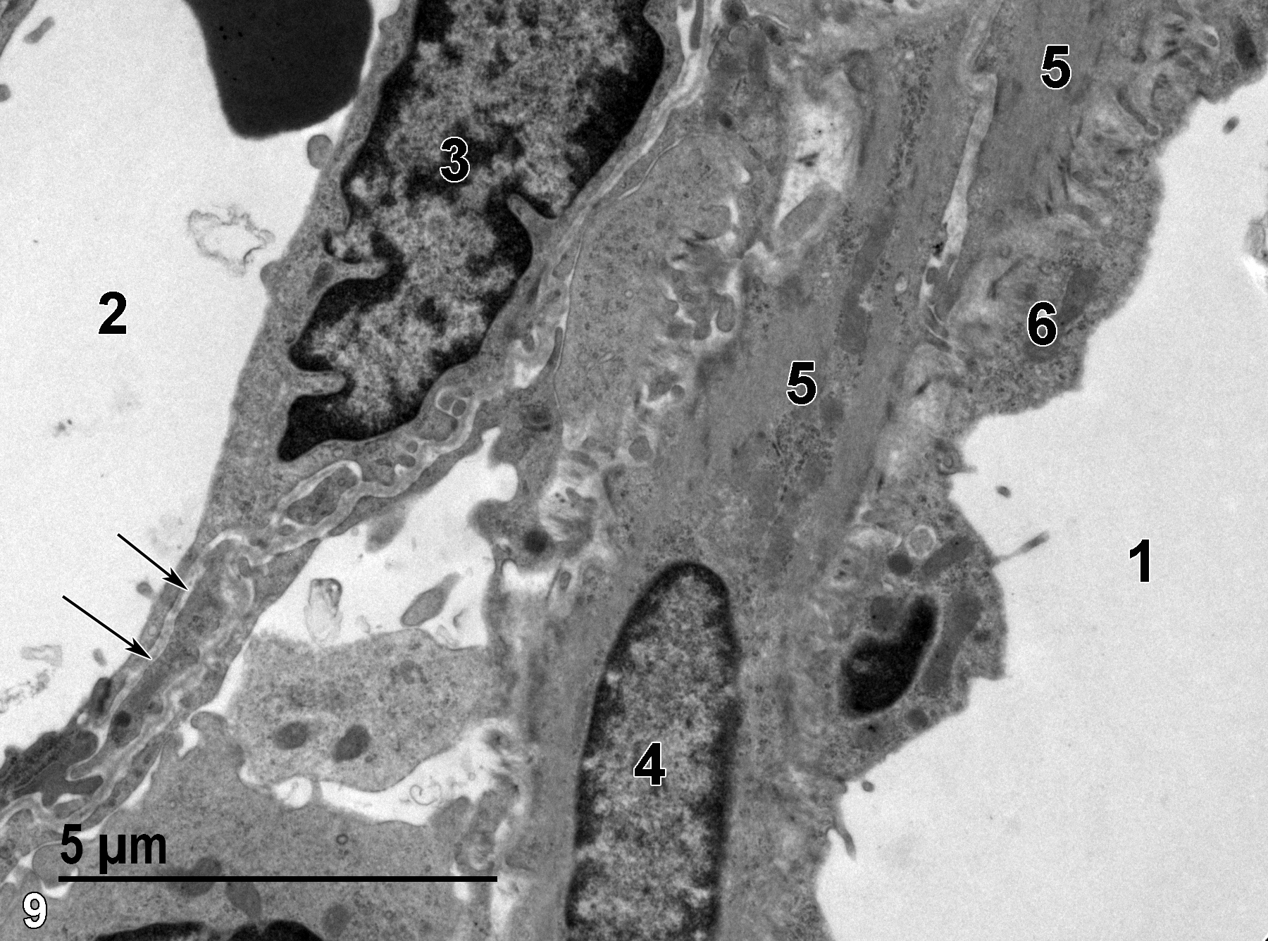
Figure 9. A higher magnification view of Figure 8 shows the portal vein lumen (2) lined by an endothelial cell (3), with an underlying basal lamina (arrows). The portal artery lumen (1) is lined with an endothelial cell layer (6) subtended by smooth muscle cells (5), one with an elongate nucleus with prominent marginated heterochromatin (4). 9300x.
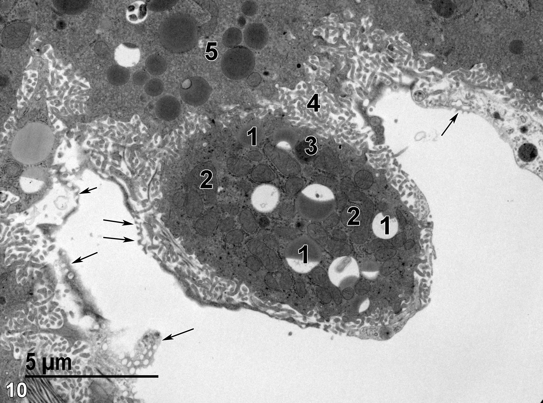
Figure 10. An Ito cell in the space of Disse with lipid droplets (1), some of which appear to be partially extracted during processing for transmission electron microscopy, as well as mitochondria (2) of various sizes and conformations. A single lysosome (3) is present. The microvillous surface (4) protruding into the space of Disse adjacent to a hepatocyte (5) is shown, along with the thin fenestrated endothelial cell lining the sinusoid (arrows). 6800x.
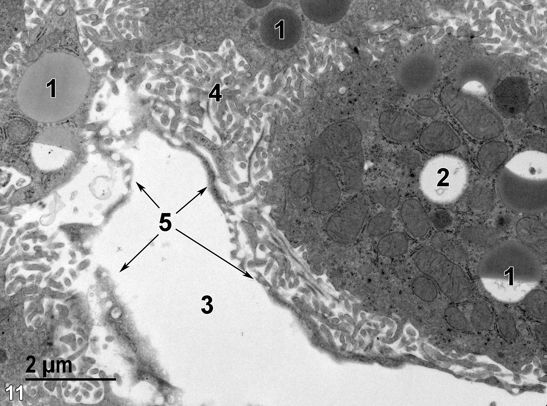
Figure 11. A higher magnification image of the Ito cell shown in Figure 10 shows the variation in lipid content and lipid staining of lipid bodies (1), including one presumed lipid body (2) that has been totally extracted. The sinusoidal lumen (3) is contained within the fenestrated endothelial cell (5, arrows). The microvilli (4) of the adjacent hepatocyte occupy the space of Disse between the hepatocyte and the Ito cell. 11000x.
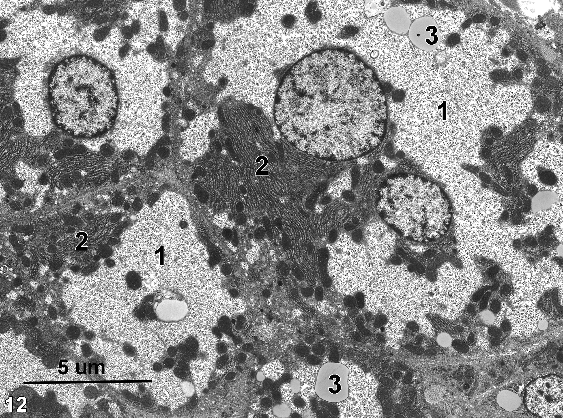
Figure 12. Hepatocytes from a well-fed rat. A large proportion of the hepatocyte cytoplasm consists of glycogen lakes (1). The granular glycogen deposits have pushed the stacks of rough endoplasmic reticulum and mitochondria (2) to the margins of the hepatocytes. Lipid bodies (3) are also present in the glycogen lakes. Magnification not known.
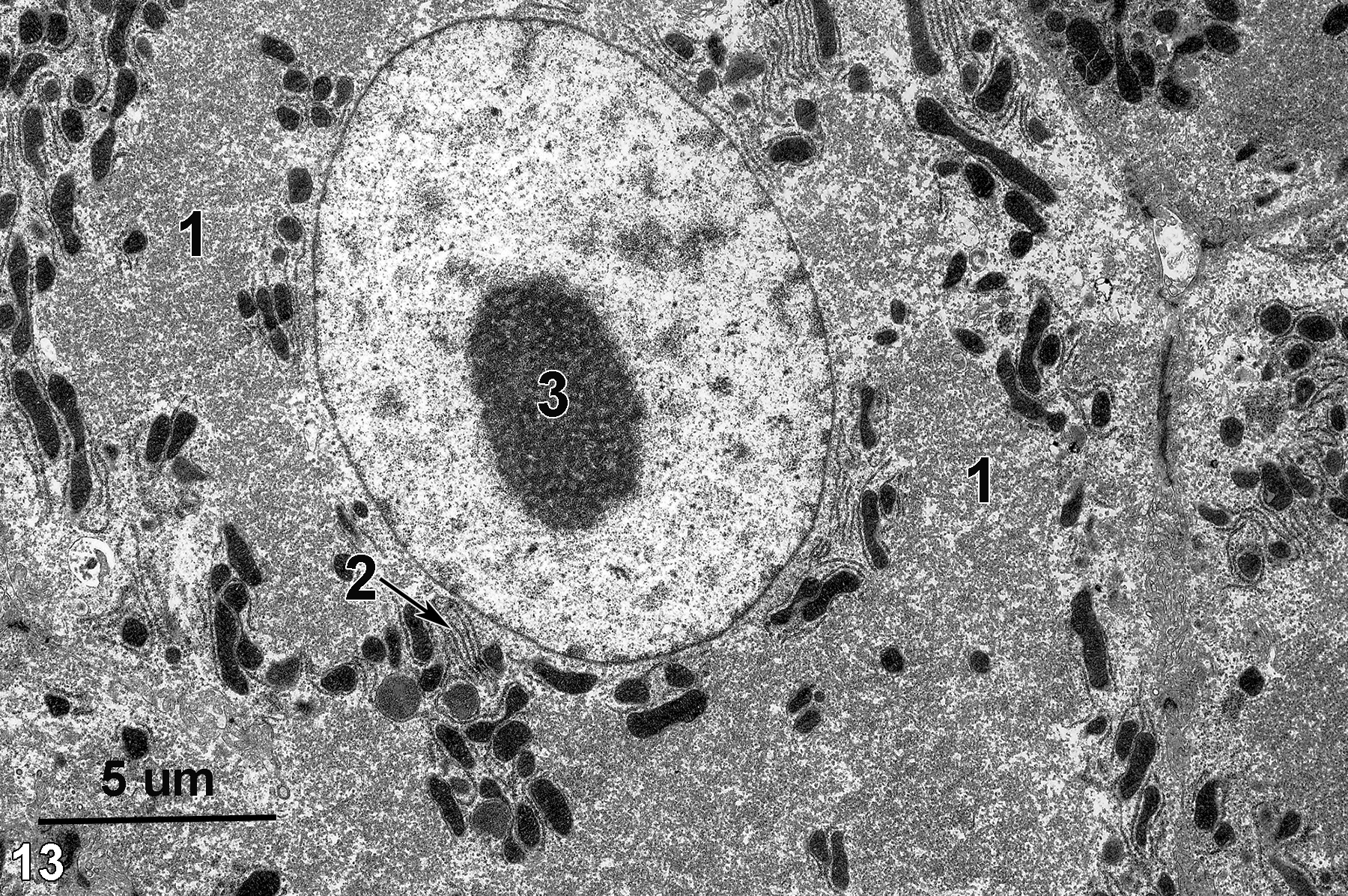
Figure 13. A hepatocyte with a typically round nucleus with a prominent nucleolus (3). A stack of rough endoplasmic reticulum (2, arrow) is seen in a perinuclear position. Mitochondria are mostly perinuclear or at the periphery of the cytoplasm due to the large amount of smooth endoplasmic reticulum (1) occupying the cytoplasm of this hypertrophied hepatocyte. Magnification not known.
| Eurell JA, Frappier BL, eds. 2006. Dellmann’s Textbook of Veterinary Histology. 6th ed. Ames, IA: Blackwell Publishing. |
| Rhodin JAG. 1974. Histology: A Text and Atlas. New York: Oxford University Press. |
| Ross MH, Kaye GI, Pawlina W. 2003. Histology: A Text and Atlas. 4th ed. Philadelphia: Lippincott Williams & Wilkins. |
| Weiss L, ed. 1988. Cell and Tissue Biology: A Textbook of Histology. 6th ed. Baltimore: Urban & Schwarzenberg. |
All Images


