

Urinary System
Kidney
Narrative
Kidneys are responsible for the ultrafiltration of blood plasma, followed by the reabsorption of most of the ultrafiltrate. This process is mediated by nephrons, the functional units of the kidney as shown in Figure 1. The ultrafiltrate is formed in the renal corpuscle (glomerulus), and then materials are added and subtracted from the urine as it passes along the tubule system that is composed of the nephron.
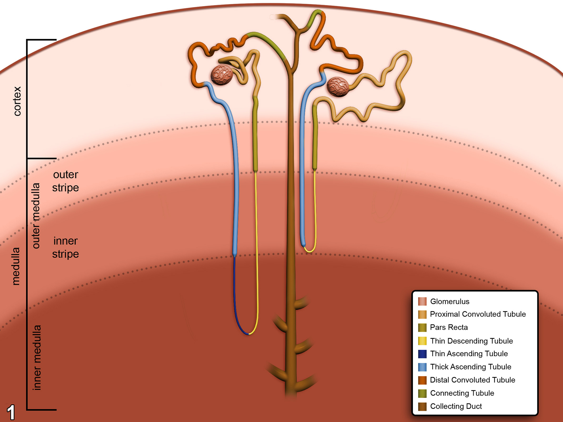
Figure 1. A diagram of the nephron system of the kidney, with the four zones within the kidney that the nephron traverses identified.
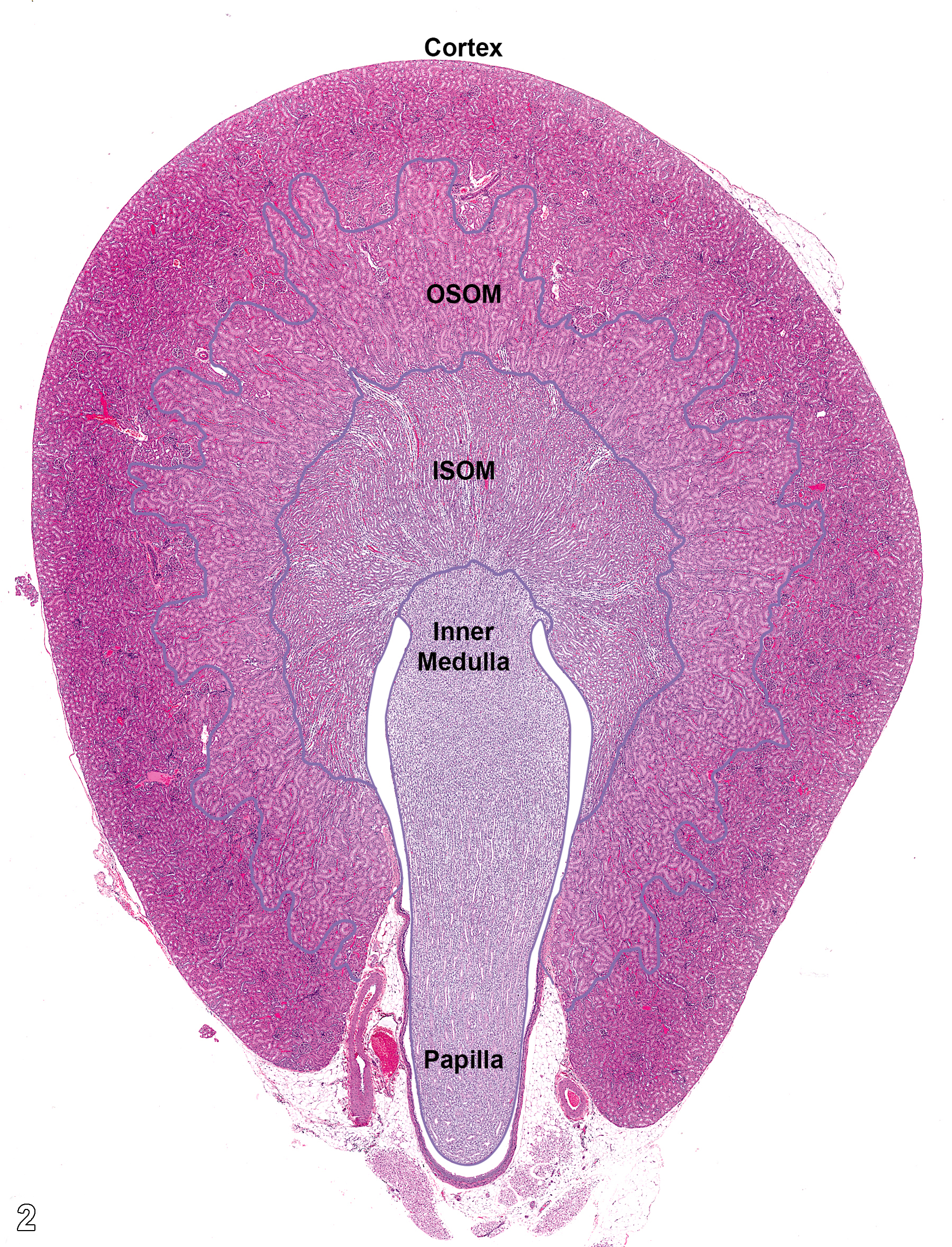
Figure 2. A histological transverse section of the rodent kidney, with the various zones that contain elements of the nephron or collecting ducts. ISOM = inner stripe of the outer medulla; OSOM = outer stripe of the outer medulla.
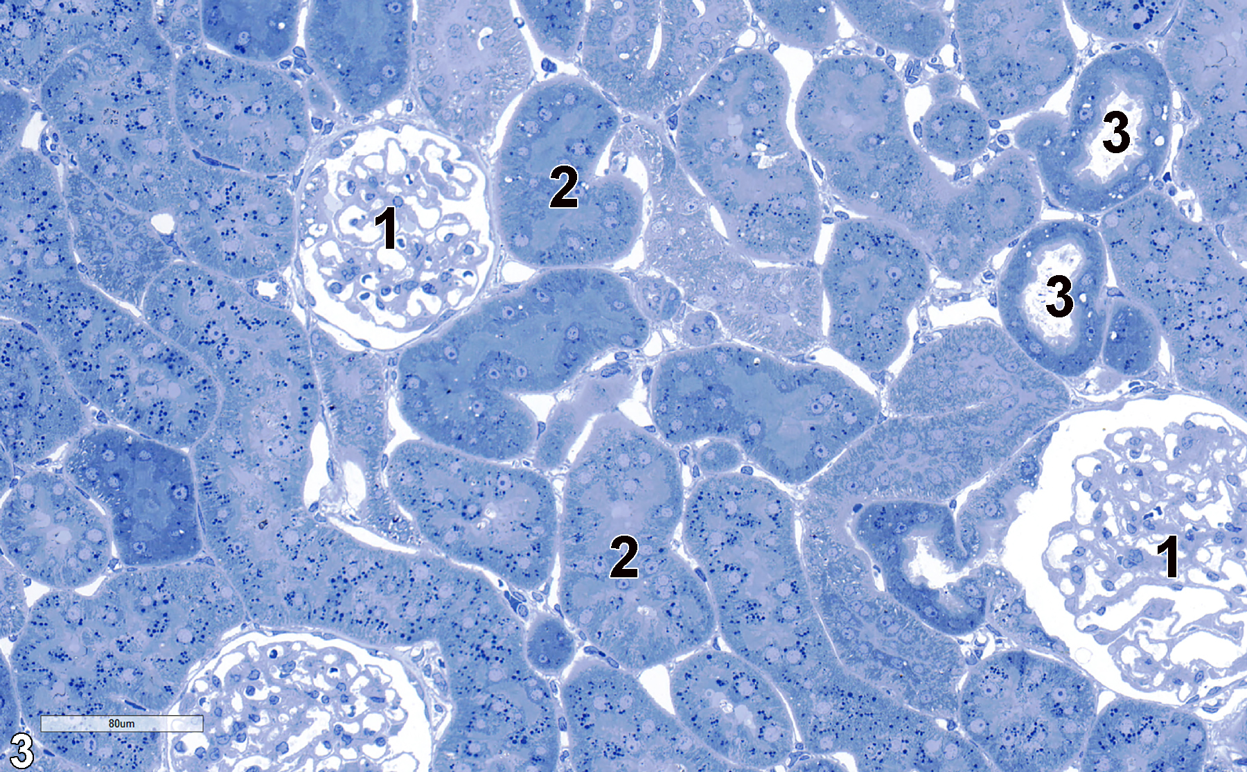
Figure 3. A toluidine blue O-stained semithin section (0.5 micrometer thick) of the cortical region of the kidney. Glomeruli (1), proximal convoluted tubules (2), and distal convoluted tubules (3) are shown. 25x.
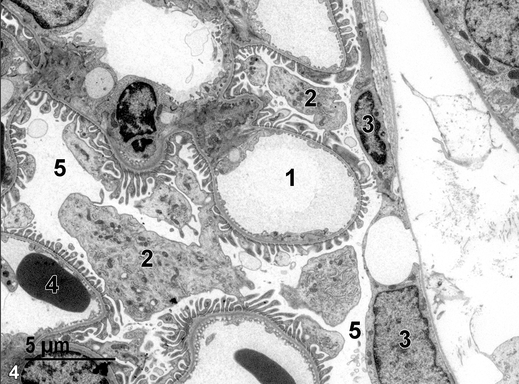
Figure 4. A low magnification view of a portion of a glomerulus, showing a capillary (1), podocytes (2) , nuclei of the parietal layer of Bowman’s capsule (3), an erythrocyte in a capillary (4), and the urinary space (5). Magnification not provided.
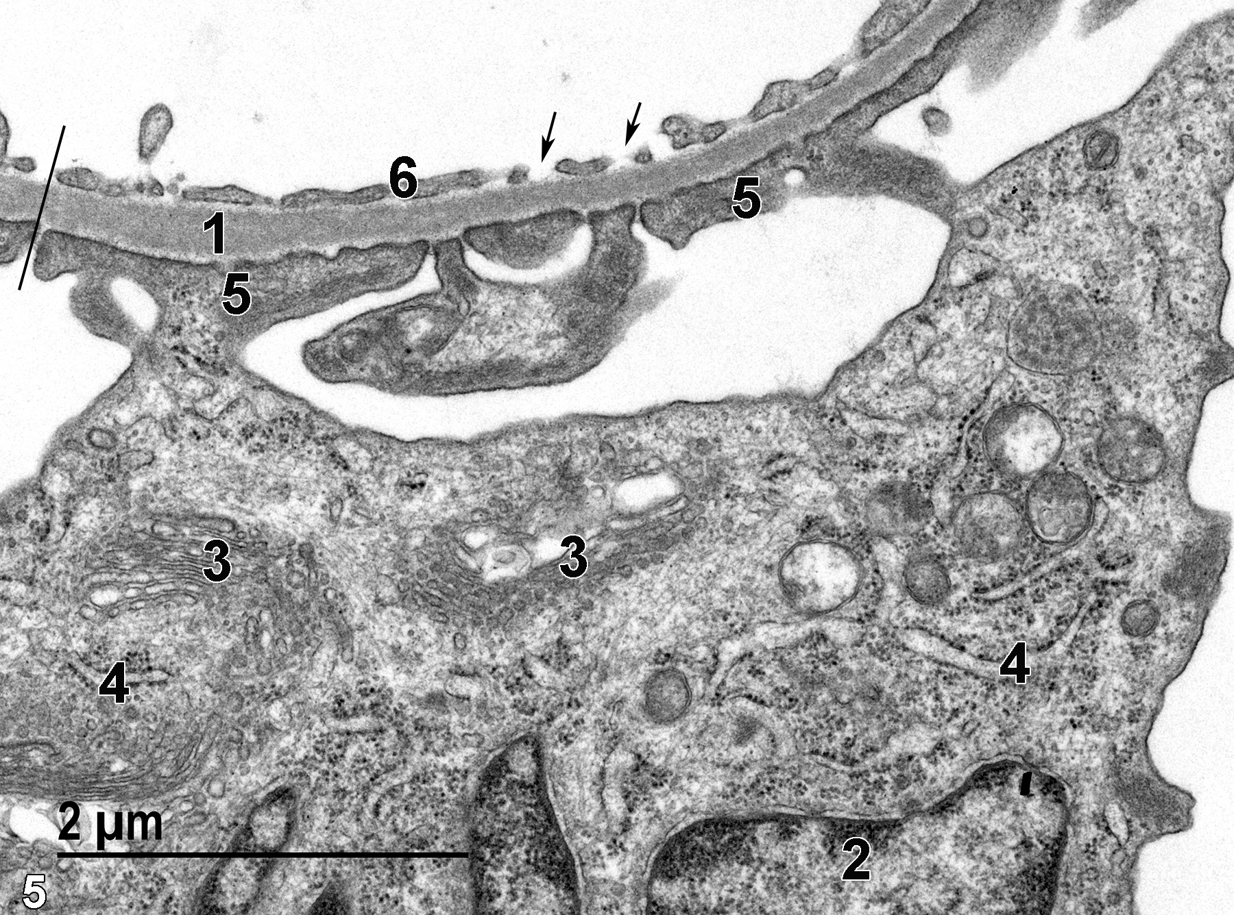
Figure 5. A higher magnification of a portion of a glomerulus showing a podocyte nucleus (2), Golgi bodies within the podocyte (3), rough endoplasmic reticulum (4), and foot processes (5) of the podocyte (pedicles) adjacent to the basal lamina (1), overlaid by the fenestrated endothelium of a capillary (6, arrows). The black line crossing the capillary endothelium, basal lamina, and the pedicles defines the filtration membrane of the glomerular system. Magnification not provided.
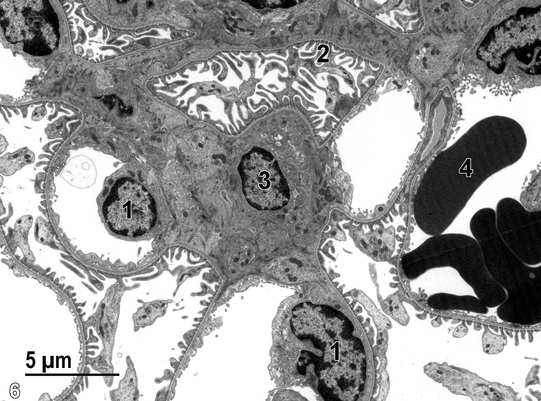
Figure 6. Another low magnification view of a portion of the glomerulus showing nuclei (1) of capillary endothelial cells, pedicles (2) of podocytes, a nucleus of mesangial cell (3), and an erythrocyte (4). Magnification not provided.
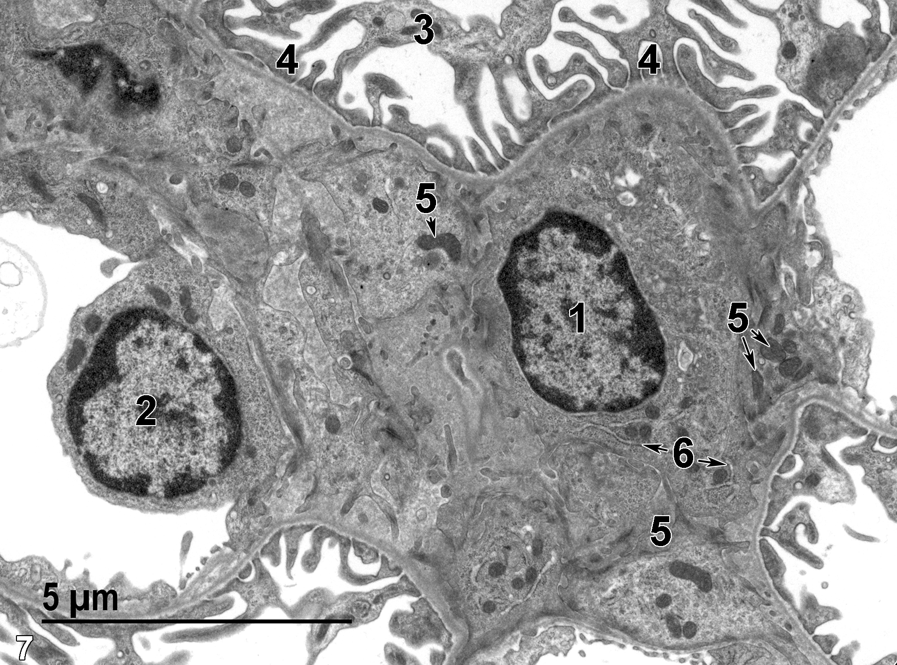
Figure 7. An enlarged view of Figure 6. The mesangial cell contains a nucleus with marginated heterochromatin (1), mitochondria (5, arrows), and rough endoplasmic reticulum (6, arrows). The nucleus (2) of a capillary endothelial cell is shown, along with pedicles (4) of a podocyte (3). Magnification not provided.
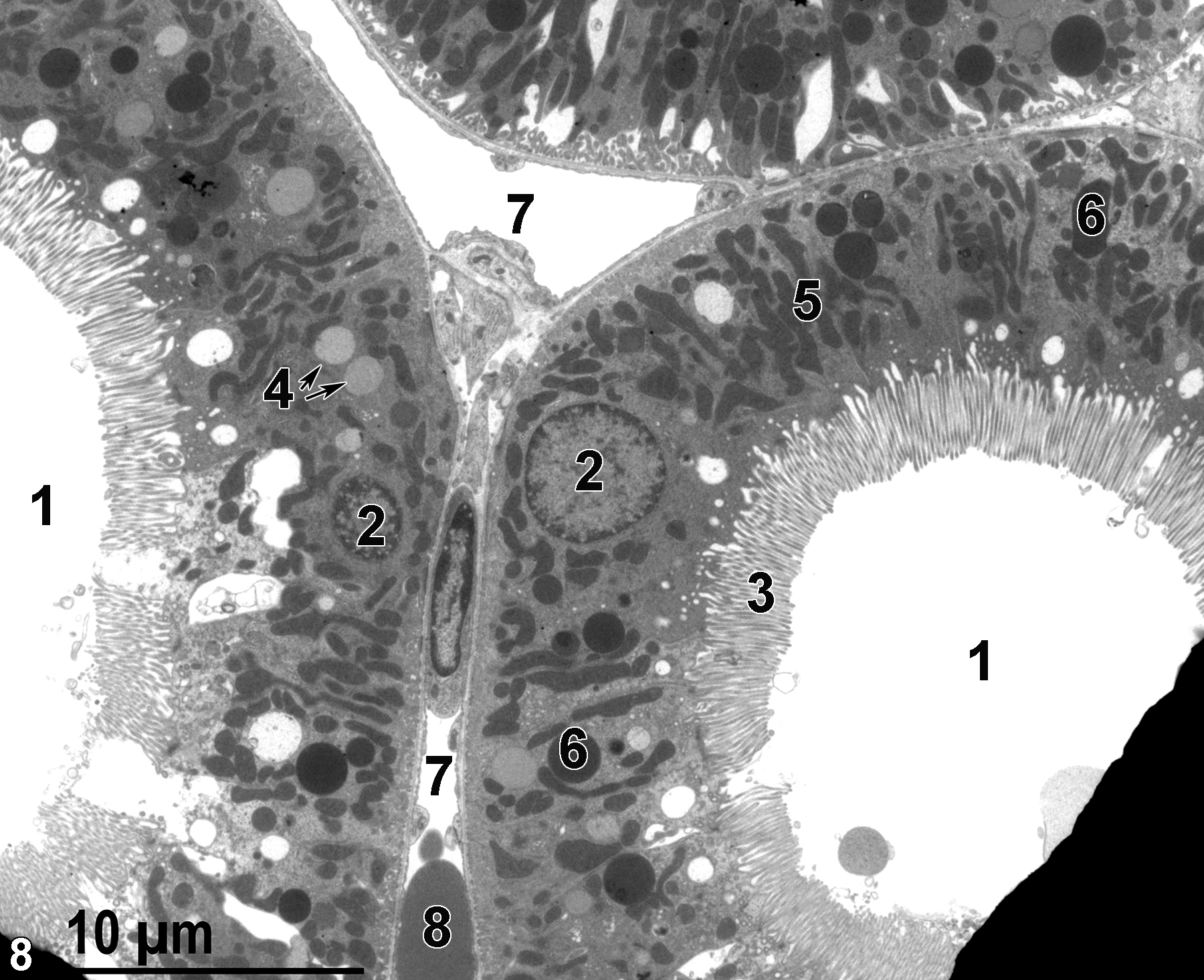
Figure 8. A low magnification image from a kidney perfusion that prevented the proximal convoluted tubules from collapsing, as happens with whole-body perfusions or immersion-fixation of kidneys. Electron-opaque grid bars are visible at the lower left and right of the image. The patent lumens of two proximal convoluted tubules (1) are lined with long microvilli (3). The epithelial cells of the tubules contain nuclei (2), mitochondria (5), lysosomes (6), and lipid bodies (4, arrows). Two peritubular capillary lumens (7) are shown, one containing an erythrocyte (8). Magnification not provided.
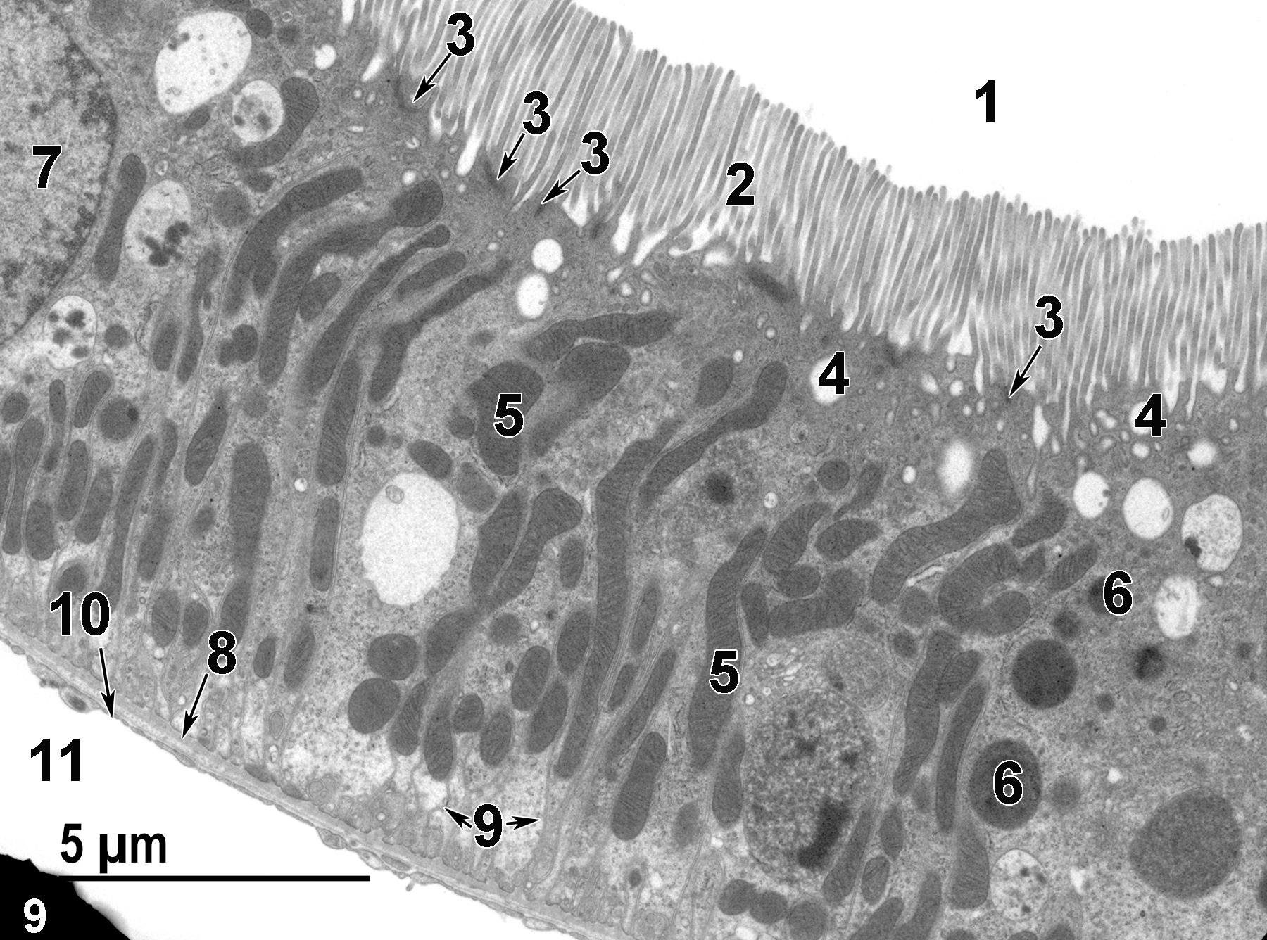
Figure 9. A higher magnification view of a proximal convoluted tubule from a perfused kidney showing the tubule lumen (1), microvilli (2), junctional complexes (3, arrows), apical vacuoles (phagosomes) (4), elongated mitochondria (5), lysosomes (6), a tubule epithelial cell nucleus (7), and basal infoldings of the epithelial cell membrane (9, arrows). The epithelial cells are underlaid by a basal lamina (8, arrow) that is, in turn, underlaid by the fenestrated endothelium of a peritubular capillary (10) and the capillary lumen (11). The edge of a grid bar is visible at the lower left of the image. Magnification not provided.
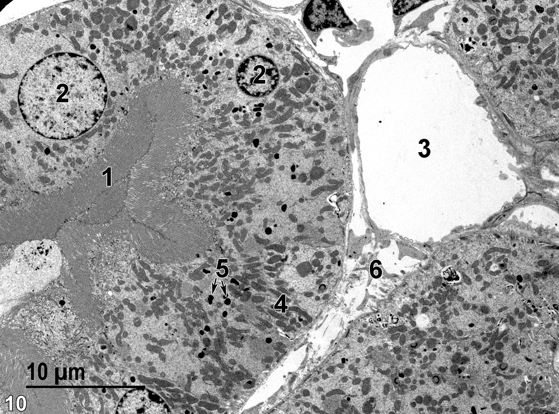
Figure 10. An image showing a proximal convoluted tubule from a kidney fixed by whole body perfusion, resulting in the collapse of the tubule lumen. The epithelial cell microvilli (1) have collapsed into the tubule lumen, occluding it. Lysosomes (5, arrows) are scattered throughout the cytoplasm, as are mitochondria (4). Epithelial cell nuclei (2) are round with prominent marginated heterochromatin. A capillary (3) is adjacent to an area of connective tissue (6). Magnification not provided.
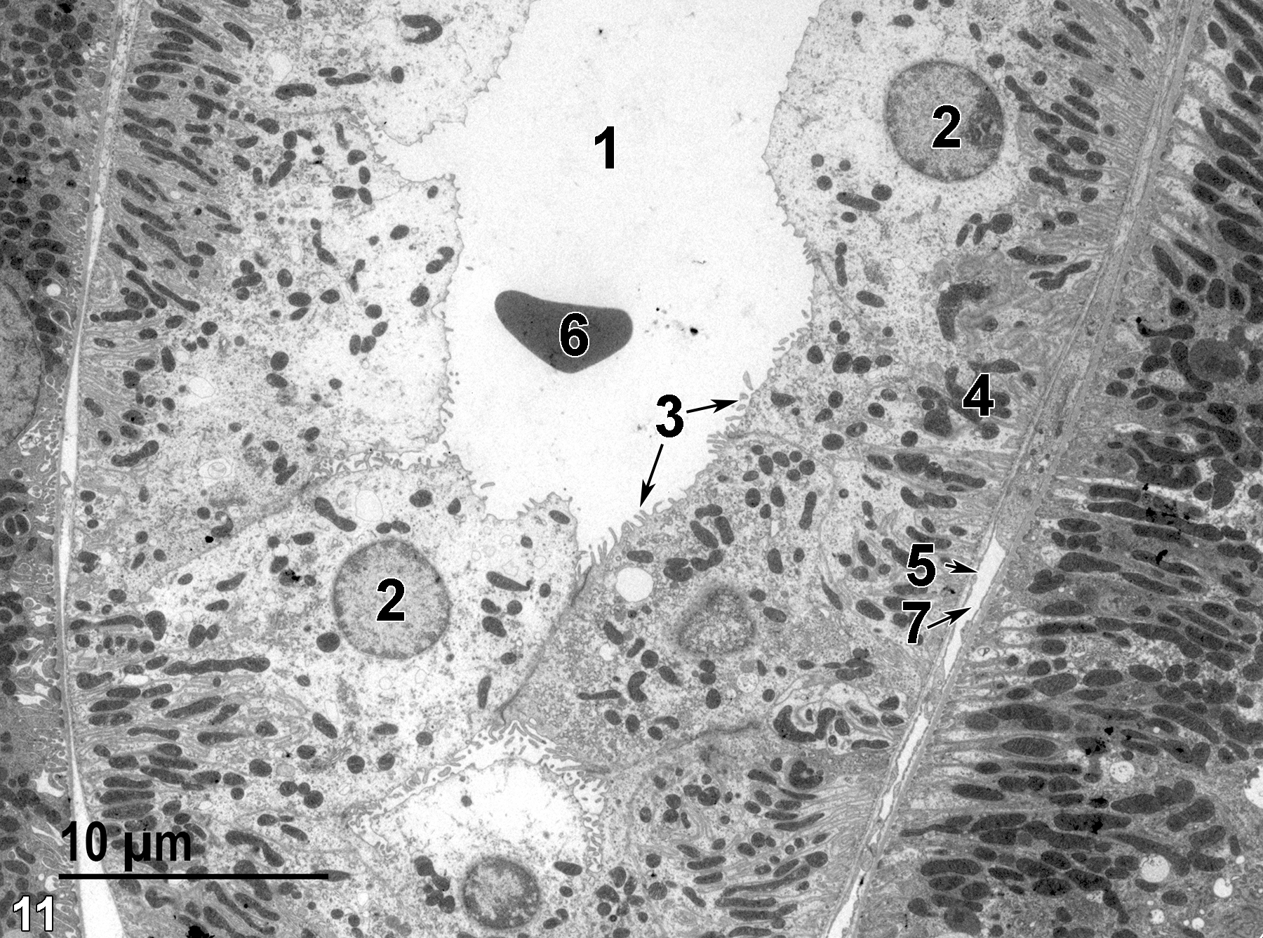
Figure 11. A distal convoluted tubule from the kidney cortex. The epithelial cells of the tubule have round nuclei (2) with marginated heterochromatin and short microvilli (3, arrows). The tubule lumen (1) contains one erythrocyte (6). Mitochondria are abundant, particularly in the basal area (4) of the epithelial cells. The peritubular capillary lumen (7, arrow) is lined with a fenestrated endothelium (5, arrow). Magnification not provided.
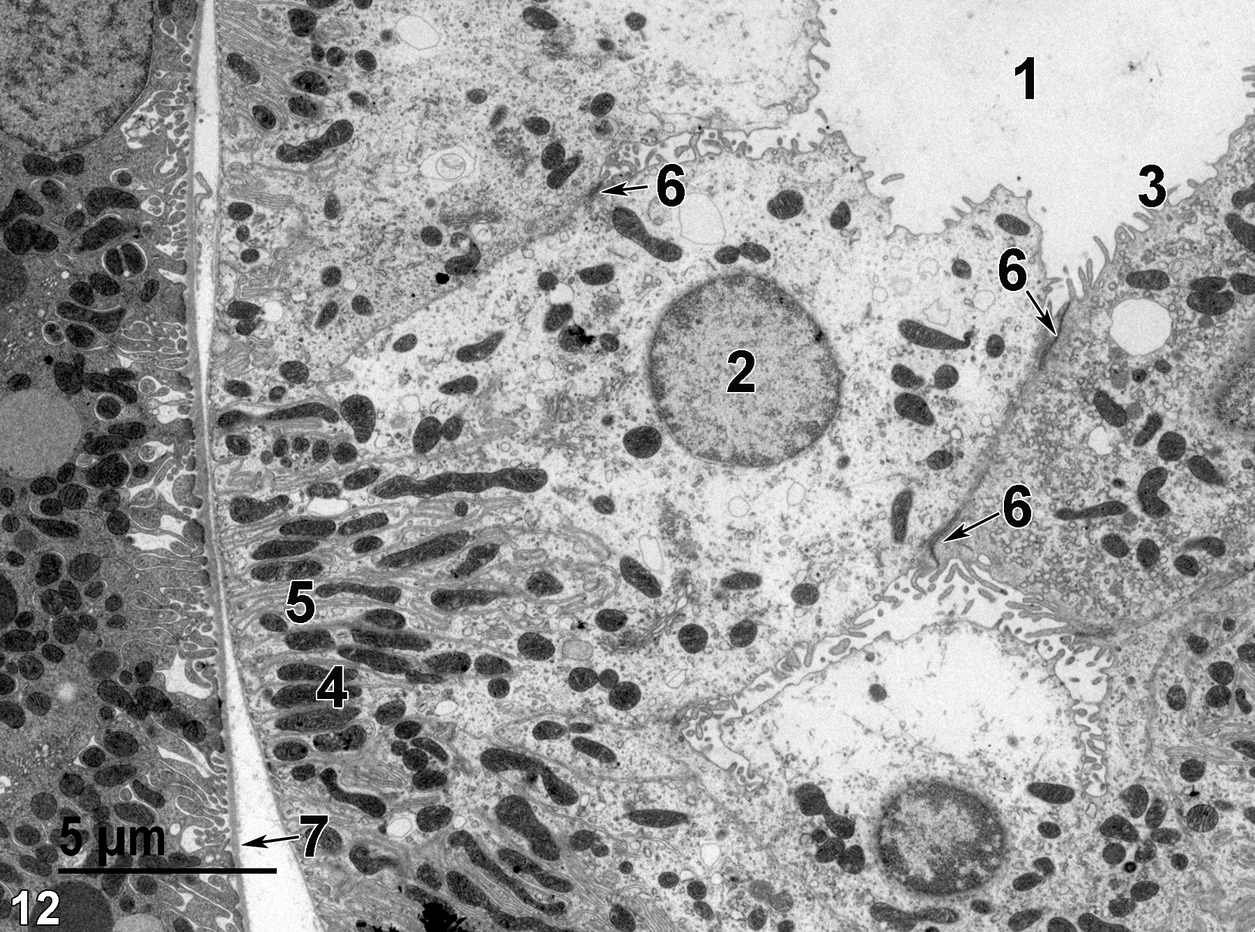
Figure 12. A higher magnification of Figure 11 showing the tubule lumen (1), a nucleus (2), short microvilli (3), elongated mitochondria (4) in the basal region of the epithelial cell intermixed with basal infoldings (5) of the plasma membrane. Junctional complexes (6, arrows) are found at the apical border of the epithelial cells. A basal lamina (7, arrow) is shown at the base of the epithelial cells. Magnification not provided.
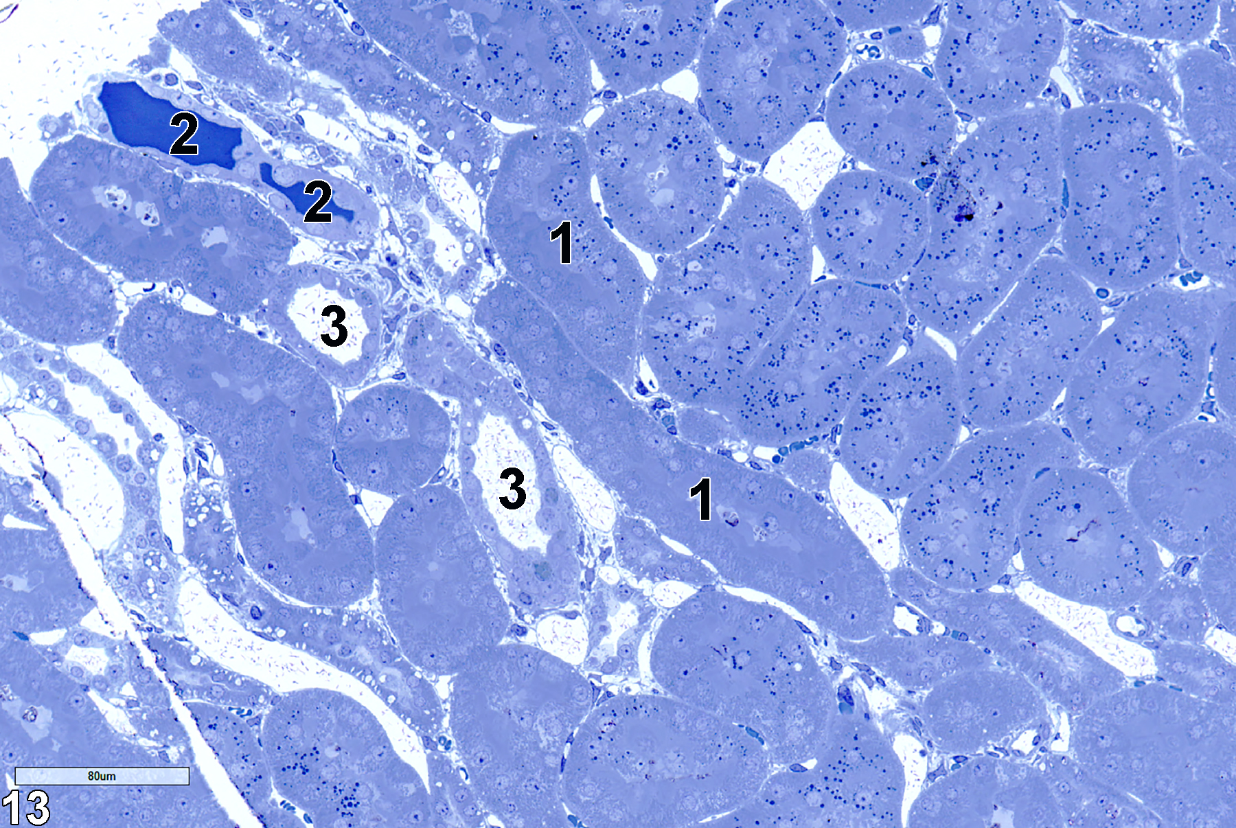
Figure 13. A toluidine blue O-stained section of the outer stripe of the outer medulla of a kidney. This area contains thick limbs of the loop of Henle (1), thin limbs of the loop of Henle (3), and capillaries with erythrocytes (2). 25x.
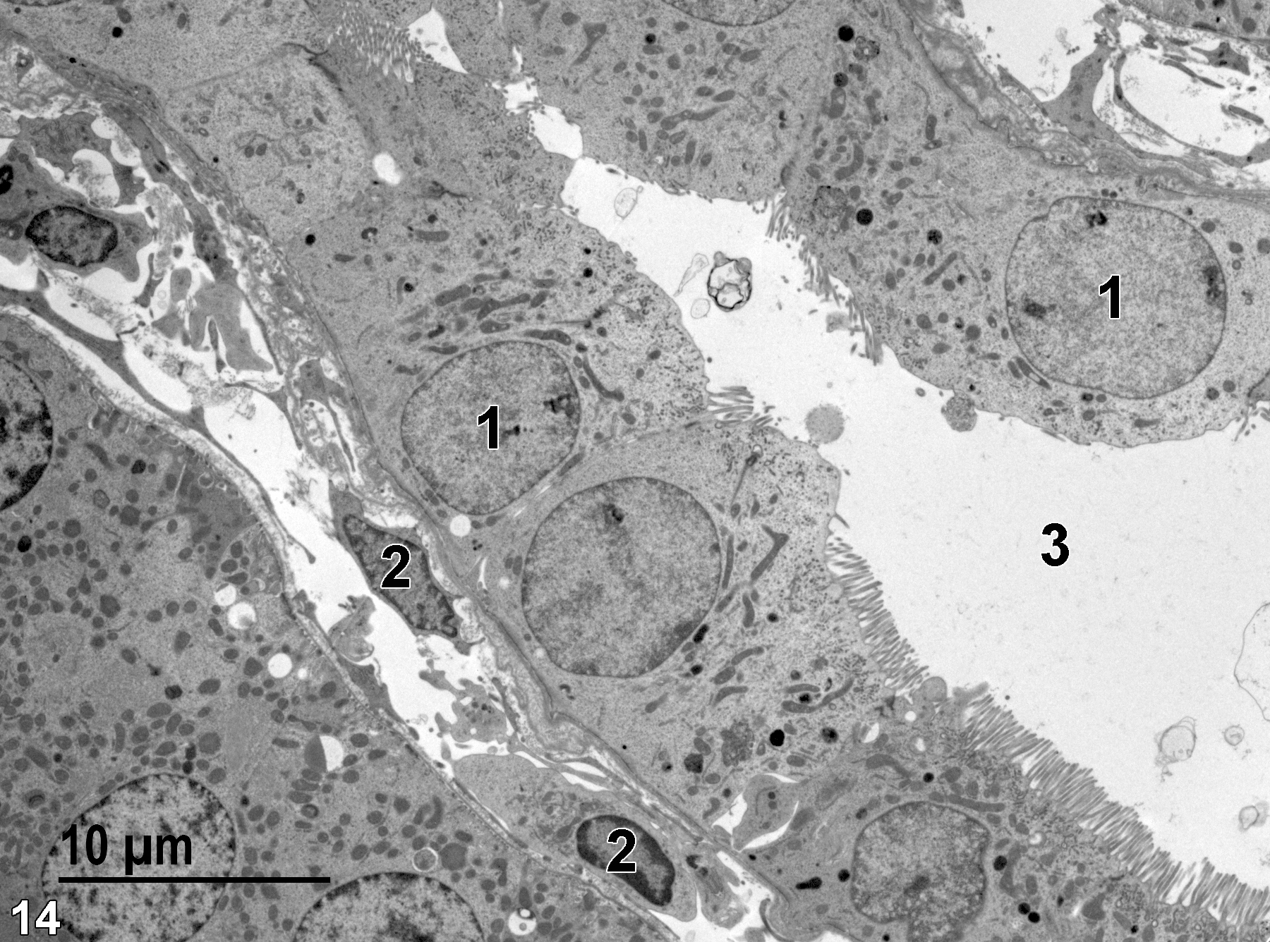
Figure 14. A thick limb has a single layer of epithelial cells with round nuclei (1) surrounding the tubule lumen (3). Nuclei of adjacent connective tissue cells (2) are shown. 2900x.
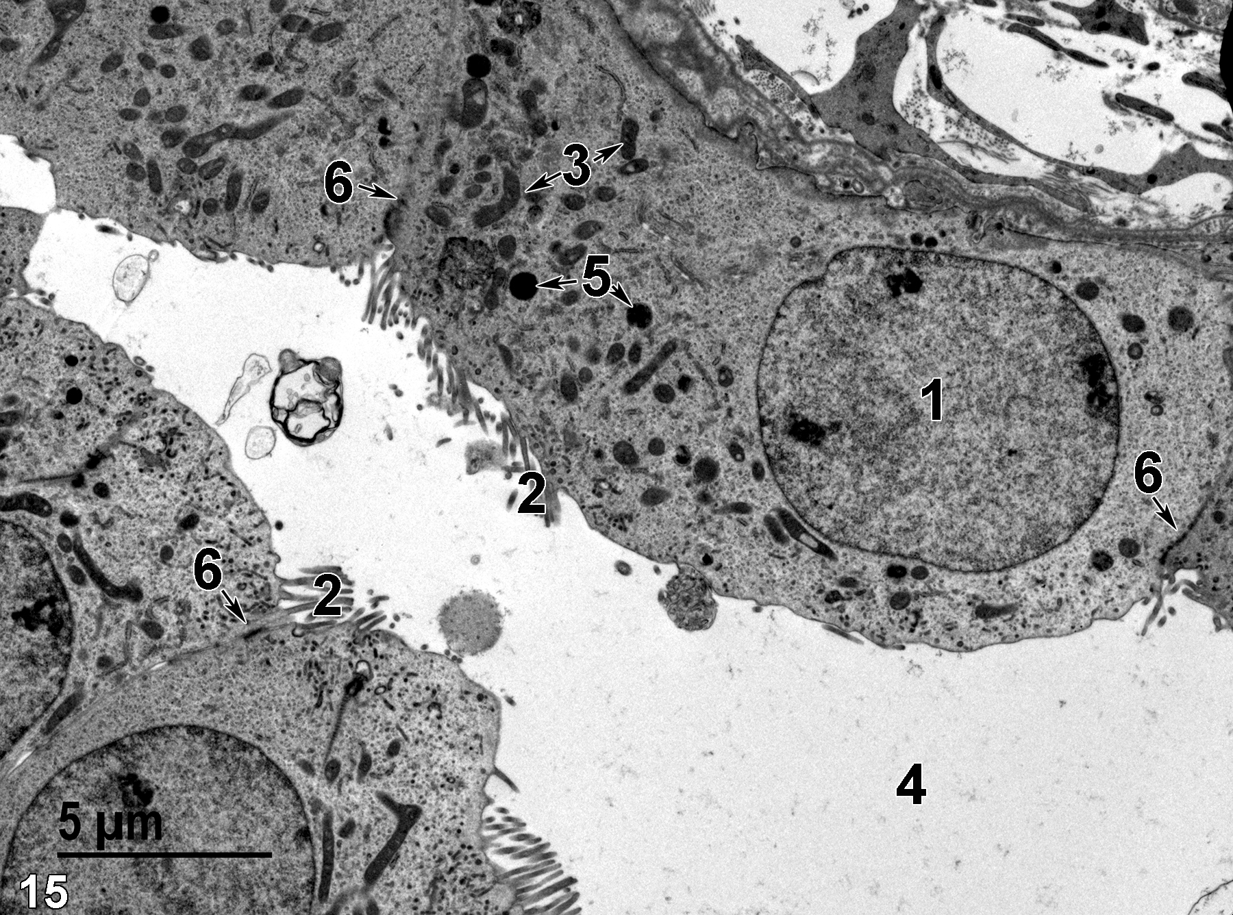
Figure 15. A higher magnification image of the thick limb shown in Figure 14 showing clusters of microvilli (2), a nucleus (1), lysosomes (5, arrows), mitochondria (3, arrows), the tubule lumen (4), and junctional complexes (6, arrows). 4800x.
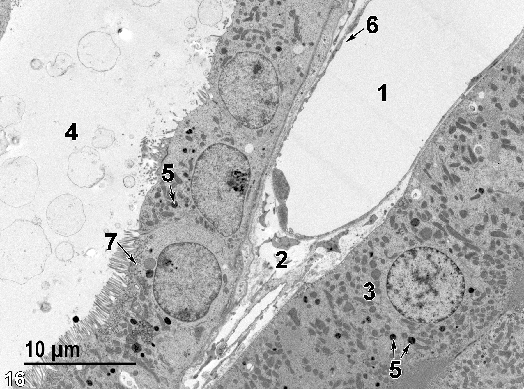
Figure 16. A lower magnification image showing a thick limb tubule lumen (4), epithelial cells (3) with lysosomes (5, arrows), and apical vesicles (7, arrow). A capillary (1) lined with fenestrated endothelial cells (6, arrow) is adjacent to connective tissue elements (2). 2900x.
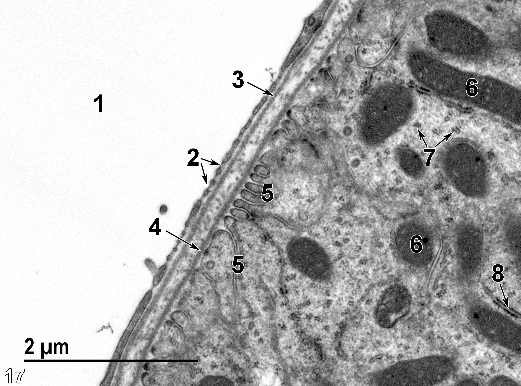
Figure 17. A high magnification image of the base of a thick limb epithelial cell showing mitochondria (6), rough endoplasmic reticulum (8, arrow), polyribosomes (7, arrows), and basal infoldings of the plasma membrane (5). The epithelial cell is underlaid with a basal lamina (4, arrow). The peritubular capillary lumen (1) is contained within the fenestrated endothelium (2, arrows) which is subtended by a basal lamina (3, arrow). In between the basal lamina of the capillary and the basal lamina of the epithelial cell is a layer containing collagen fibrils. 23000x.
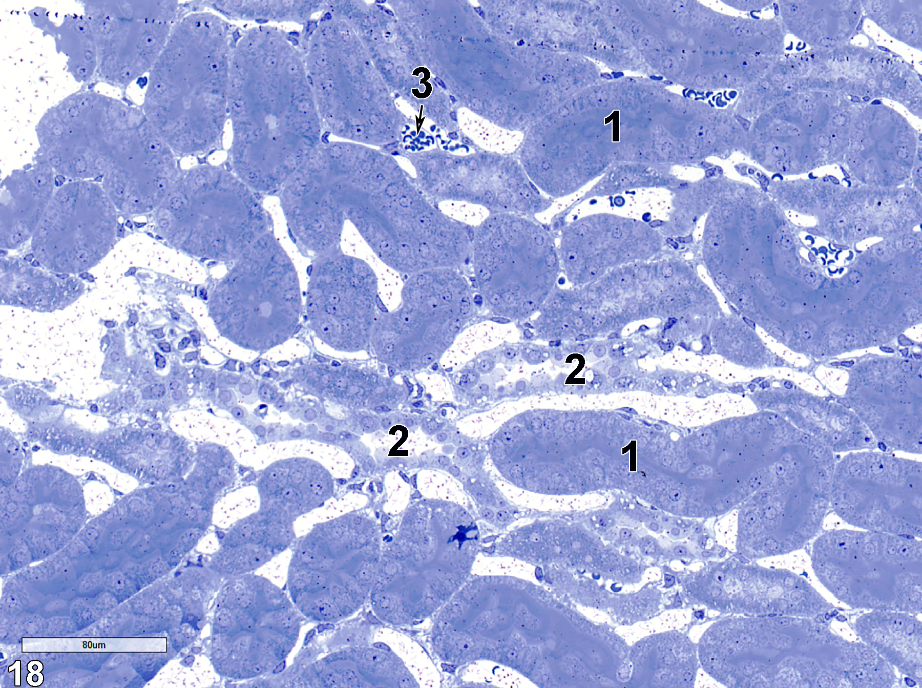
Figure 18. A toluidine blue O-stained section showing thick limbs of Henle (1), thin limbs of Henle (2), and a capillary containing erythrocytes (3, arrow). 25x.
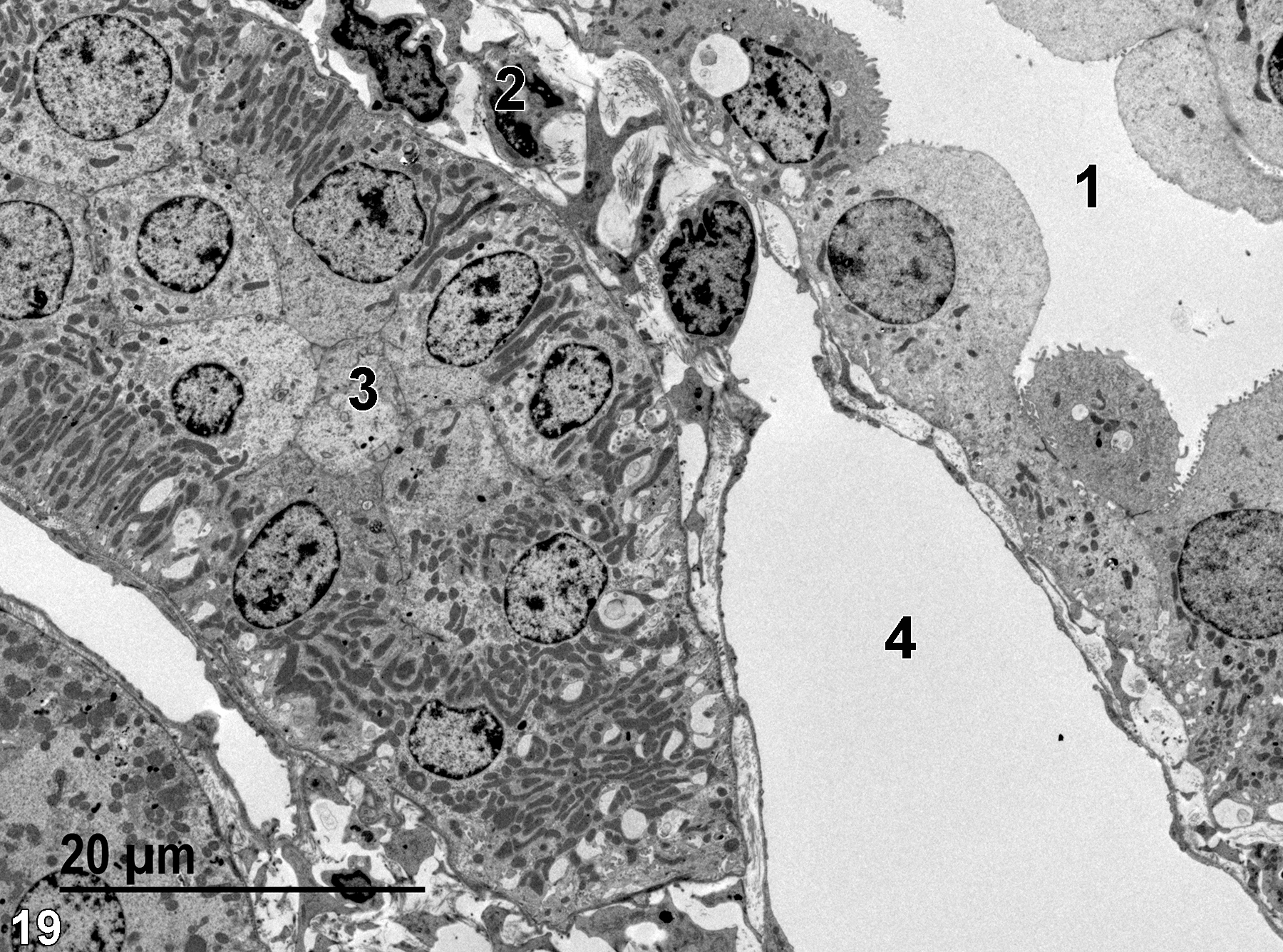
Figure 19. An image showing a capillary lumen (4), a thin limb of the loop of Henle lumen (1), and a thick limb of the loop of Henle (3). Connective tissue (2) is adjacent to the capillary and thin limb. 1900x.
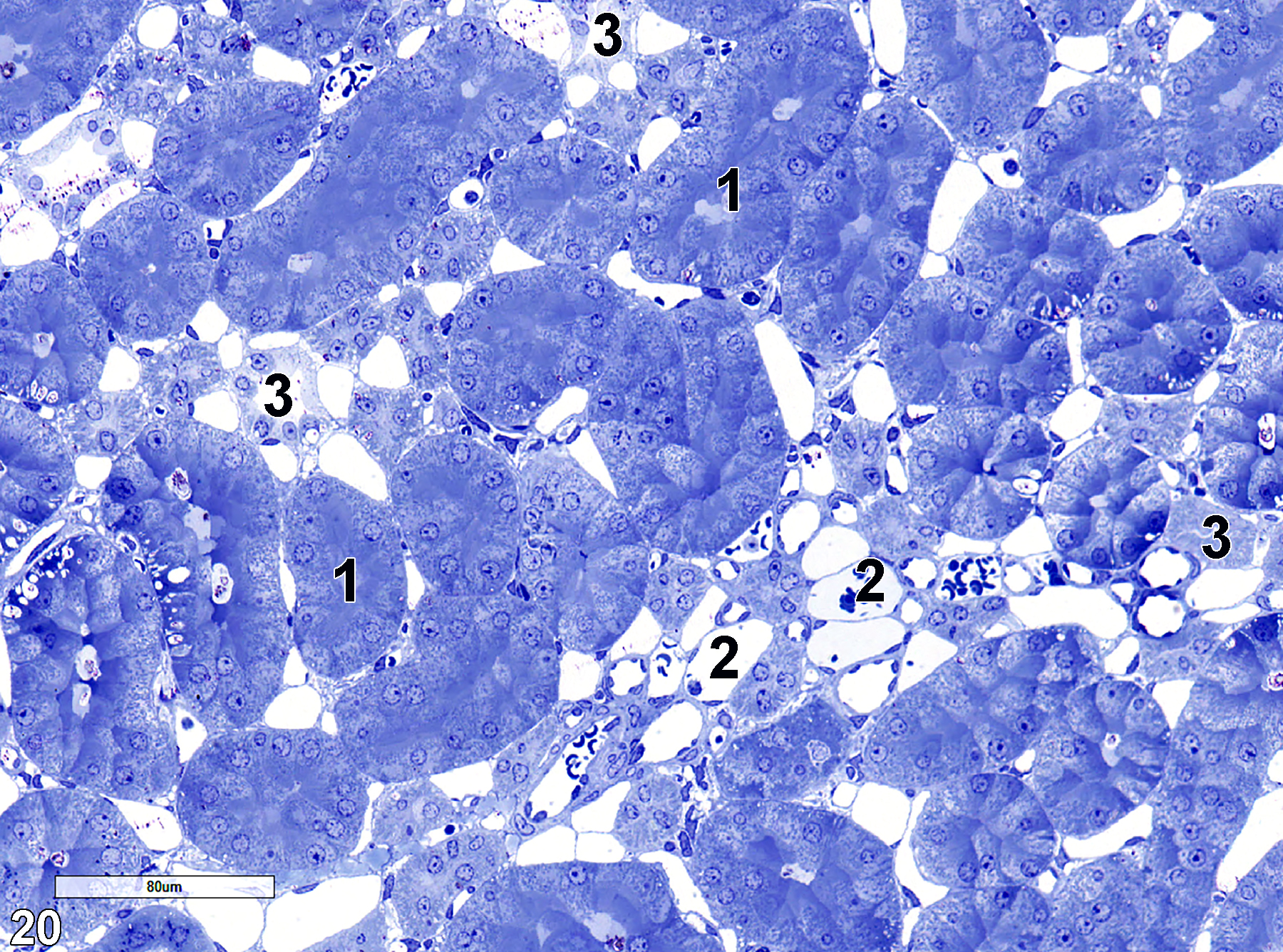
Figure 20. A toluidine blue O-stained semithin section of inner medulla tissue showing collecting ducts (1), capillaries (2), and thin limbs of loops of Henle (3). 25x.
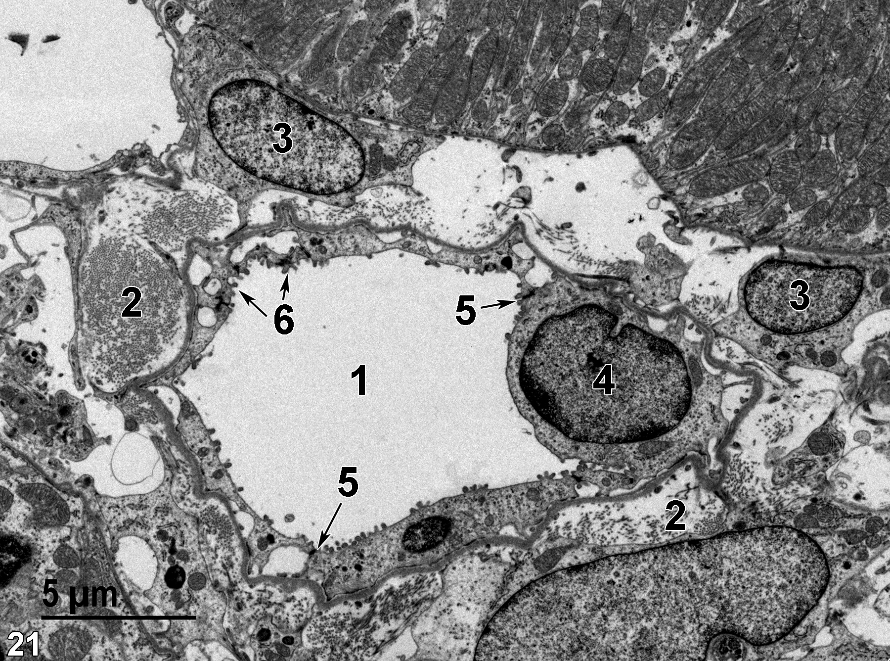
Figure 21. An image showing the lumen of a thin limb (1), collagen of connective tissue (2), nuclei of connective tissue cells (3), a nucleus of a thin limb epithelial cell (4), junctional complexes between adjacent tubule epithelial cells (5, arrows), and microvilli of the thin limb epithelial cells (6, arrows). 4800x.
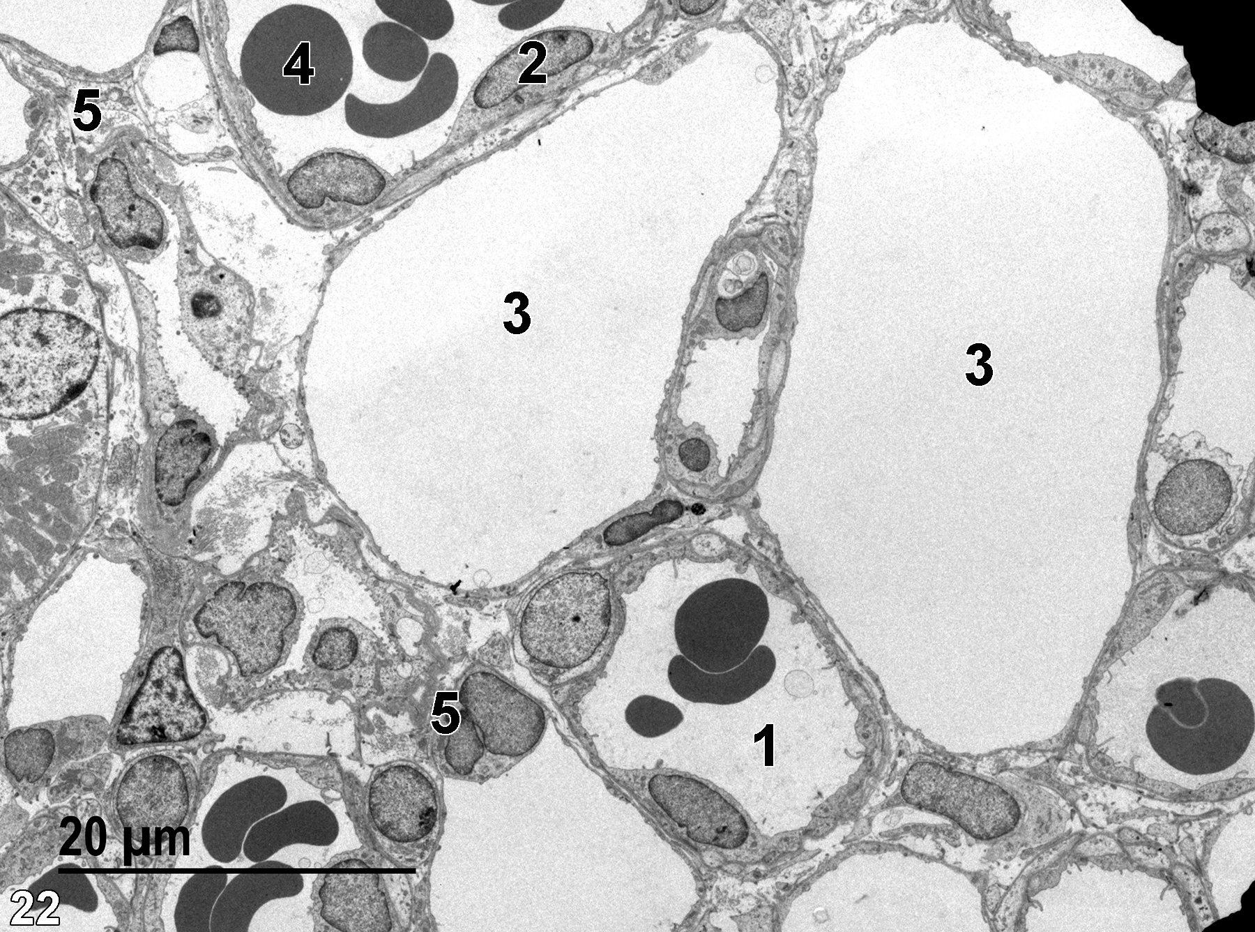
Figure 22. An image showing an arteriole lumen (1), another arteriole with a labeled endothelial cell nucleus (2), along with a labeled erythrocyte (4). Two elements of the venous vasa recta (3) are shown. A small area of connective tissue (5) is shown. 1900x.
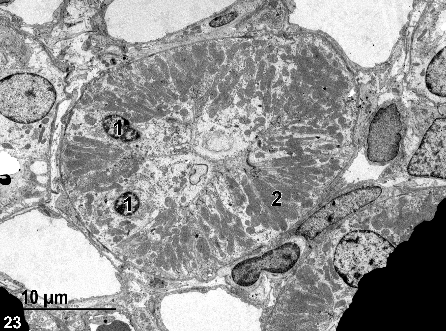
Figure 23. A transverse section of a collecting tubule showing two nuclei (1) and mitochondria (2). Electron-opaque grid bars are seen at the lower left and lower right of the image. 2900x.

Figure 24. A toluidine blue O-stained semithin section showing a segment of kidney papilla. The chief features shown are collecting ducts (1), capillaries (2), and thin limbs of the loops of Henle (3). 25x.
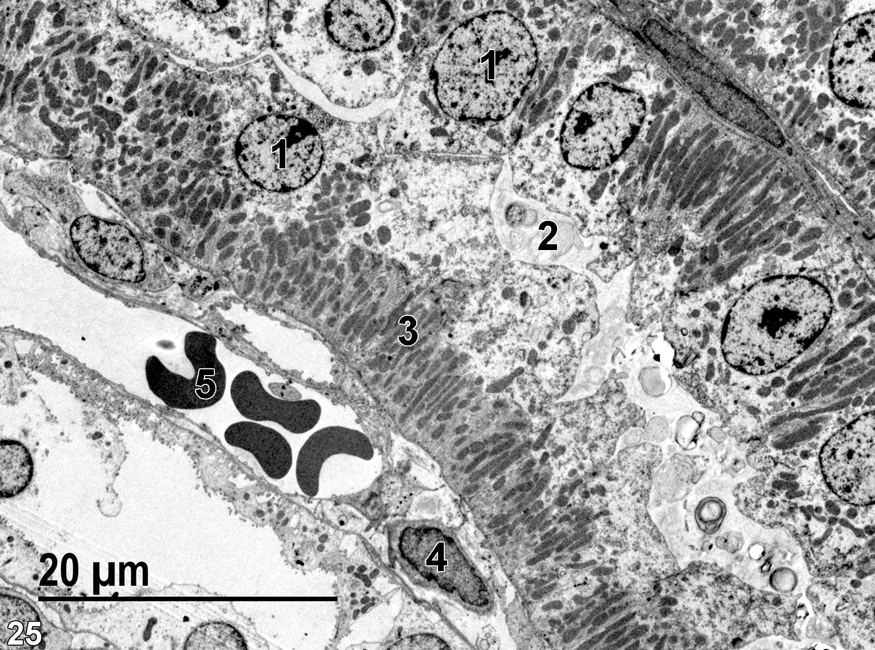
Figure 25. A low magnification view of a collecting duct, showing nuclei (1), the luminal space containing membranous debris (2), elongated mitochondria of the epithelial cells (3), a nucleus of a connective tissue cell (4), and an erythrocyte in a peritubular capillary (5). 1900x.
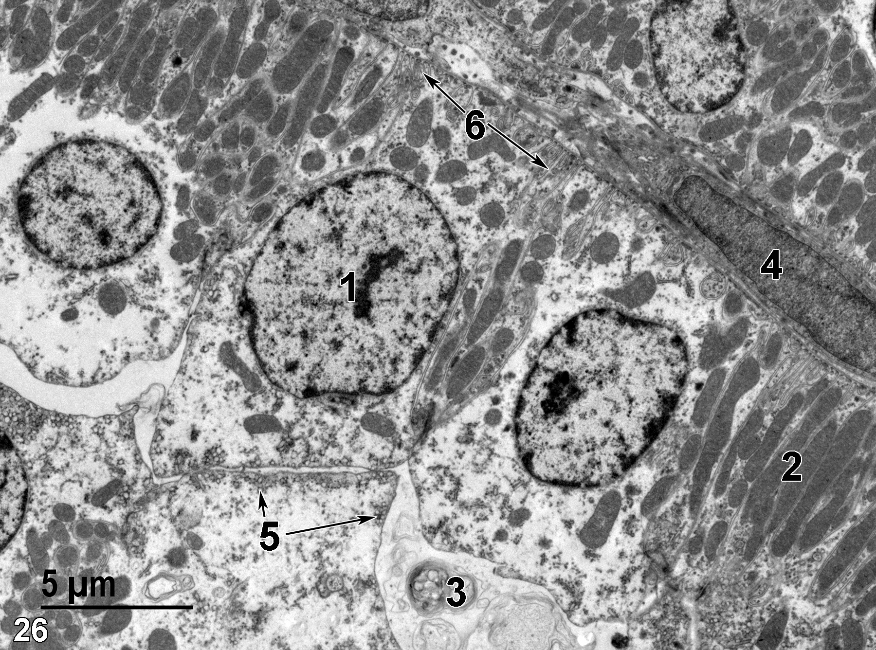
Figure 26. A higher magnification view of a collecting duct showing elongated mitochondria (2), the membranous debris (3) within the duct lumen, the nucleus of a connective tissue cell (4), apical vesicles in an epithelial cell (5, arrows), the nucleus (1), and basal infoldings (6, arrows) of an epithelial cell. 4800x.
| Allen F, Tisher CC. 1976. Morphology of the ascending thick limb of Henle. Kidney Int 9:8-22. |
| Cesta MF, Malarkey DE, Herbert R, Brix A, Sills RC, eds. (2014) National Toxicology Program’s Nonneoplastic Lesion Atlas. Research Triangle Park, NC: National Institute of Environmental Health Sciences. Available: https://ntp.niehs.nih.gov/nnl/ |
| Griffith LD, Bulger RE, Trump BF. 1967. Ultrastructure of the functioning kidney. Lab Invest 16:220−246. |
| Moffat DB. 1967. The fine structure of the blood vessels of the renal medulla with particular reference to the control of the medullary circulation. J Ultrastructure Res 19:532−545. |
| Rhodin JAG. 1974. Histology: A Text and Atlas. New York: Oxford University Press. |
| Schwartz MM, Venkatachalam MA. 1974. Structural differences in thin limbs of Henle: physiological implications. Kidney Int 6:103−208. |
| Verlander JW. 2006. Urinary system. In Dellman’s Textbook of Veterinary Histology (Eurell JA, Frappier BL, eds.). 6th ed. Ames, IA: Blackwell Publishers, 212−232. |
All Images


