

Reproductive System, Female
Uterus
Narrative
The uterus consists of bilateral horns, each having three layers, the endometrium (mucosa/submucosa), the myometrium (muscularis), and the perimetrium (serosa). The endometrium consists of a functional layer that changes depending on the stage of the estrous cycle observed. At the surface of this layer is an epithelium that varies in thickness depending on the estrous stage. The subepithelial layer below that (stroma) contains uterine glands, vascular elements, connective tissue, fibroblasts that produce the connective tissue, macrophages, mast cells, and occasional leukocytes such as eosinophils, neutrophils, and lymphocytes. Below the functional layer, the myometrium contains smooth muscle cells, connective tissue, fibroblasts, and macrophages. Finally, the perimetrium consists of loose connective tissue, smooth muscle cells, vascular elements, and neural elements.
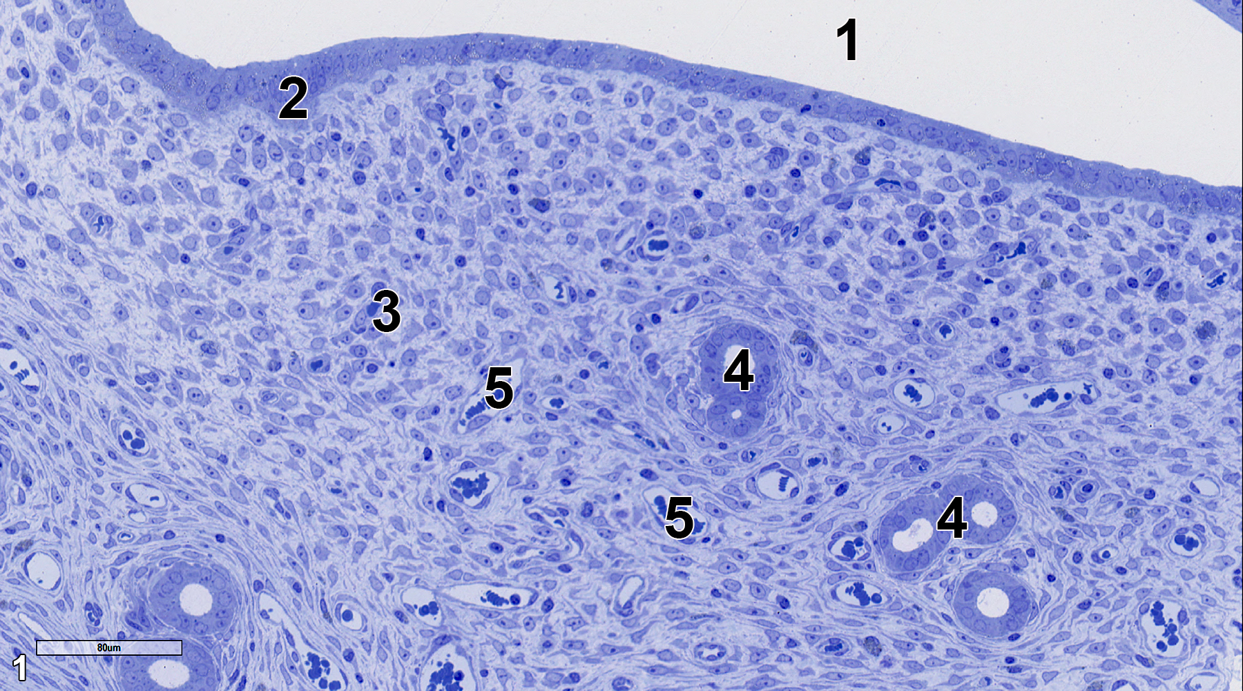
Figure 1. A toluidine blue O-stained semithin section (0.5 micrometer thick) of the uterus' epithelial and stromal (subepithelial) layers. The uterine lumen (1) is lined with an epithelium of cuboidal cells (2) subtended by a sub-epithelium or a stromal layer (3) with uterine glands (4) and many vascular elements (5). 25x.
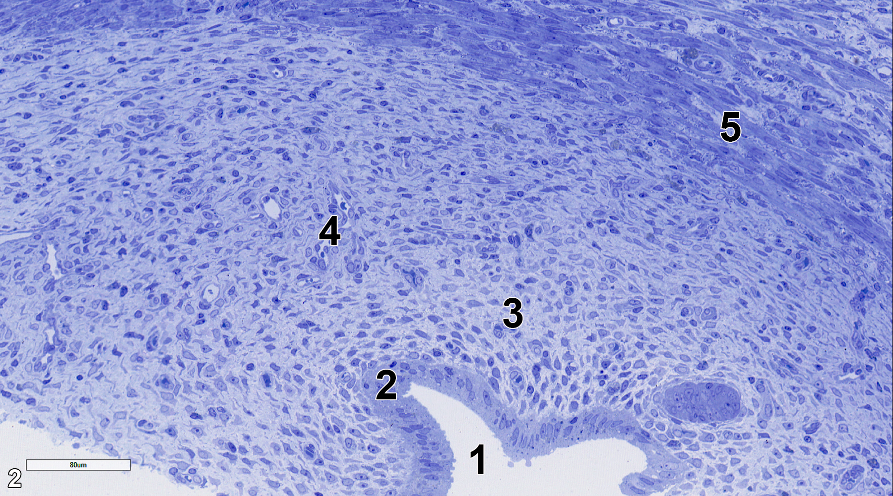
Figure 2. Another semithin section of uterine tissue. Lining the uterine lumen (1) is an epithelial layer (2), then the stromal subepithelial layer (3), which contains fibrous tissue and blood vessels (4), and then the muscularis (5), which consists of smooth muscle cells and connective tissue. 25x.
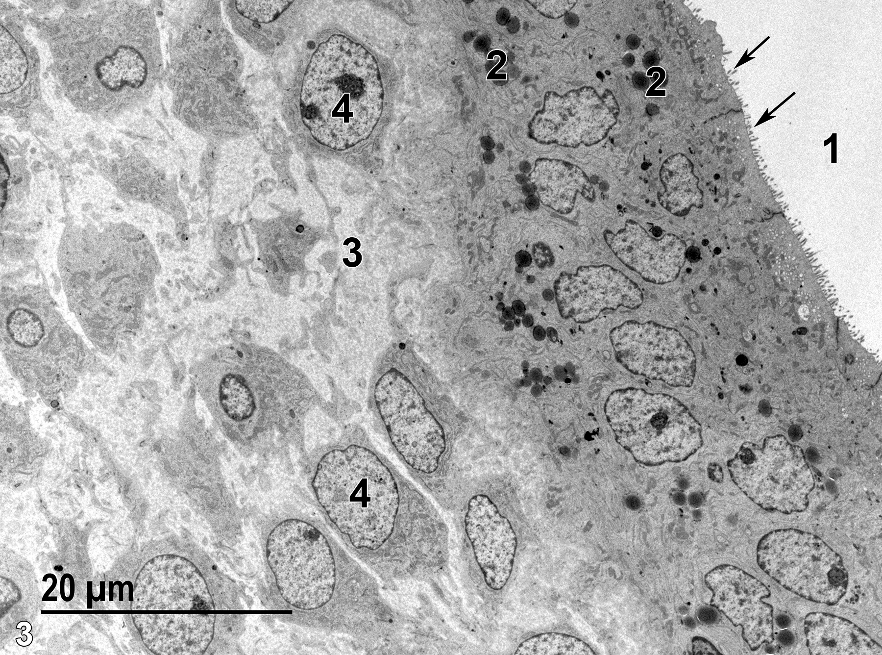
Figure 3. A low magnification electron micrograph from an area comparable to Figure 1. The uterine lumen (1) is lined with cuboidal epithelial cells with microvilli on the luminal side (arrows). The cells also contain electron-dense secretory granules (2). The stromal layer is filled with connective tissue (3) produced by fibroblasts (4). 1900x.
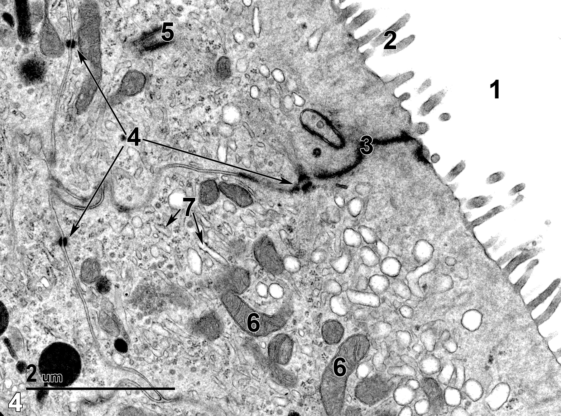
Figure 4. A higher magnification view of an epithelial cell, with microvilli (2) extending into the uterine lumen (1), a long junctional complex (3), and desmosomes (4) holding adjacent epithelial cells together (arrows). A single centriole (5) is present, as well as a number of mitochondria (6) and rough endoplasmic reticulum elements (7, arrows). 18500x.
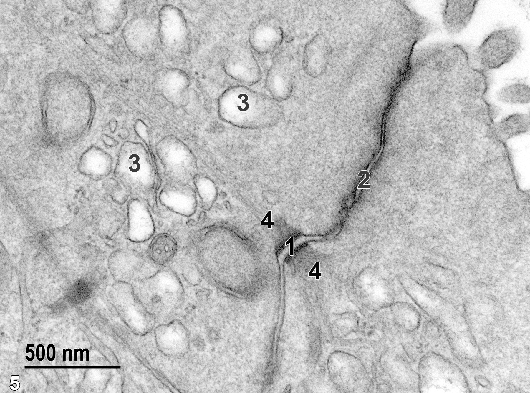
Figure 5. A higher magnification view of Figure 4, showing two components of the junctional complex, an intermediate junction (2) and a desmosome (1) with attached tonofilaments (4). Apical vesicles (3) are also present. 49000x.
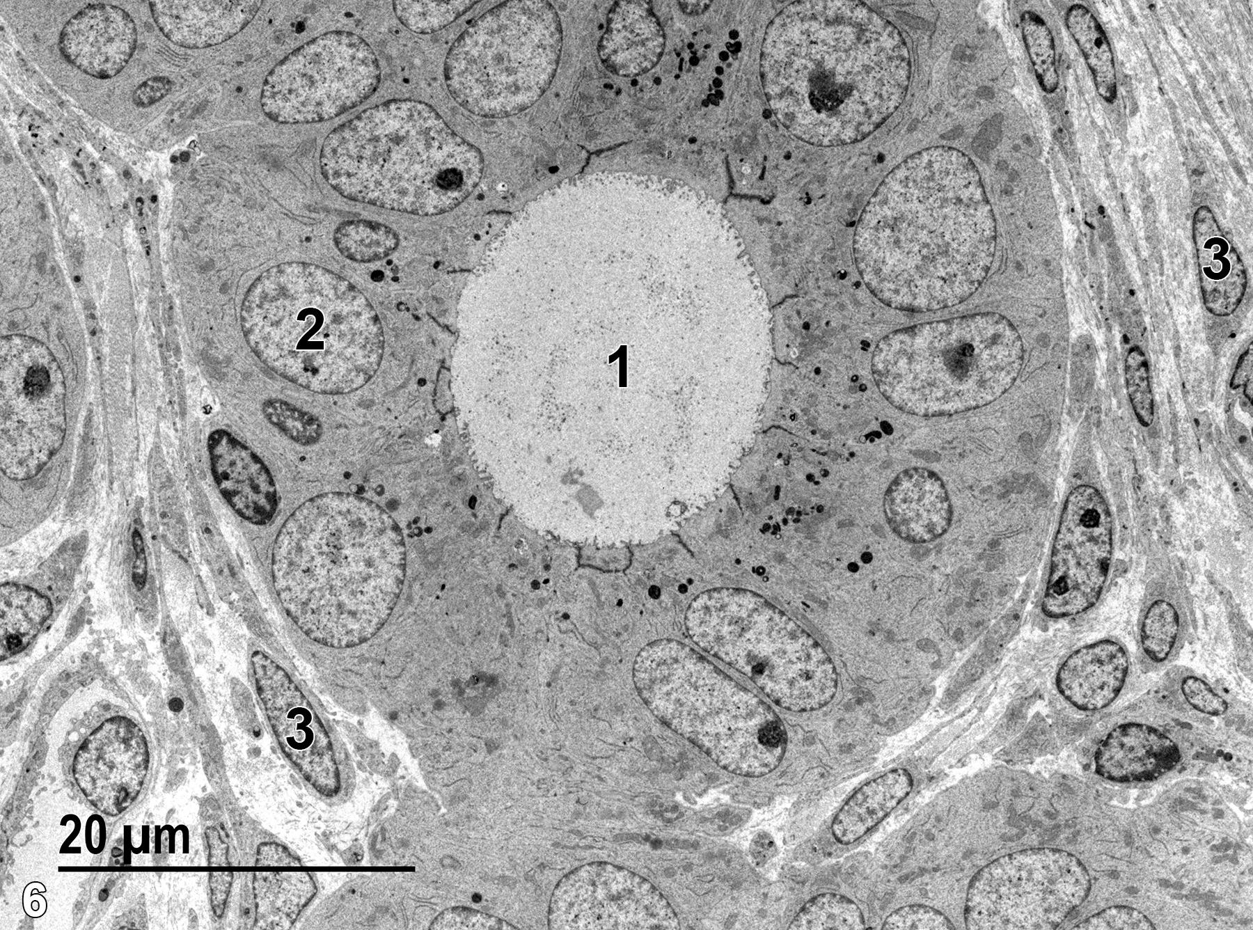
Figure 6. A uterine gland with cuboidal epithelial cells (2). The epithelial cells have microvilli extending into the glandular lumen (1) and electron-dense apical secretory granules. The electron-dense linear structures at the apical end of the glandular epithelial cells are junctional complexes. Connective tissue, which is composed of collagen and elastin fibrils and numerous fibroblasts (3), surrounds the gland. 1900x.
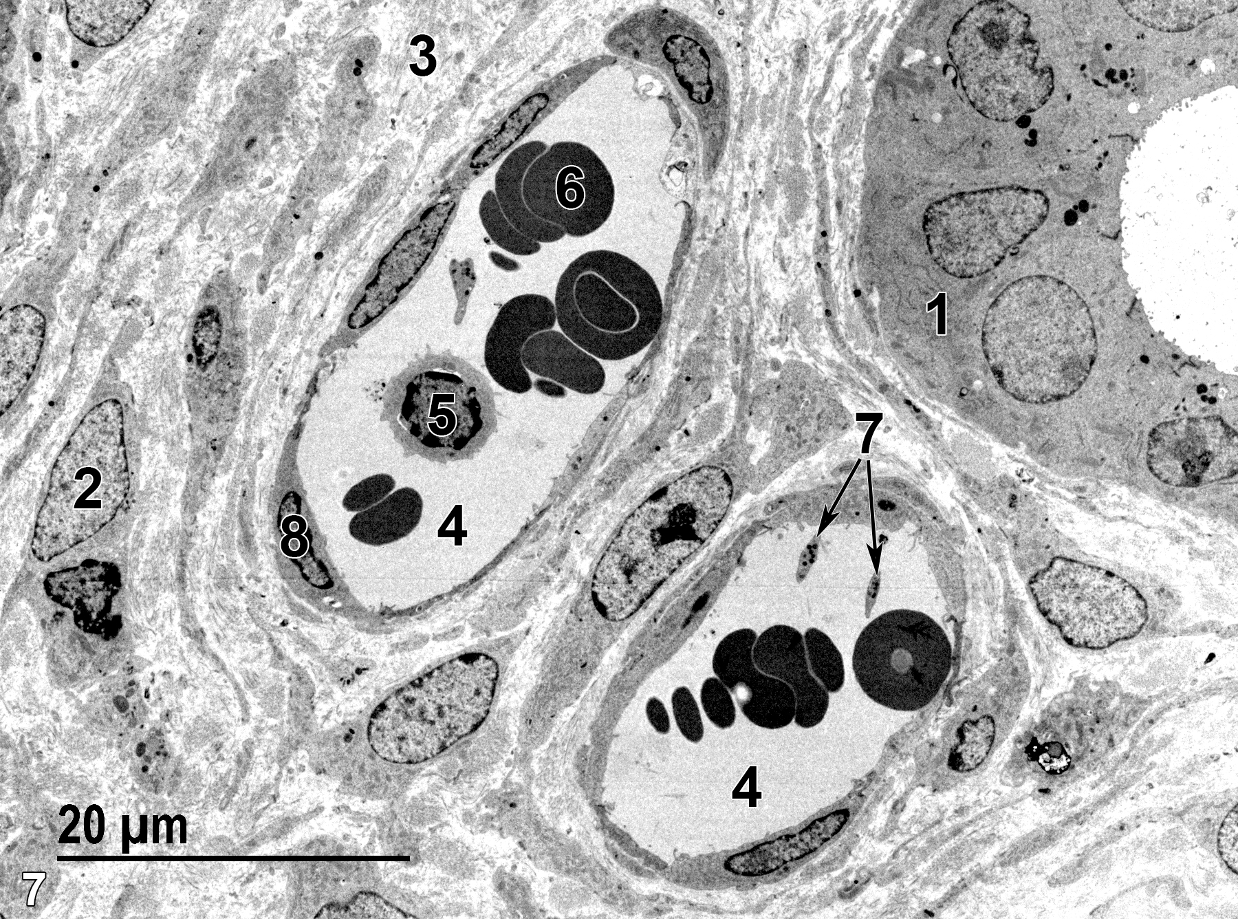
Figure 7. Another electron micrograph of the subepithelial (stromal) layer, with a single uterine gland (1) and two capillaries. Surrounding the capillaries is a collagenous matrix (3) containing numerous fibroblasts, one of which is labeled (2). The capillary lumens (4) are surrounded by a thin endothelium with elongated nuclei (8). A lymphocyte (5), erythrocytes (6), and platelets (7, arrows) can be seen within the capillary lumens. 1900x.
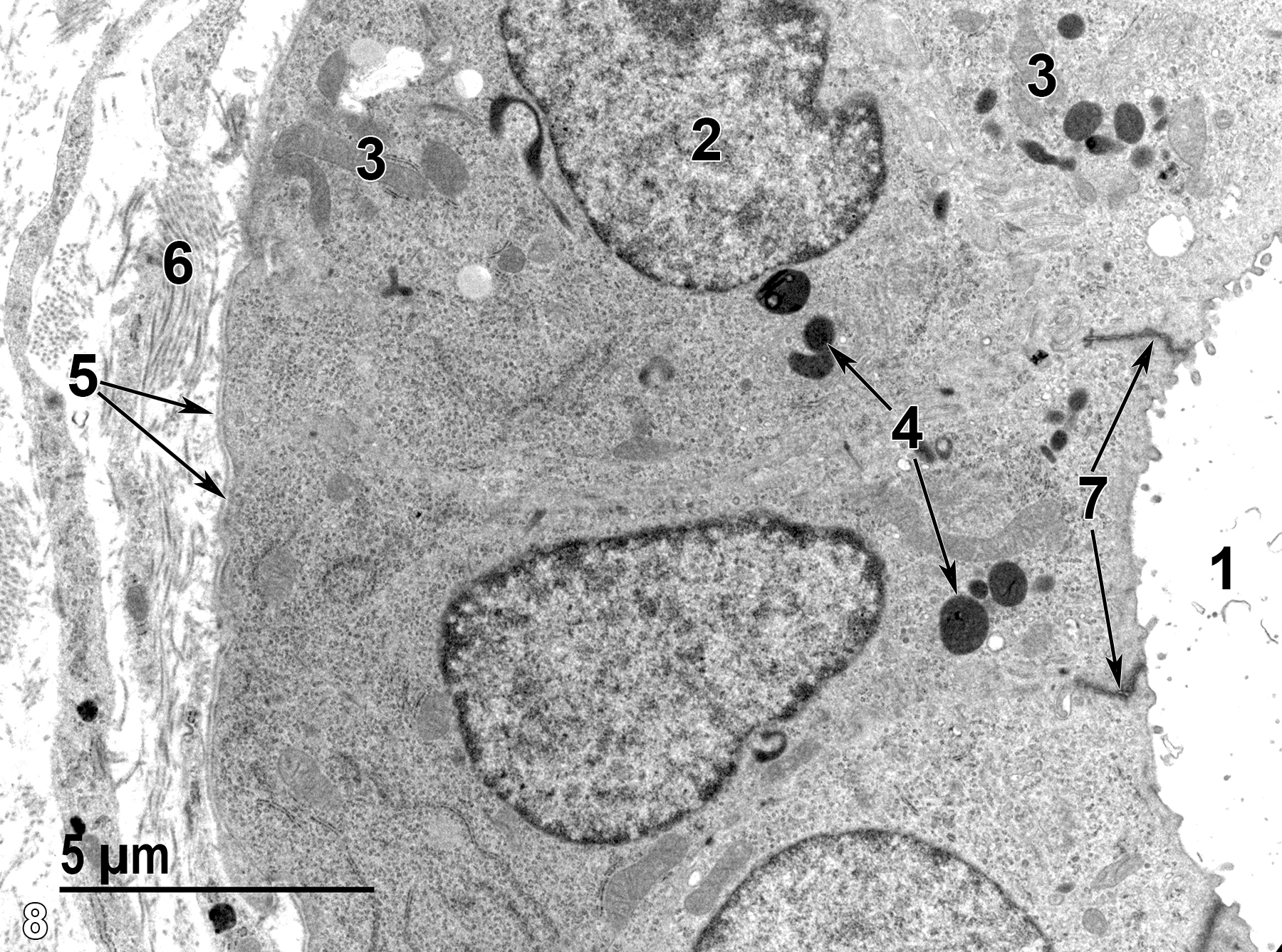
Figure 8. A higher magnification view of a uterine gland showing the glandular lumen (1) lined by epithelial cells (2) with short microvilli. The cells have pleomorphic mitochondria (3), apical secretory granules (4, arrows), and a basal lamina on the cell surface (5, arrows) adjacent to the surrounding collagenous matrix (6). The epithelial cells are connected by junctional complexes (7) at their apical surfaces (arrows). 6800x.
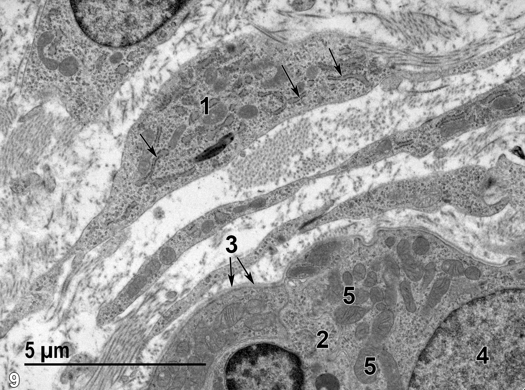
Figure 9. An area at the border of the sub-epithelium (stroma) and the muscularis layer. One of several fibroblasts (1) shows the three major characteristics of this cell type: large amounts of rough endoplasmic reticulum (arrows); an elongate cellular profile; and no basal lamina. A single smooth muscle cell (2) of the muscularis is shown. It has a basal lamina (3, arrows), a single nucleus (4), and a relative paucity of rough endoplasmic reticulum. A number of mitochondria (5) are evident. Collagen fibrils can be seen among the two cell types. 9300x.
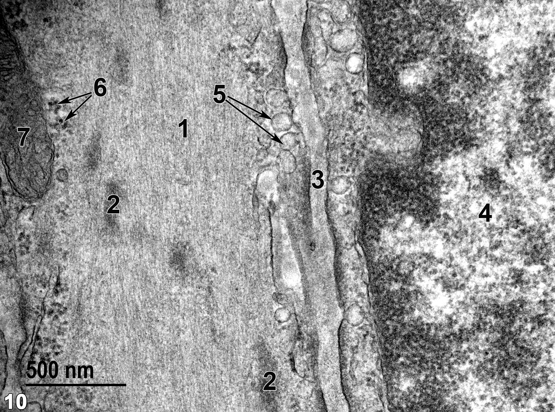
Figure 10. A relatively high magnification view of two adjacent smooth muscle cells. Actin and myosin filaments (1) comprise the proteinaceous filaments seen in the cell to the left. Periodic dense bodies (2) are evident among the actin and myosin filaments. Basal lamina components (3) are located between the adjacent smooth muscle cells. The cell to the right in the image contains a nucleus (4) with marginated chromatin. Both cells have pinocytotic vesicles (5, arrows) near the cell membrane that are characteristic of smooth muscle cells. The cells also contain scattered ribosomes (6, arrows) and a mitochondrion (7). 49000x.
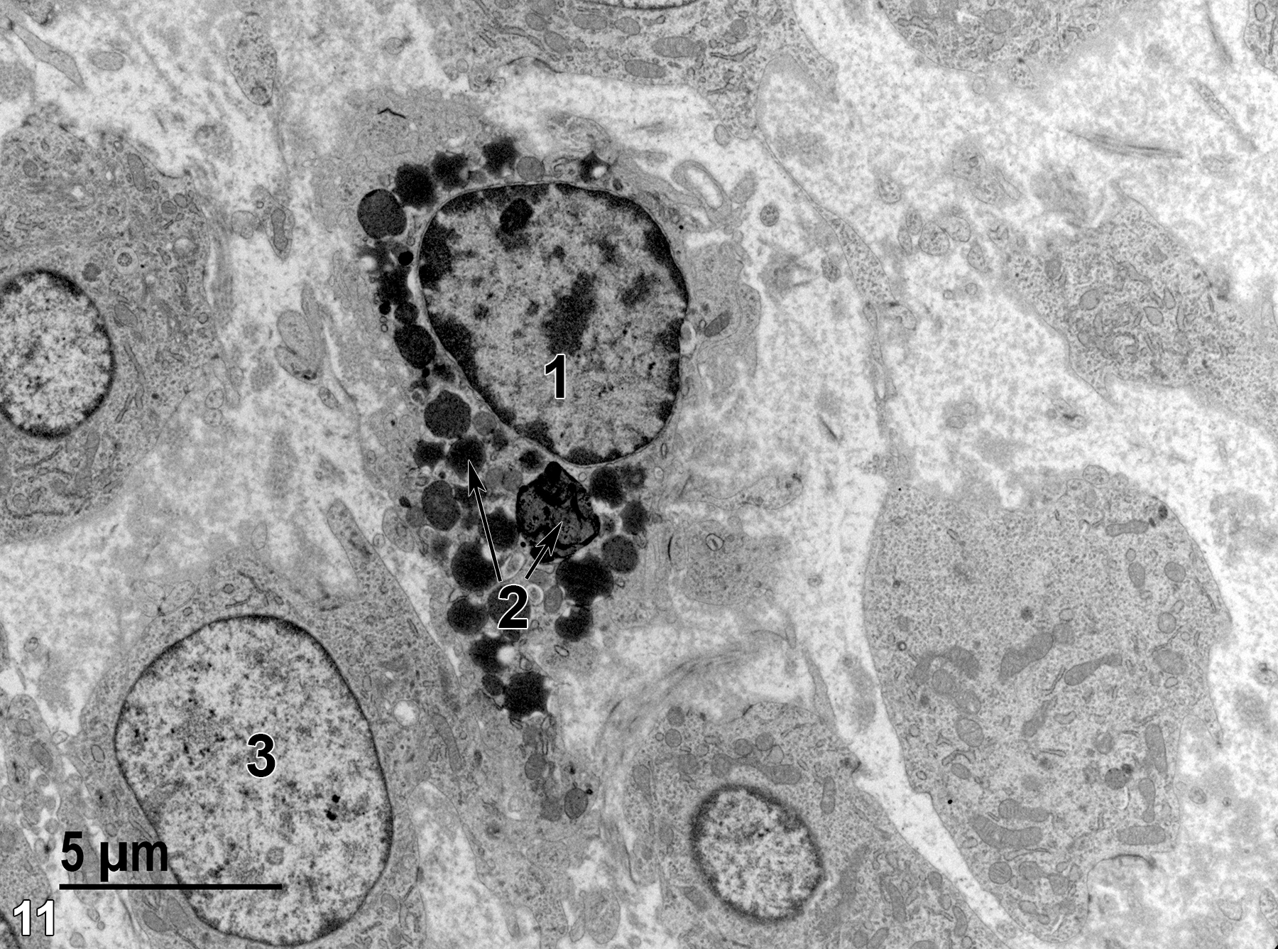
Figure 11. A single macrophage (1) with numerous lysosomes (2, arrows) of various sizes. These cells are primarily found within the stromal layer among fibroblasts (3) but also occur in the muscularis layer. 4800x.
| Cross PC, Mercer KL. 1993. Cell and Tissue Ultrastructure: A Functional Perspective. New York: W.H. Freeman and Company. |
| Dellmann HD, Eurell J, eds. 1998. Textbook of Veterinary Histology. 5th ed. Philadelphia: Lippincott Williams & Wilkins. |
| Rhodin JAG. 1974. Histology: A Text and Atlas. New York: Oxford University Press. |
| Weiss L, ed. 1988. Cell and Tissue Biology: A Textbook of Histology. 6th ed. Baltimore: Urban & Schwarzenberg. |
All Images


