

Alimentary System
Tongue
Narrative
The tongue is a muscular organ covered by a mucosa composed of a stratified squamous epithelium that is non-keratinized on its ventral surface and keratinized on its dorsal surface, which has the thicker epithelium. The dorsal surface has lingual papillae of several types. The most common type are filiform papillae that consist of conical elongate projections, with a convex part facing the anterior aspect and a concave part facing the posterior aspect. The posterior side has hard keratinization that ends in a hard pointed spine. Filiform papillae have no taste buds. The next most common type of papillae are the fungiform papillae that project above the general level of the mucosa and are found in the anterior two-thirds of the tongue with the filiform papillae. They possess taste buds, are more vascularized, and have more nervous tissue than the filiform papillae. They are broad at the tip, hence the term fungiform. Circumvallate papillae are located in the posterior one-third of the tongue and are larger than the fungiform papillae. They have the largest concentration of taste buds of any of the lingual papillae. Finally, there are foliate papillae located on the lateral borders of the tongue at the border between the anterior and posterior aspects. This region is characterized by parallel clefts, with the foliate papillae consisting of folds of mucous membranes between the clefts. These papillae have large numbers of taste buds. The papillae have a keratinized epithelial layer above the stratified squamous epithelial cell layer, which has smaller basal cells subtended by a basal lamina. Beneath the basal lamina is the lamina propria, which is composed of connective tissue with blood vessels, collagen, fibroblasts, and occasional mast cells. Taste buds found on all lingual papillae, except filiform papillae, are oval clusters of specialized epithelial cells that extend across the thickness of a papilla epithelial layer. Sensory cells are the most numerous type in taste buds and extend from the basal lamina to the apical taste pores. They have tight junctions with adjacent cells and microvilli at their apexes. Less numerous supporting cells have no microvilli or tight junctions and extend from the basal lamina to the taste pore. Finally, the basal cells of taste buds are located near the basal lamina and serve as stem cells for the other two taste bud cell types.
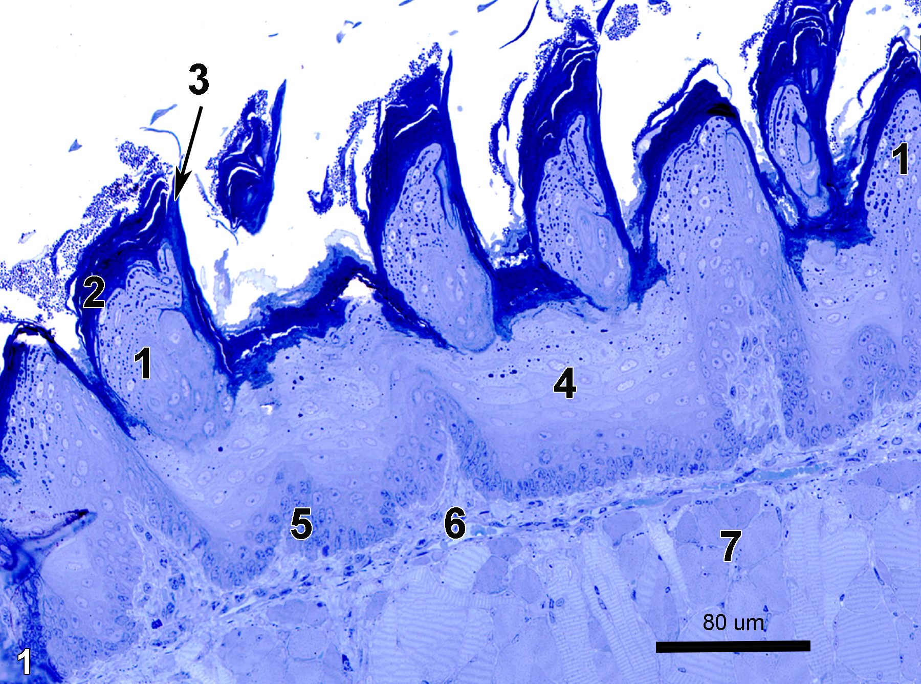
Figure 1. A semithin section (0.5 micrometer thick) of a toluidine blue O-stained section of the dorsal surface of the anterior portion of the tongue showing filiform papillae (1). The filiform papillae have a keratinized layer on the surface on both the convex side facing in the anterior direction (2) and also over the posterior portion, which is concave (3, arrow). The posterior side is composed of hard keratin extending into a spine. Below the keratinized surface are the stratified squamous epithelial cell layers (4), with a layer of basal cells (5). The lamina propria (6), which is composed of connective tissue, is located between the basal lamina of the epithelium and the striated muscle cell layer (7). 25x.
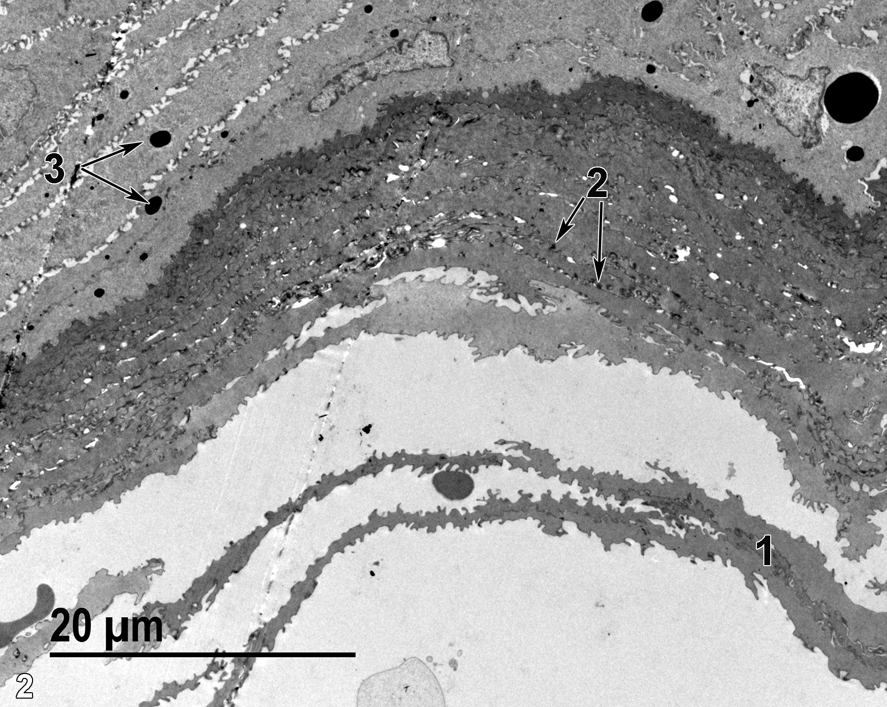
Figure 2. An ultrastructural view of the dorsal side of the tongue’s epithelial cell surface. Several cell layers of the keratinized epithelium (1) slough off of the epithelial surface. Small keratohyalin granules (2, arrows) can be seen in the superficial epithelial cells, whereas larger keratohyalin granule precursors (3, arrows) can be seen in more basal epithelial cells. 1900x.
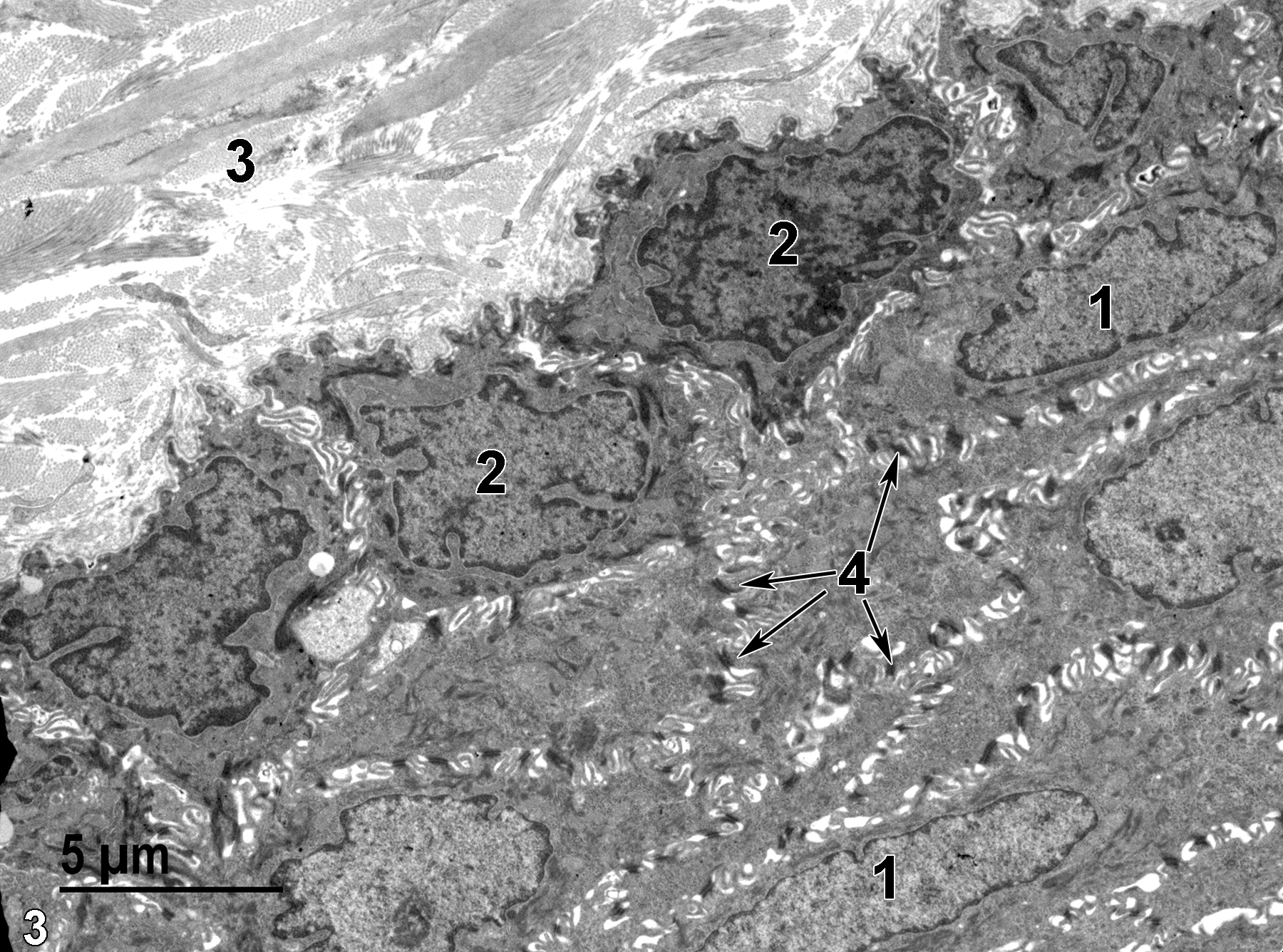
Figure 3. An electron micrograph of the basal side of the epithelial layer of the tongue. Stratified squamous epithelial cells have elongated nuclei (1) compared with the more rounded nuclei (2) of the basal cells. Bundles of collagen (3) are the predominant feature of the lamina propria. Note the large numbers of desmosomes (4, arrows) that hold the spinose projections of the adjacent cells together. 4800x.
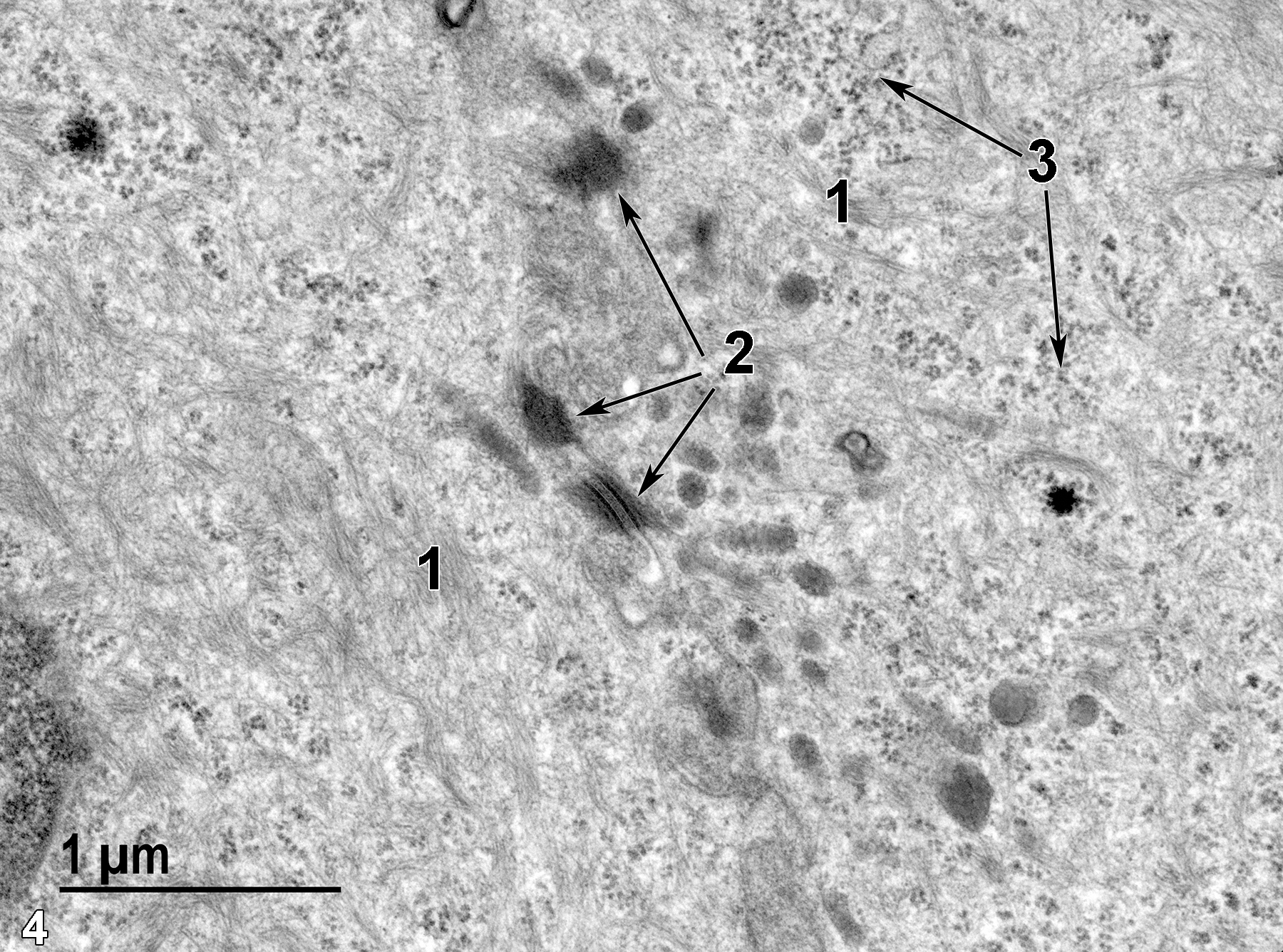
Figure 4. A high magnification view of the cytoplasm of an epithelial cell with large amounts of intermediate filament bundles (1), desmosomes joining adjacent cells (2, arrows), and large accumulations of clusters of free ribosomes (3, arrows). 30000x.
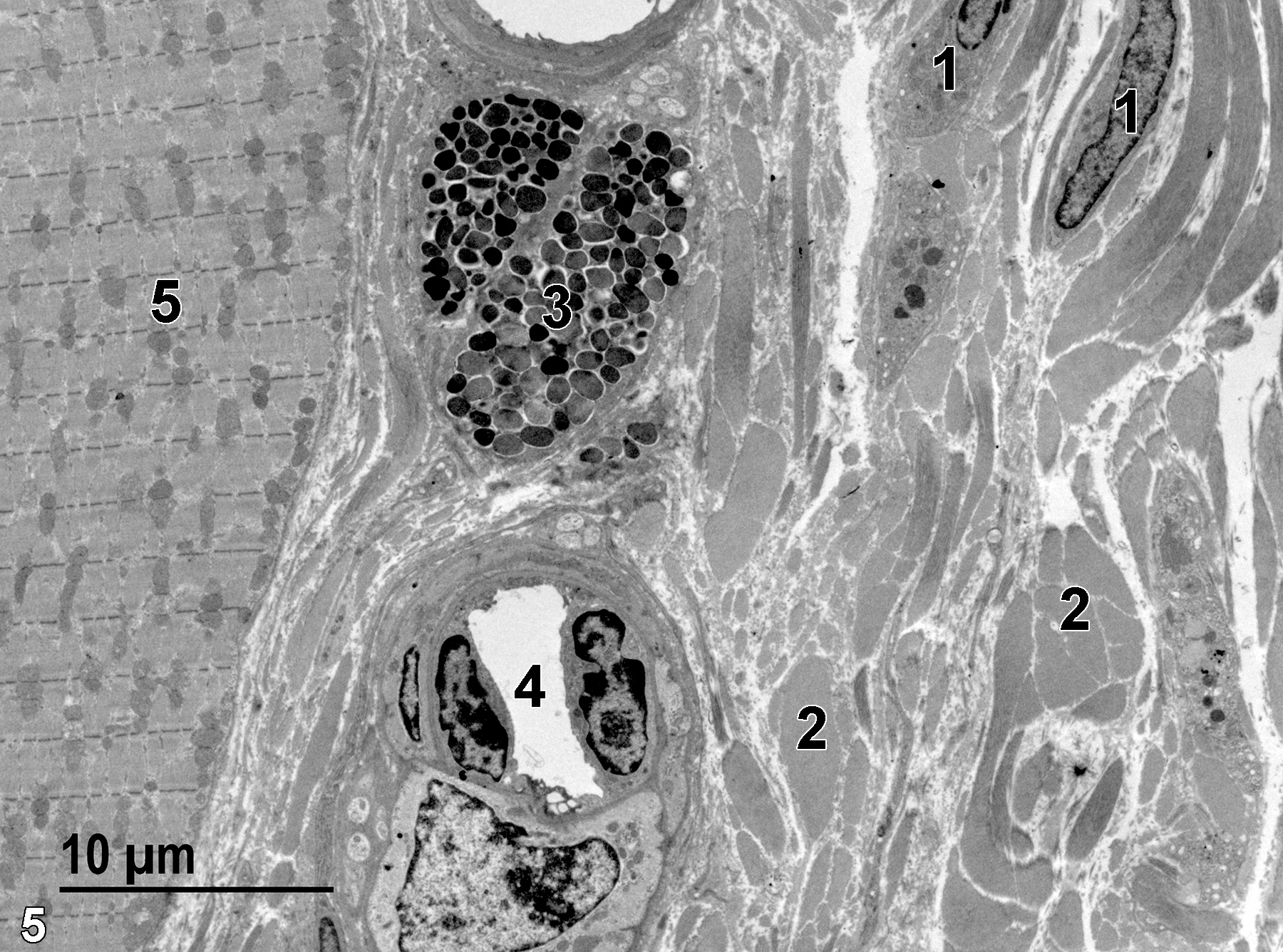
Figure 5. A portion of the lamina propria with fibroblasts (1), large bundles of collagen fibers (2), a mast cell (3), and a capillary (4). A single striated muscle cell (5) is shown. 2900x.
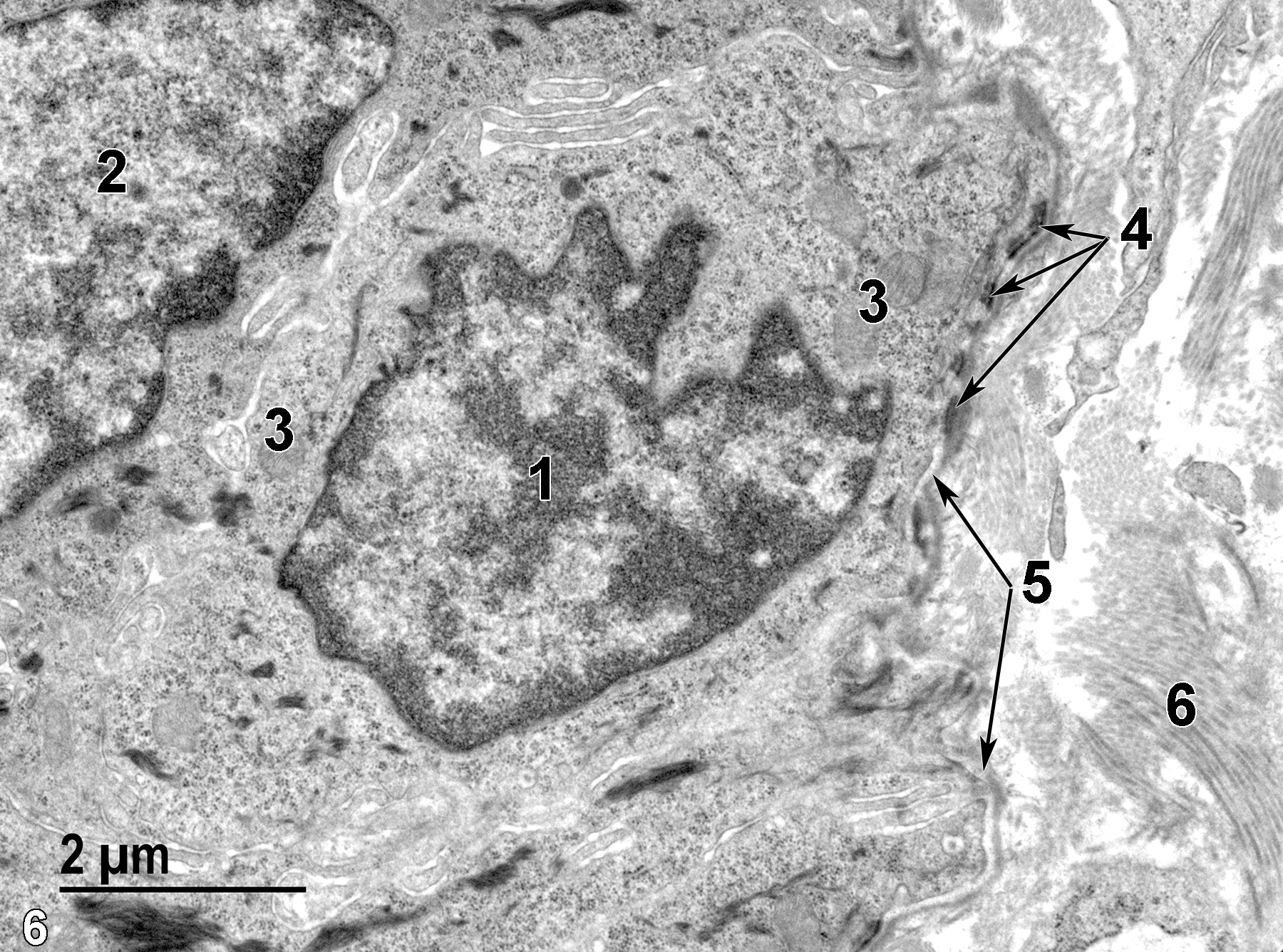
Figure 6. A high magnification view of a basal cell of the epithelial cell layer. The nucleus of the basal cell (1) is more rounded than that of the elongated nucleus of the stratified epithelial cell (2). The basal cell has several mitochondria (3), as well as numerous hemidesmosomes (4, arrows) along the basal lamina (5, arrows) that separates the basal cell layer from the underlying collagen (6) of the lamina propria. 13000x.
| Dellmann HD, Eurell J, eds. 1998. Textbook of Veterinary Histology. 5th ed. Philadelphia: Lippincott Williams & Wilkins. |
| Rhodin JAG. 1974. Histology: A Text and Atlas. New York: Oxford University Press. |
| Ross MH, Kaye GI, Pawlina W. 2003. Histology: A Text and Atlas. 4th ed. Philadelphia: Lippincott Williams & Wilkins. |
| Weiss L, ed. 1988. Cell and Tissue Biology: A Textbook of Histology. 6th ed. Baltimore: Urban & Schwarzenberg. |
All Images


