

Endocrine System
Pituitary Gland
Narrative
The pituitary gland (hypophysis) is an endocrine gland located below the ventral surface of the forebrain. The secretory portions consist of an anterior lobe, the adenohypophysis, and the posterior lobe, or neurohypophysis. The adenohypophysis consists of the pars distalis (represented in the following images), pars tuberalis, and pars intermedia. Rathke’s cleft separates the pars distalis from the pars intermedia. The neurohypophysis consists of the pars nervosa and infundibulum (Remick and Brown 2018).
The adenohypophysis produces hormones (detailed in the following) that influence the adrenal cortex, testes, ovary, thyroid, and mammary glands, as well as general growth. The neurohypophysis secretes a hormone that stimulates the contraction of the uterus (oxytocin) and another hormone that is antidiuretic (antidiuretic hormone) (Cross and Mercer 1993).
The pituitary gland sample shown in the figures came from a perfusion-fixed rat, which may account for the expanded capillaries.
Adenohypophysis
The adenohypophysis (pars distalis) consists of loose cords of secretory cells surrounded by sinusoids (capillaries). The cords of cells have basal laminae and collagen fibrils around them. There are numerous intercellular spaces and capillaries within the tissue.
Acidophilic cells make up approximately 40% of the cells within the adenohypophysis. Two types of cells are acidophilic - somatotrophs and mammotrophs. Somatotrophs produce growth hormone, have large round nuclei, round or oval mitochondria, well-developed rough endoplasmic reticulum and Golgi, free ribosomes, and many secretory granules of similar size (300-350 nanometers diameter) with tight membranes around their electron-dense cores. Mammotrophs produce prolactin and are found mostly in the posterolateral part of the adenohypophysis. They contain few small, round mitochondria, little rough endoplasmic reticulum, and sparse secretory granules that are elliptical in shape and range from 600-900 nanometers in diameter.
Basophilic cells account for approximately 10% of the adenohypophysis cells. Two types of secretory cells have basophilia: gonadotrophs and thyrotrophs. Gonadotrophs are the largest cells found in the adenohypophysis and have numerous secretory granules that vary in size from 100-300 nanometers and may have nuclei with infoldings of the nuclear envelope. They produce follicle-stimulating hormone and luteinizing hormone. Thyrotrophs produce thyrotrophic hormone. They are large cells with somewhat stellate shapes, large nuclei, large and abundant mitochondria, sparse rough endoplasmic reticulum, and small secretory granules with tight membranes around a moderately electron-dense core. Some of the granules have an electron-lucent halo around them. The granules are small (100-160 nanometers in diameter).
Chromophobe cells make up the remaining 50% of the adenohypophysis secretory cells. They are small cells with sparse cytoplasm and only a few secretory granules. Corticotroph cells secrete adrenocorticotrophic hormone and are stellate or irregular in shape, with long cytoplasmic processes that end near sinusoids. They have electron-lucent cytoplasm, few small mitochondria, well-developed Golgi and rough endoplasmic reticulum, and sparse granules (200 nanometers in diameter) arranged in a single layer near the plasma membrane. Precursor cells are stem cells that are believed to differentiate into the other cell types in the pituitary gland. These cells are small, with little cytoplasm and few small secretory granules.

Figure 1. A semithin section (0.5 micrometer thick) of a toluidine blue O-stained area of the adenohypophysis (anterior lobe of the pituitary gland). There is a thin capsule of connective tissue (1, arrow) surrounding loose cords of endocrine secretory cells (4), capillaries (2), and intercordal spaces (3, short arrows). 25x.
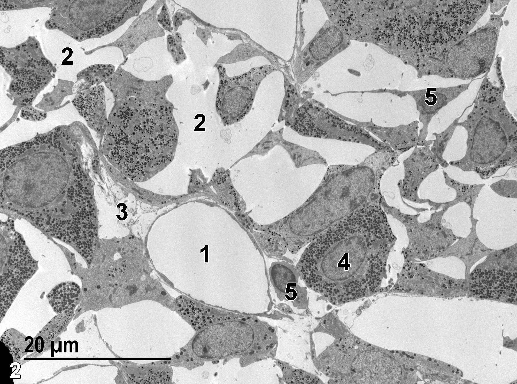
Figure 2. A low-power electron micrograph of a capillary lined with a thin endothelium (1), intercellular spaces (2), an intercordal space (3) with connective tissue elements, a somatotroph cell with a single nucleus (4), and numerous secretory granules of similar sizes. Two precursor cells (5) with scant cytoplasm and rare secretory granules are present. 1900x.
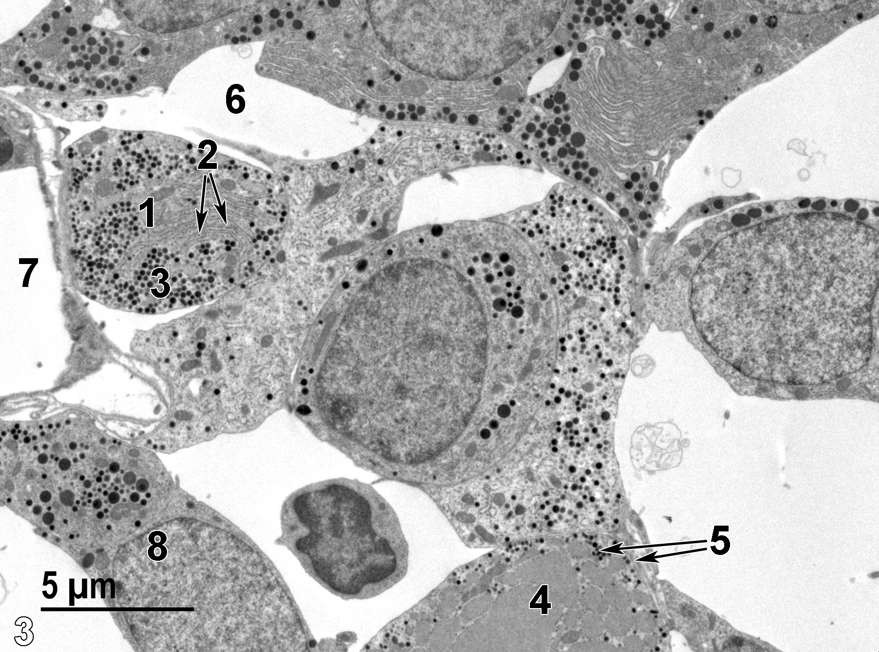
Figure 3. A higher magnification view of the adenohypophysis. A probable somatotroph cell (1) is present with numerous fairly uniform-sized secretory granules (3) and a well-developed field of stacked rough endoplasmic reticulum (2, short arrows). A thyrotroph cell with many large round mitochondria (4) and small secretory granules is present (5, long arrows). An intercellular space (6) is shown, as well as a capillary (7) lined with a thin endothelium. Finally, a cell with highly variable secretory granule sizes is consistent with a gonadotroph cell (8). 4800x.
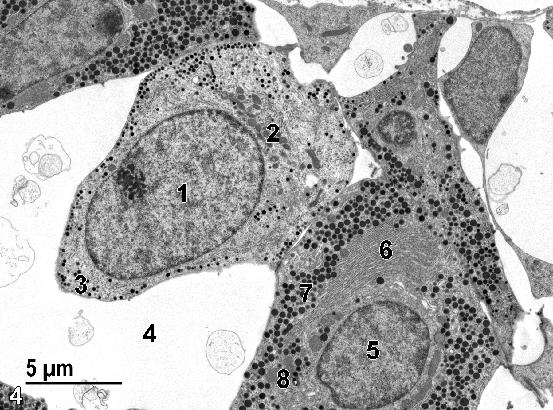
Figure 4. Another higher magnification image showing a corticotrophic cell (1) with a cluster of small mitochondria (2) and a thin band of small secretory granules (3) around the periphery of the cell. An intercellular space (4) with some membranous debris is shown. A somatotroph cell is present with a round nucleus (5), stacks of rough endoplasmic reticulum (6), numerous secretory granules (7), and sparse mitochondria (8). 4800x.
| Cross PC, Mercer KL. 1993. Cell and Tissue Ultrastructure: A Functional Perspective. New York: W.H. Freeman and Company. |
| Dellmann HD, Eurell J, eds. 1998. Textbook of Veterinary Histology. 5th ed. Philadelphia: Lippincott Williams & Wilkins. |
| Remick AK, Brown DL. 2018. Chapter 31: Pituitary gland. In Boorman’s Pathology of the Rat (Suttie AW, ed). 2nd ed. London: Academic Press; 631–648. |
| Rhodin JAG. 1974. Histology: A Text and Atlas. New York: Oxford University Press. |
| Weiss L, ed. 1988. Cell and Tissue Biology: A Textbook of Histology. 6th ed. Baltimore: Urban & Schwarzenberg. |
All Images


