

Reproductive System, Male
Epididymis
Narrative
The epididymis consists of a single convoluted duct (ductus epididymis). The ductus epididymis, in combination with connective tissue and muscle, forms four regions of the epididymis: the initial segment; the head (caput) at the upper pole of the testis; the body (corpus) following the medial posterior of the testis; and the tail (cauda) near the lower pole of the testis where it continues as the ductus deferens. The epididymal duct is lined with a pseudostratified columnar epithelium of multiple cell types, including principal cells, basal cells, clear cells, apical cells, and halo cells. These cell types vary in number and appearance depending on the segment of the epididymis (De Grava Kempinas and Klinefelter 2015). The columnar principal cells have stereocilia (long microvilli) at their apical aspects that extend into the duct lumen and numerous pinocytotic vesicles at the cell surface, large amounts of smooth endoplasmic reticulum, large vacuoles, Golgi bodies, mitochondria, and sparse rough endoplasmic reticulum. Principal cells in the corpus have abundant supranuclear lipid. The basal aspect of the columnar epithelial cells (principal cells) has a single, mostly rounded nucleus with fairly dispersed heterochromatin, large amounts of rough endoplasmic reticulum, and lysosomes. The basal epithelial cells have somewhat flattened nuclei that are smaller than those of the columnar cells and have more marginated heterochromatin than that seen in the columnar cells. The height of the epithelium is decreased in the cauda compared with the caput and corpus. Clear cells are prevalent in the corpus. The epithelial cells are separated from loose connective tissue and smooth muscle cells surrounding the ductus epididymis by a thin basal lamina. The smooth muscle cells beneath the basal lamina of the epithelium have flattened elongated nuclei, prominent pinocytotic vesicles along the plasma membrane, and cytoplasm filled with actin and myosin filaments. Collagen fibers are in clusters around the smooth muscle cells and then in larger quantities at the outside of the muscle cell layers.
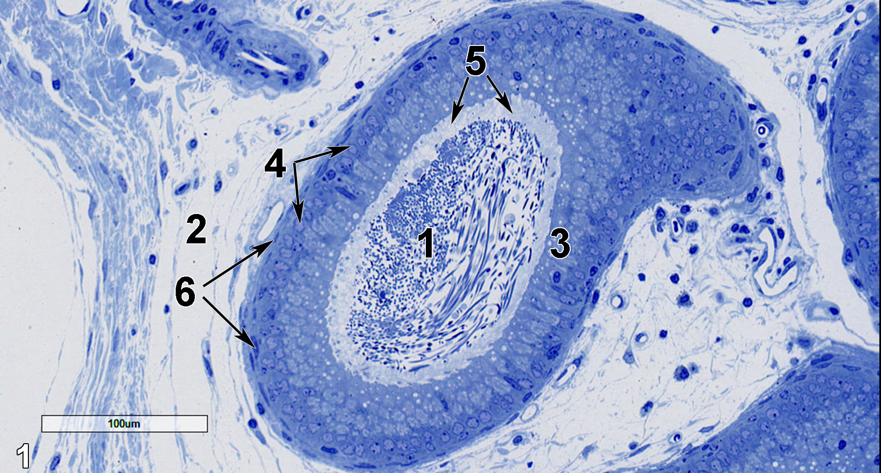
Figure 1. A semithin section (0.5 micrometer thick) of a toluidine blue O-stained portion of the ductus epididymis. Numerous spermatozoa (1) are seen in the duct lumen. The duct is surrounded by a matrix of loose connective tissue (2). A row of columnar epithelial cells (3) has lightly stained round nuclei at their basal aspect (4, arrows). The smaller and more darkly stained nuclei of the basal epithelial cells are difficult to discern in this image. Stereocilia (5, arrows) form a lightly stained layer above the columnar epithelial cells, extending into the duct lumen. The smooth muscle cells surrounding the duct have elongated and flattened nuclei (6, arrows) surrounding the duct have elongated and flattened nuclei. 20x.
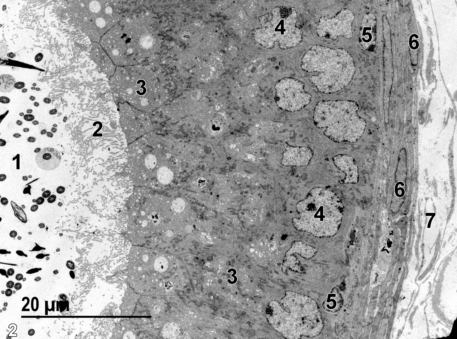
Figure 2. A low magnification electron micrograph of the duct structure showing the lumen with spermatozoa (1), the stereocilia (2) at the apical surface of the columnar epithelial cells, the apical aspect of the columnar cells with numerous vesicles and vacuoles (3), and the basally located nuclei (4) of the columnar cells with fairly dispersed heterochromatin. The basal epithelial cells have smaller nuclei (5) with more visible marginated heterochromatin. The nuclei of the smooth muscle cells (6) are elongated and flattened. Outside of the smooth muscle cell layers are large bundles of collagen fibers (7). 1900x.
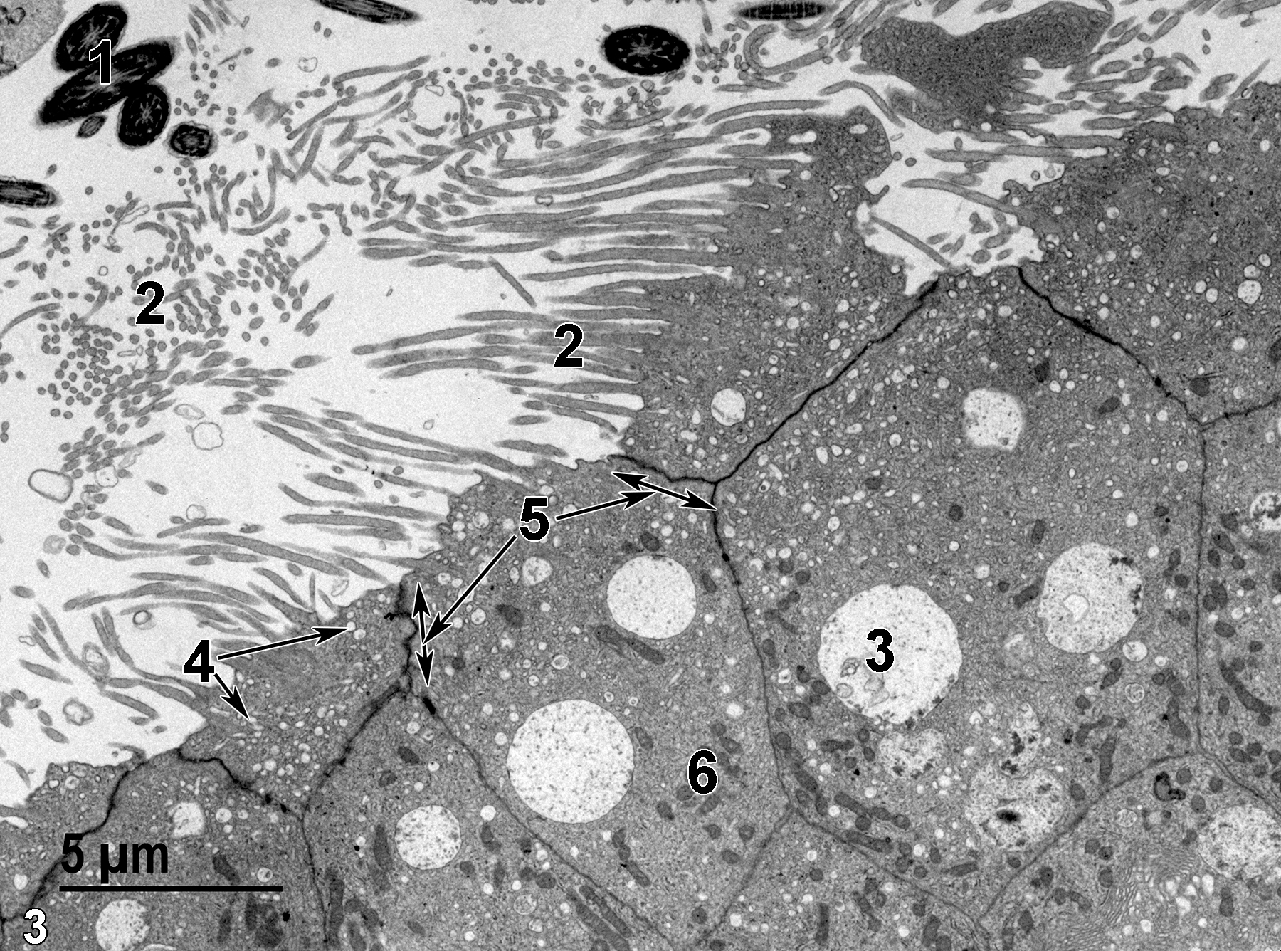
Figure 3. A higher magnification view of the apex of columnar epithelial cells. The lumen of the duct contains several midpieces of spermatozoa (1) and stereocilia (2). The epithelial cells have large vacuoles with small amounts of membranous debris (3), numerous pinocytotic vesicles (4, arrows), and prominent junctional complexes binding the adjacent cell surfaces together (5, arrows and double-headed arrows). Mitochondria (6) are fairly numerous. 4800x.
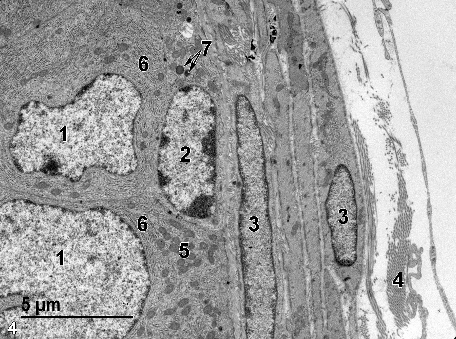
Figure 4. The basal aspect of the epithelium with two nuclei of columnar epithelial cells (1) and one nucleus of a basal epithelial cell (2). Note the flattened aspect of the basal cell nucleus and the evident marginated heterochromatin. The underlying smooth muscle cell nuclei (3) are flattened and elongated. Collagen fibers (4) are present between the smooth muscle cells and outside the duct, forming the loose connective tissue between duct segments. The columnar epithelial cells contain more mitochondria (5) than those found in the basal epithelial cells. Large amounts of rough endoplasmic reticulum (6) are found in the basal aspect of the columnar epithelial cells. Lysosomes are evident in the basal epithelial cell (7, arrows). 6800x.
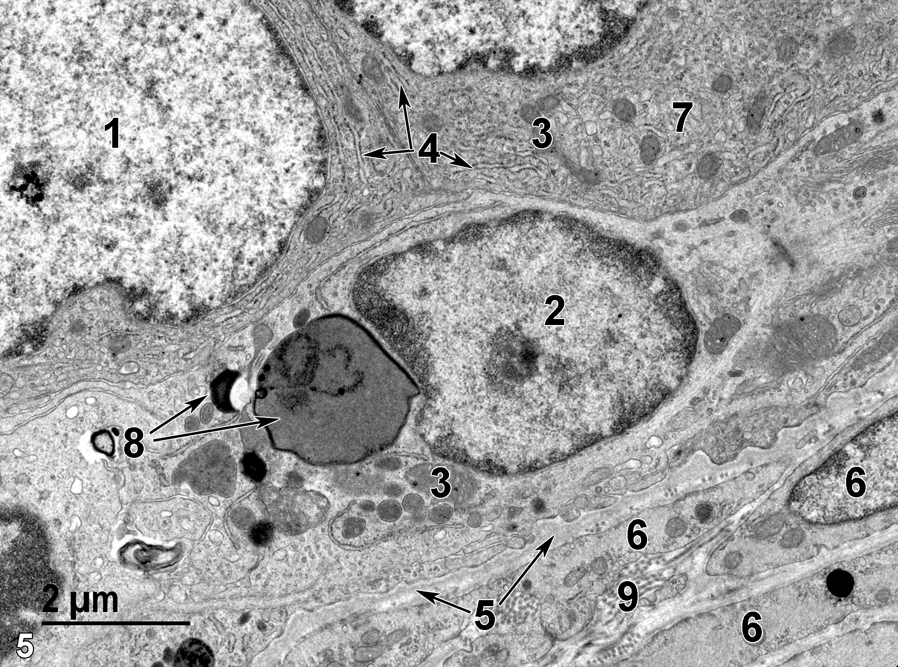
Figure 5. A higher magnification view of the base of the duct. Two columnar cells are shown, one with a large nucleus (1) with dispersed heterochromatin. A single basal epithelial cell has a smaller nucleus with marginated chromatin (2), mitochondria (3), and several lysosomes showing variable size and electron density (8, arrows). The columnar cells contain prominent rough endoplasmic reticulum (4, arrows), mitochondria (3), and some areas of smooth endoplasmic reticulum (7). The basal epithelial cell layer is separated from the smooth muscle cells (6) by a thin basal lamina (5, arrows). Collagen fibers (9) are found between the smooth muscle cells. 11000x.
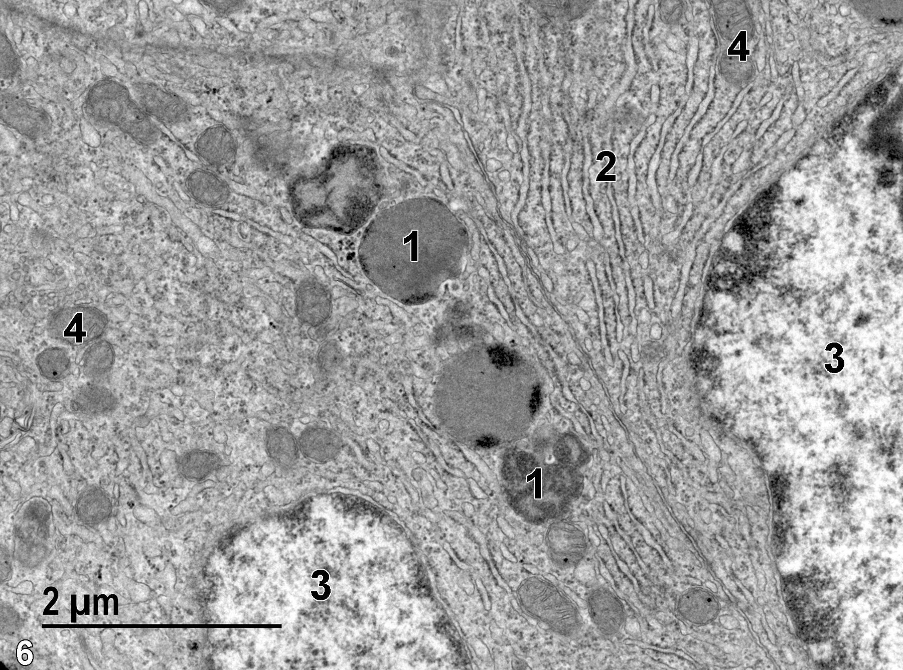
Figure 6. A higher magnification view of the base of two columnar epithelial cells showing lysosomes (1), prominent rough endoplasmic reticulum (2), large nuclei with sparse heterochromatin (3), and mitochondria (4). 18500x.
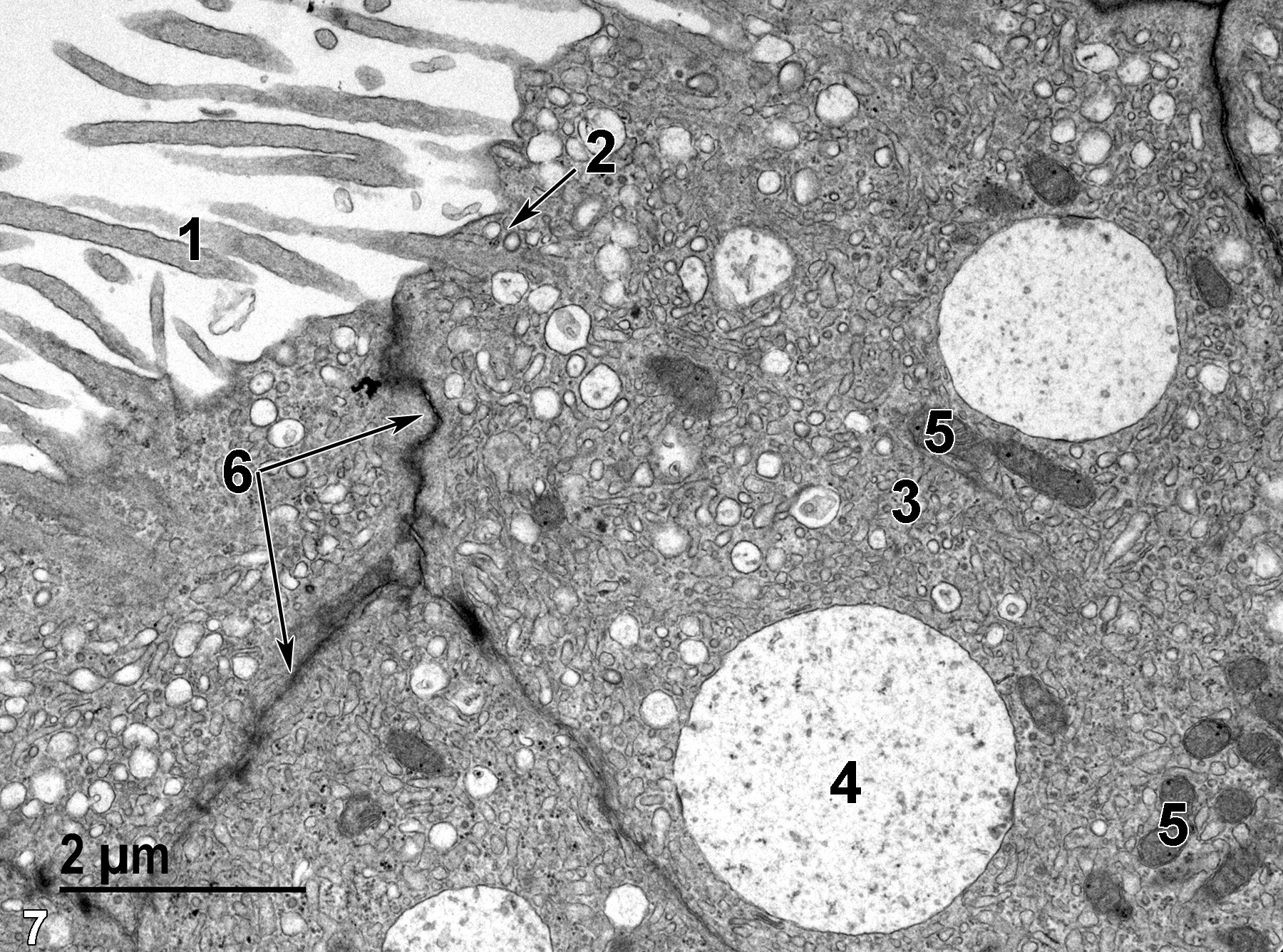
Figure 7. The apical area of columnar epithelial cells with stereocilia (1), pinocytotic vesicles (2, arrow), smooth endoplasmic reticulum vesicles (3), large vacuoles (4), mitochondria (5), and prominent junctional complexes (6, arrows). 13000x.
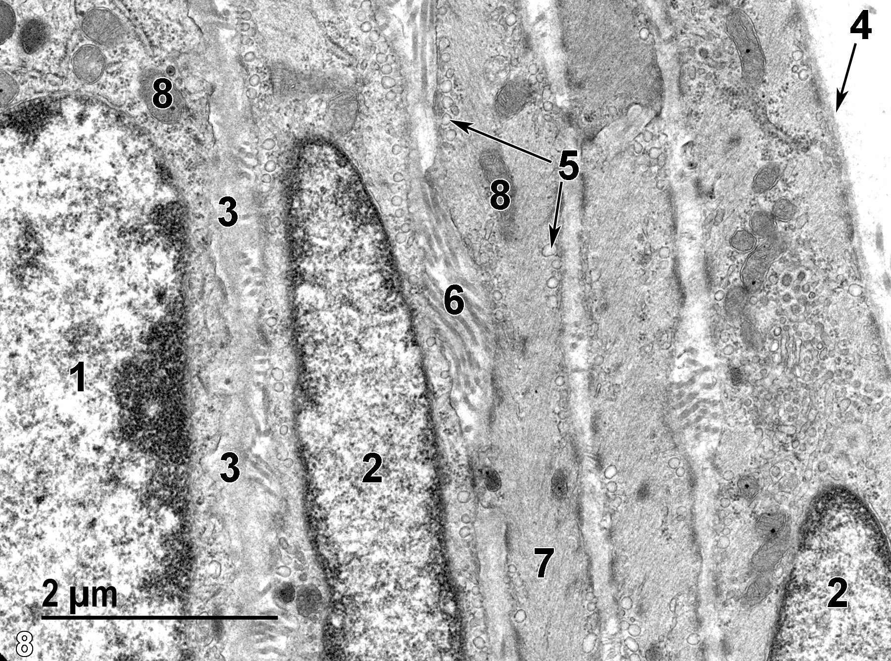
Figure 8. A high magnification view of the basal aspect of a duct. The basal epithelial cell has a few mitochondria (8) and a flattened nucleus (1) that is distinct from the flattened and elongated nuclei (2) characteristic of smooth muscle cells. A thin basal lamina (3) separates the epithelial cells from the smooth muscle cells. Each smooth muscle cell has its own basal lamina (4, arrow), numerous pinocytotic vesicles along the plasma membrane (5, arrows), cytoplasm filled with myosin and actin filaments (7), and mitochondria (8). Collagen (6) is found in clusters between the smooth muscle cells. 18500x.
| De Grava Kempinas W, Klinefelter GR. 2015. Interpreting histopathology in the epididymis. Spermatogenesis 8;4(2):e979114. |
| Dellmann HD, Eurell J, eds. 1998. Textbook of Veterinary Histology. 5th ed. Philadelphia: Lippincott Williams & Wilkins. |
| Rhodin JAG. 1974. Histology: A Text and Atlas. New York: Oxford University Press. |
| Ross MH, Kaye GI, Pawlina W. 2003. Histology: A Text and Atlas. 4th ed. Philadelphia: Lippincott Williams & Wilkins. |
| Weiss L, ed. 1988. Cell and Tissue Biology: A Textbook of Histology. 6th ed. Baltimore: Urban & Schwarzenberg. |
All Images


