

Musculoskeletal System
Tendon
Narrative
Tendons are composed of dense regular connective tissue that attaches muscles to bone. The tendon is bound by a thin band of collagenous tissue (epitendineum) that is continuous with the endotendineum, which is made up of collagen fibers, fibroblasts, and elastic fibers. The endotendineum delimits fascicles of collagen. Longitudinal sections of collagen fibers reveal the repeating periodicity of 68 nm bands evident with electron microscopy.
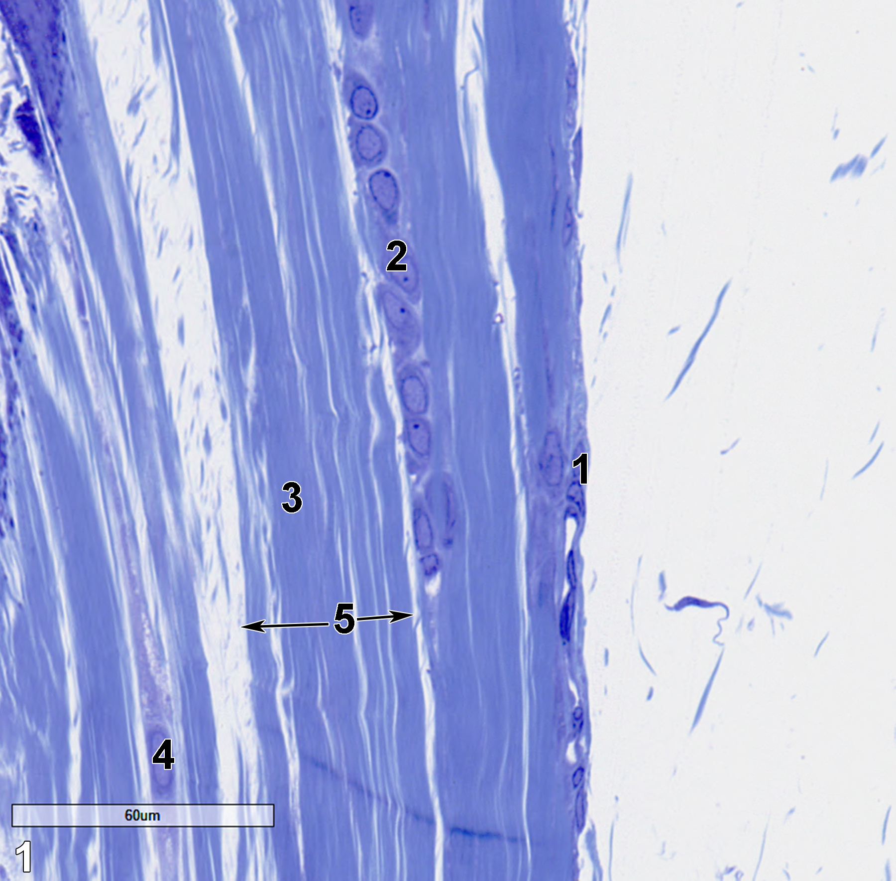
Figure 1. A semithin section (0.5 micrometer thick) of a toluidine blue O-stained view of a tendon. The thin capsule, or epitendineum (1), consists of collagenous tissue with fibroblasts. The endotendineum, (2) which is composed of collagen (3) and elastic fibers and fibroblasts (4), define the surface fascicles of a tendon (5, double arrows). 40x.
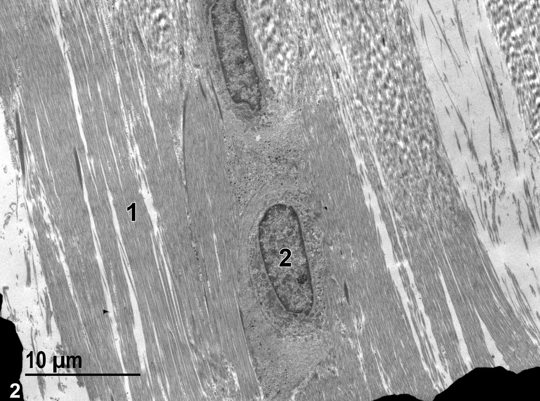
Figure 2. A low magnification electron micrograph of a longitudinal section of a tendon. Collagen bundles (1) make up a fascicle that has fibroblasts along its surface. A single fibroblast nucleus (2) is shown. 2900x.
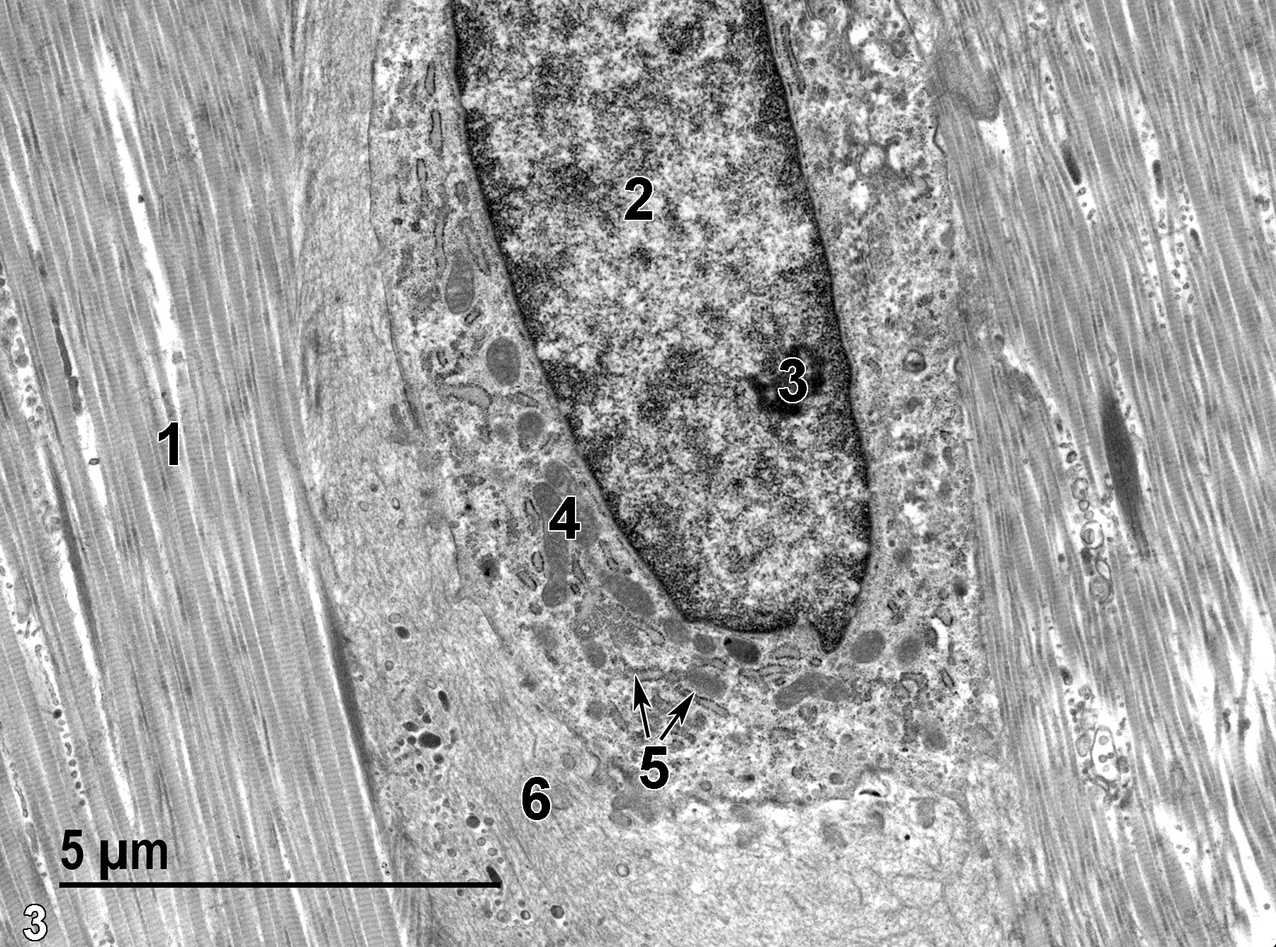
Figure 3. A higher magnification view of one of the fibroblasts in Figure 2. The fibroblast nucleus (2) contains a single nucleolus (3). The fibroblast has numerous mitochondria (4) and a prominent rough endoplasmic reticulum (5, double arrows). The fibroblast is surrounded by elastic fibers (6) and compact bundles of collagen fibers (1). 9300x.
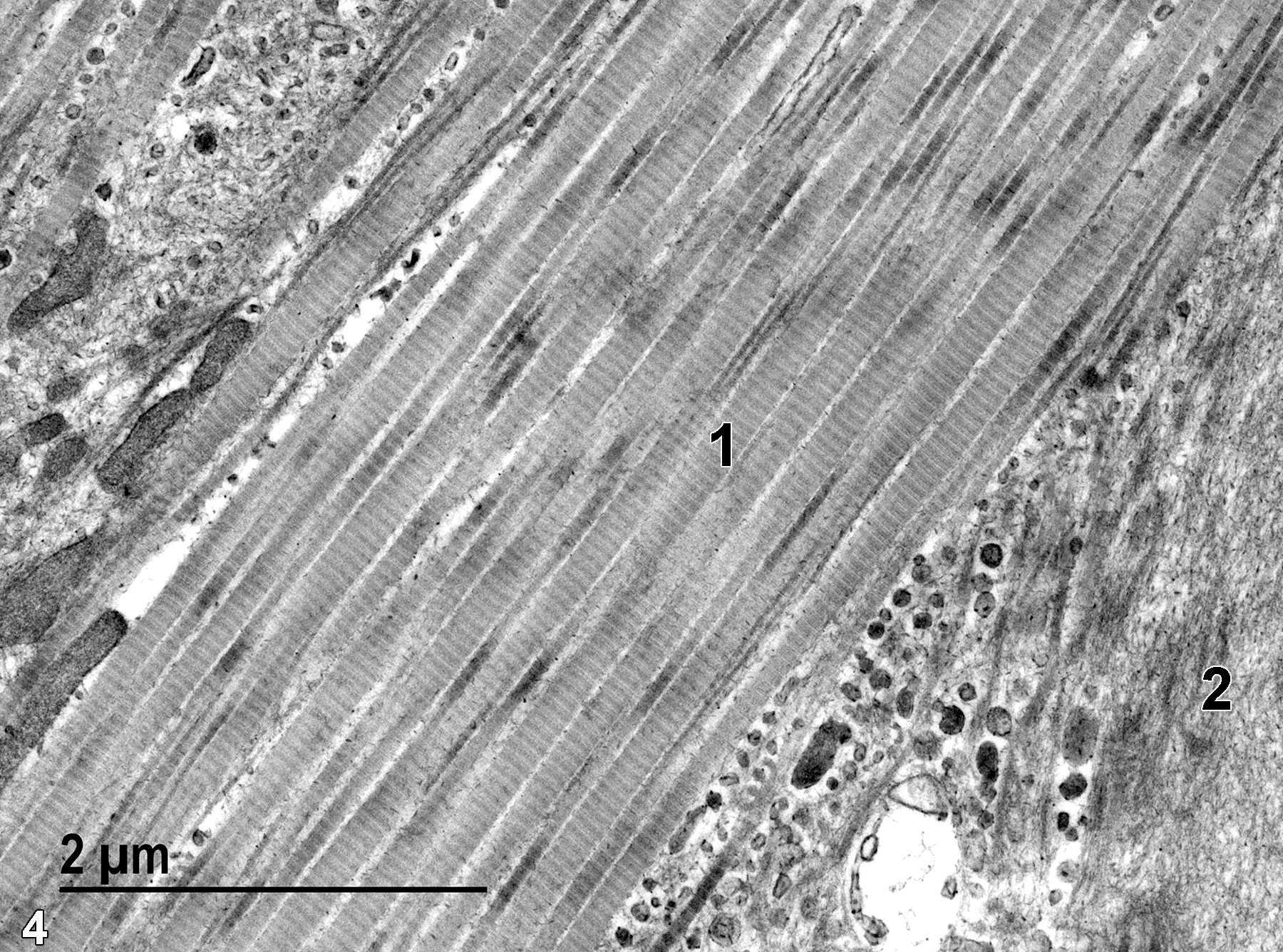
Figure 4. An even higher magnification image showing the parallel collagen fibers (1) with their characteristic 68-nm repeating banding pattern. An accumulation of elastic fibers (2) is shown. 23000x.
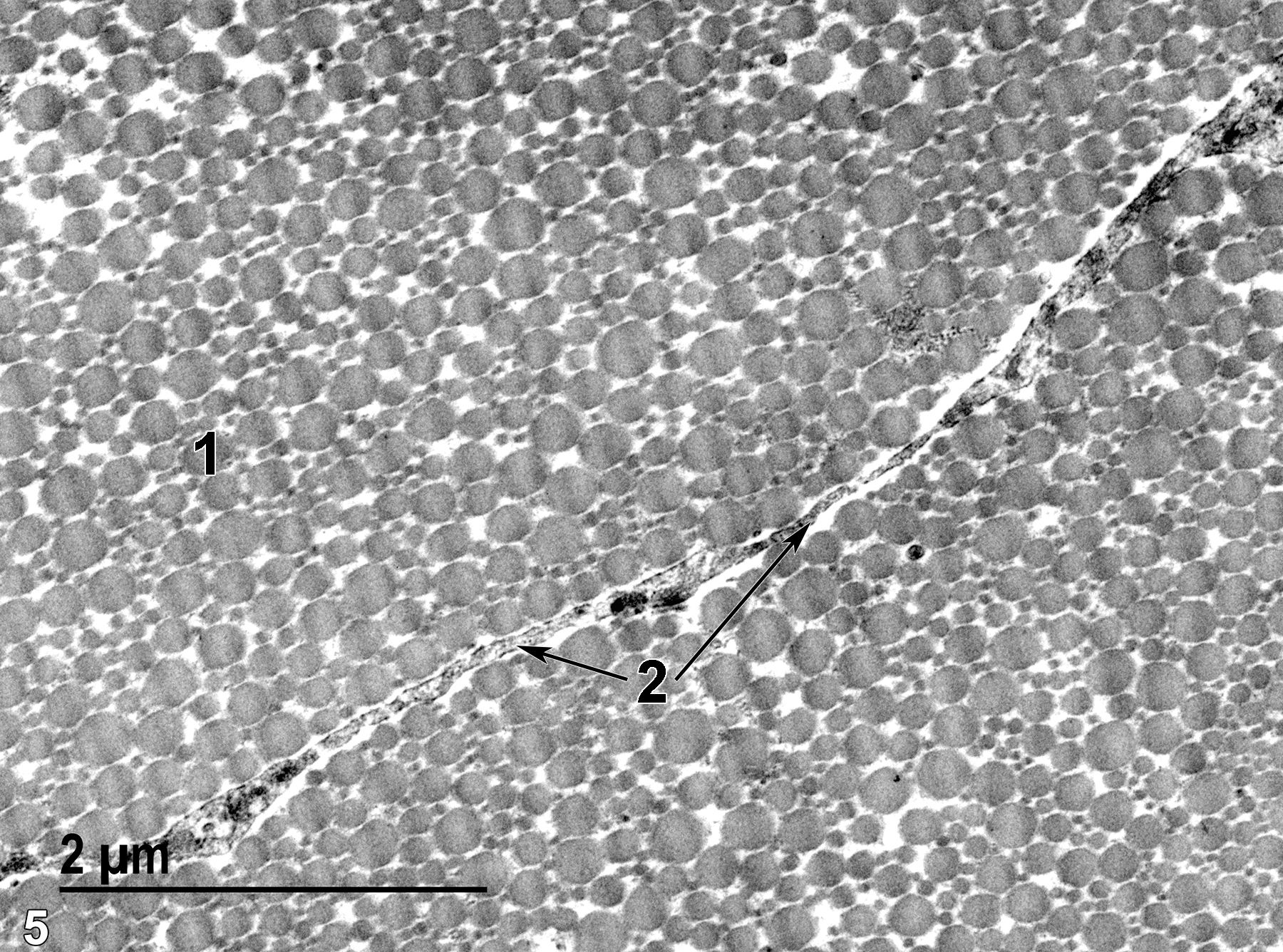
Figure 5. A high magnification image of a cross section of a tendon showing collagen fibers (1) that vary significantly in diameter. In some tendons, the collagen fiber diameters are less varied, and in these tendons, the amount of variability shown in this image might indicate tendon pathology. Cytoplasmic processes (2, double arrows) are located between adjacent fascicles of the tendon. 23000x.
| Eurell JA, Frappier BL, eds. 2006. Dellmann’s Textbook of Veterinary Histology. 6th ed. Ames, IA: Blackwell Publishing. |
| Rhodin JAG. 1974. Histology: A Text and Atlas. New York: Oxford University Press. |
| Ross MH, Kaye GI, Pawlina W. 2003. Histology: A Text and Atlas. 4th ed. Philadelphia: Lippincott Williams & Wilkins. |
| Weiss L, ed. 1988. Cell and Tissue Biology: A Textbook of Histology. 6th ed. Baltimore: Urban & Schwarzenberg. |
All Images


