

Integumentary System
Skin, Hair Follicle - Dilatation and Cyst
Narrative
Whenever present, cysts should be diagnosed and documented as present but should not be graded. Cysts in the skin should be diagnosed as “Skin – Cyst”; no modifier is necessary. Findings that are secondary to a large or ruptured cyst, such as inflammation or fibrosis, need not be diagnosed but should be described in the pathology narrative.
Elwell MR, Stedman MA, Kovatch RM. 1990. Skin and subcutis. In: Pathology of the Fischer Rat: Reference and Atlas (Boorman GA, Eustis SL, Elwell MR, Montgomery CA, MacKenzie WF, eds). Academic Press, San Diego, 261-277.
Abstract: https://www.ncbi.nlm.nih.gov/nlmcatalog/9002563Klein-Szanto AJP, Conti CJ. 2002. Skin and oral mucosa. In: Handbook of Toxicologic Pathology, 2nd ed (Haschek WM, Rousseaux CG, Wallig MA, eds). Academic Press, San Diego, 2:85-116.
Abstract: http://www.sciencedirect.com/science/book/9780123302151Peckham JC, Heider K. 1999. Skin and subcutis. In: Pathology of the Mouse: Reference and Atlas (Maronpot RR, Boorman GA, Gaul BW, eds). Cache River Press, Vienna, IL, 555-612.
Ramot Y, Nyska A, Lieuallen W, Maly A, Flake G, Kissling E, Brix A, Malarkey DE, Hooth MJ. 2009. Inflammatory and chloracne-like skin lesions in B6C3F1 mice exposed to 3,3',4,4'-tetrachloroazobenzene for 2 years. Toxicology 265:1-9.
Abstract: https://www.ncbi.nlm.nih.gov/pubmed/19737593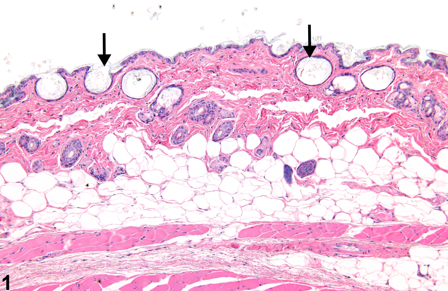
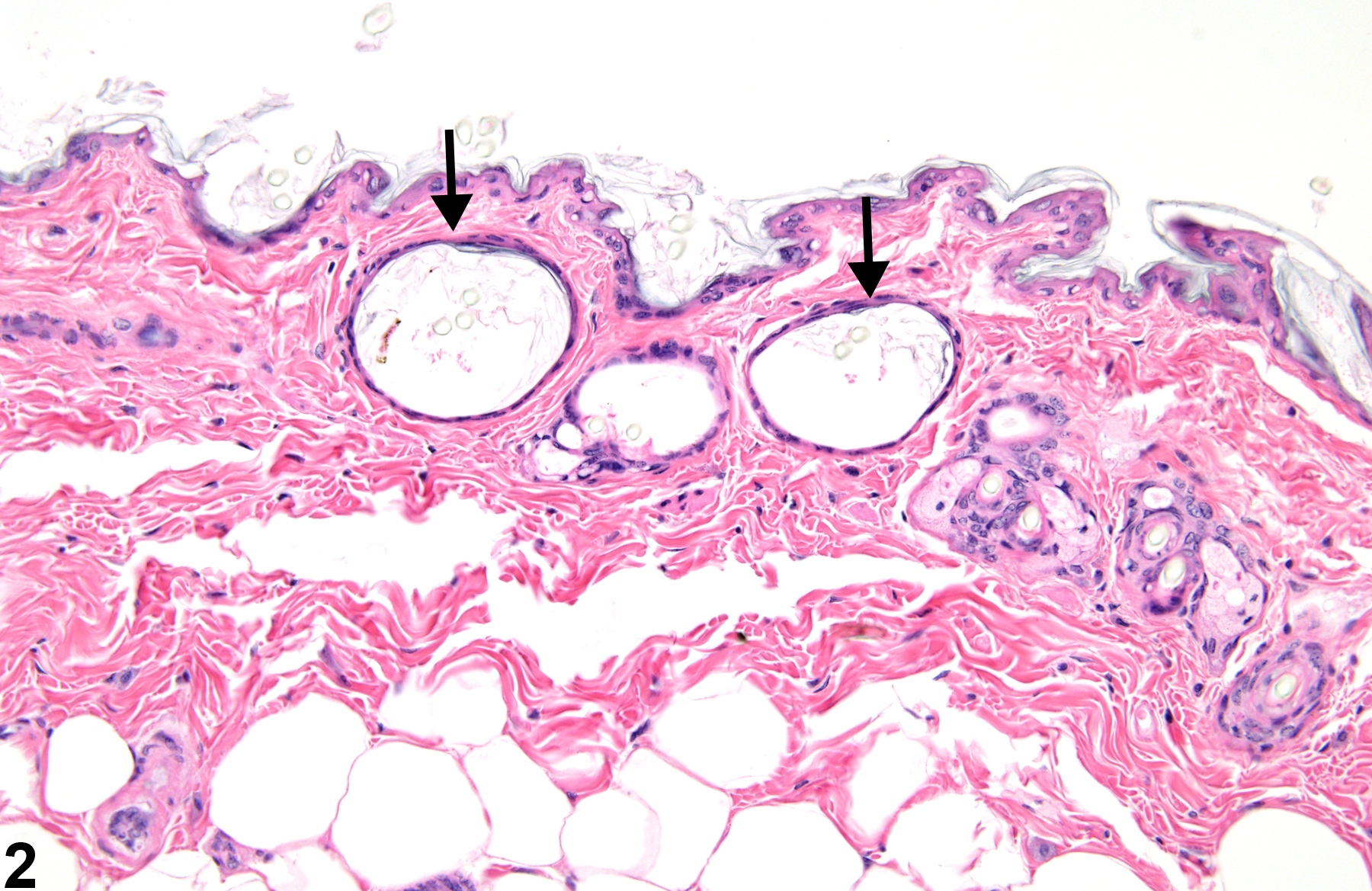
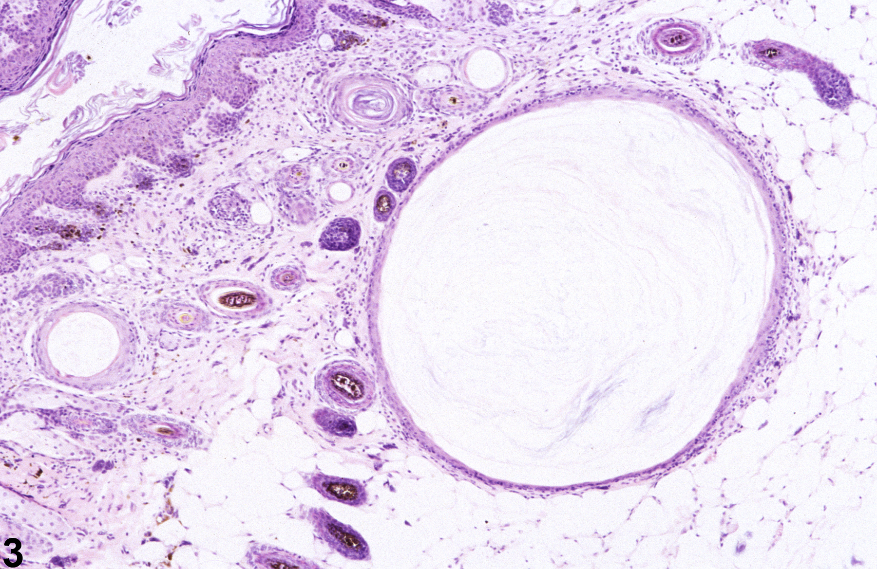
Hair follicle dilatation-ectatic hair follicles with intraluminal keratin (arrows) in a male B6C3F1 mouse from a chronic study.
All Images

Hair follicle dilatation-ectatic hair follicles with intraluminal keratin (arrows) in a male B6C3F1 mouse from a chronic study.

Hair follicle dilatation-ectatic hair follicles with intraluminal keratin (arrows) in a male B6C3F1 mouse from a chronic study.

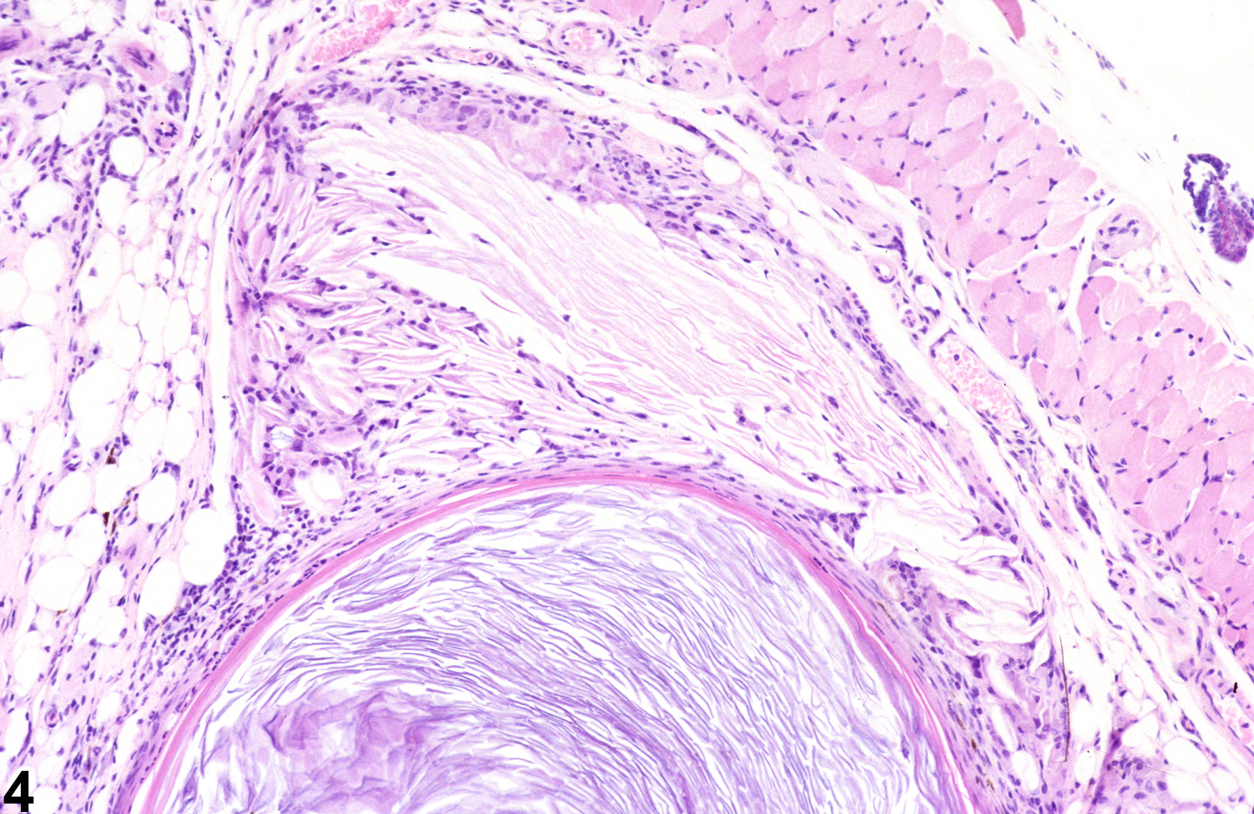
Epithelial cyst-flattened squamous epithelium surrounding a core of inspissated keratin in a male B6C3F1 mouse from a chronic study.

Epithelial cyst-ruptured cyst lining and resulting granulomatous inflammation in a female B6C3F1 mouse from a chronic study.