

Nervous System
Spinal Cord, Dorsal Root Ganglion, Neuron - Vacuolation
Narrative
In view of the known occurrence of neuronal vacuolation as an occasional incidental finding, it is important to carefully examine control and treatment groups for its presence in comparable regions. The existence of associated changes such as neuronal chromatolysis or adjacent responses by glia or satellite cells may be helpful in attributing vacuolization as a lesion.
Groves MJ, Scaravilli F. 2005. Pathology of peripheral neuron cell bodies. In: Peripheral Neuropathy, 4th ed (Dyck PJ, Thomas PK, eds). Elsevier, Philadelphia, PA, 683-732.
Rogers-Cotrone T, Burgess MP, Hancock SH, Hinckley J, Lowe K, Ehrich MF, Jortner BS. 2010. Vacuolation of sensory ganglion neuron cytoplasm in rats with long-term exposure to organophosphates. Toxicol Pathol 38:554-559
Abstract: http://www.ncbi.nlm.nih.gov/pubmed/20448080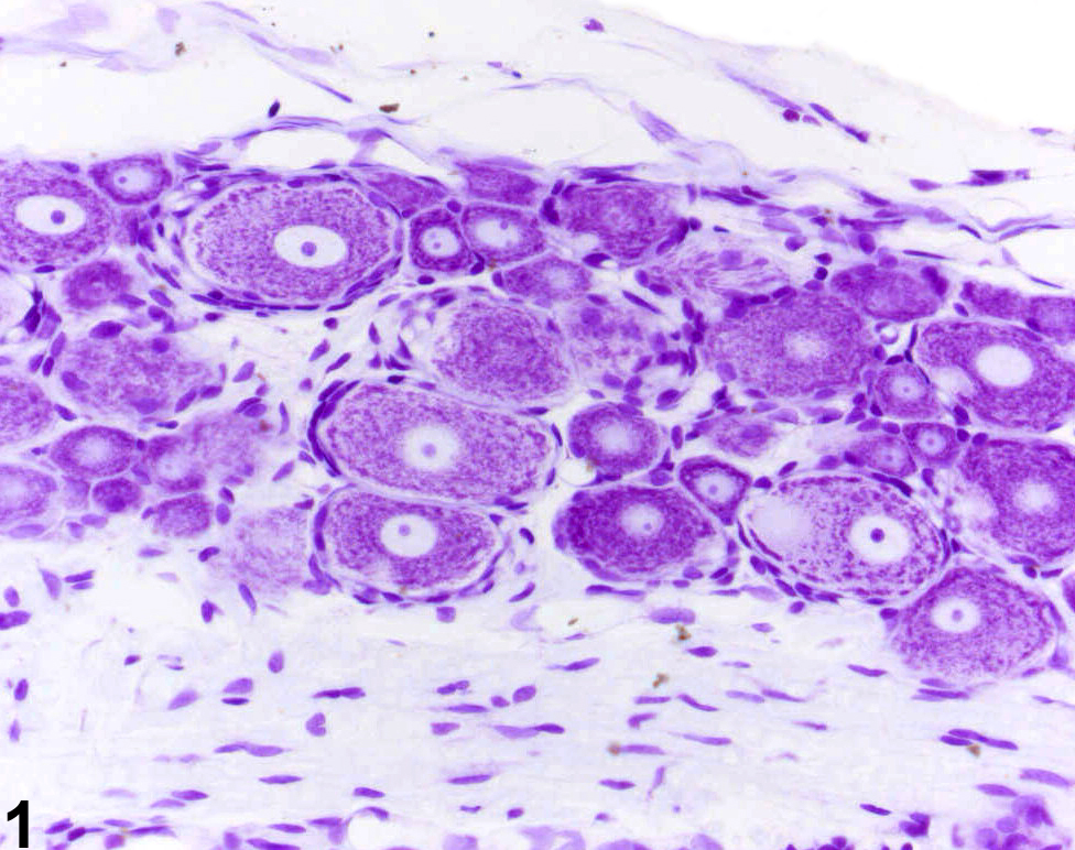
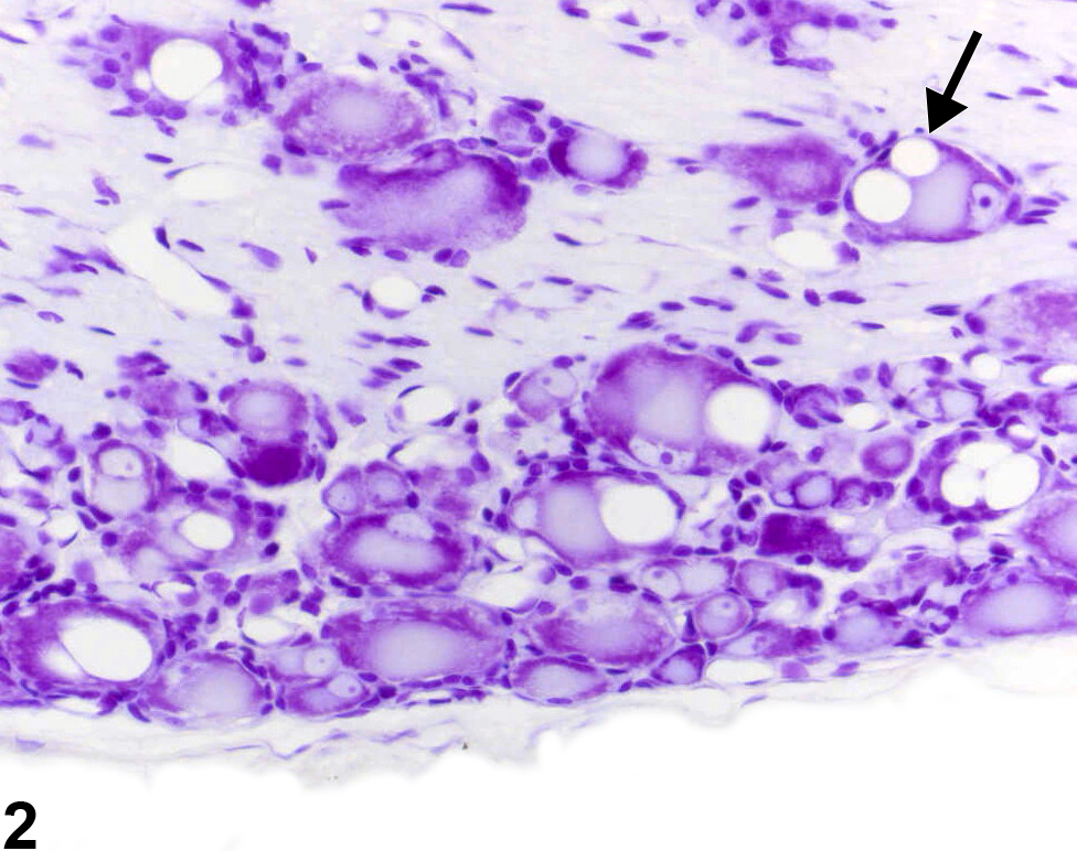
Normal appearance of rat dorsal root ganglionic neurons (cresyl violet). Image provided courtesy Dr. G. Krinke.
All Images

Normal appearance of rat dorsal root ganglionic neurons (cresyl violet). Image provided courtesy Dr. G. Krinke.

Genuine dorsal root ganglionic vacuoles, appearing as variably sized, but generally large, clear vacuoles in the perikaryon of the affected neurons, may be accompanied by chromatolysis (arrow). Image provided courtesy Dr. G. Krinke.