

Reproductive System, Male
Testis, Seminiferous Tubule - Giant Cells
Narrative
Hild SA, Reel JR, Dykstra MJ, Mann PC, Marshall GR. 2007. Acute adverse effects of the indenopyridine CDB-4022 on the ultrastructure of Sertoli cells, spermatocytes, and spermatids in rat testes: Comparison to the known Sertoli cell toxicant di-n-pentylphthalate (DPP). J Androl 28:621-629.
Abstract: http://www.ncbi.nlm.nih.gov/pubmed/17409460MacGregor GR, Russell LD, Van Beek MEAB, Hanten GR, Kovac MJ, Kozak CA, Meisrich ML, Overbeek PA. 1990. Symplastic spermatids (sys): A recessive insertional mutation in mice causing a defect in spermatogenesis. Proc Natl Acad Sci USA 87:5016-5020.
Abstract: http://www.ncbi.nlm.nih.gov/pubmed/2164218Russell LD, Hikim AP, Overbeek PA, MacGregor GR. 1991. Testis structure in the sys (symplastic spermatids) mouse. Am J Anat 192:169-182.
Abstract: http://www.ncbi.nlm.nih.gov/pubmed/1759682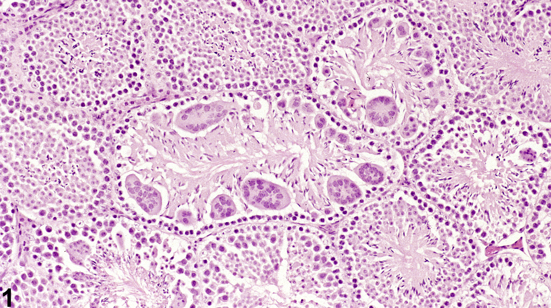
Testis, Seminiferous tubule - Giant cells in a male B6C3F1 mouse from a subchronic study. These cells are associated with germ cell degeneration.
All Images

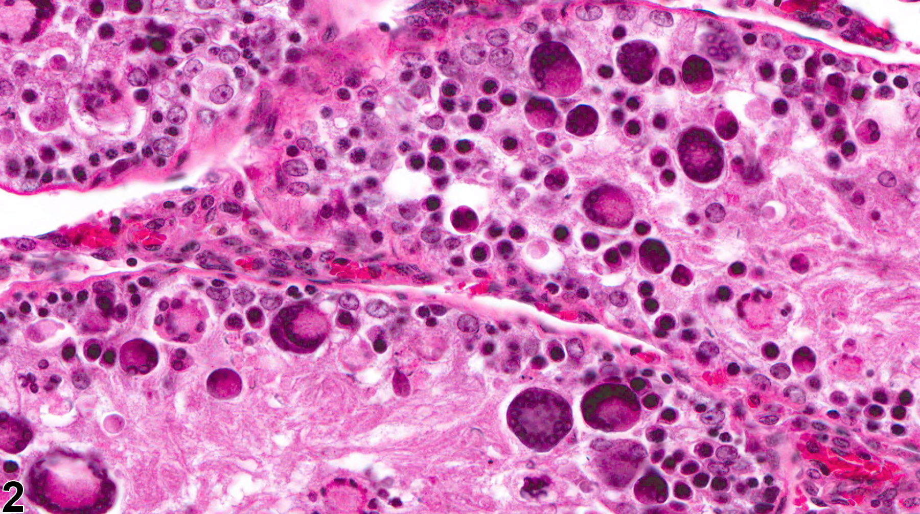
Testis, Seminiferous tubule - Giant cells in a male B6C3F1 mouse from a subchronic study. These cells are associated with germ cell degeneration.

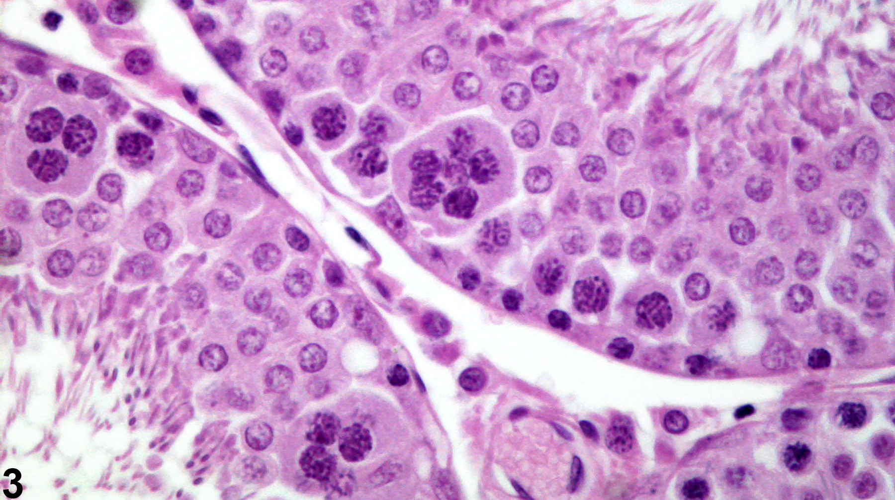
Testis, Seminiferous tubule - Giant cells in a male F344/N rat from a chronic study. These cells are associated with germ cell degeneration.
Testis, Seminiferous tubule - Giant cells in a male Harlan Sprague-Dawley rat. These cells represent spermatocytes that have coalesced into individual syncytial cells. (Photograph courtesy of Dr. D. Creasy.)
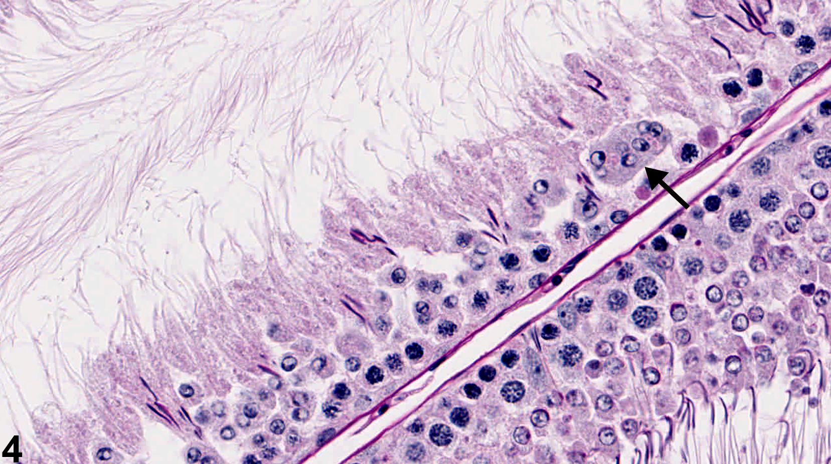
Testis, Seminiferous tubule - Giant cells in a male Harlan Sprague-Dawley rat. A multinucleated giant cell (arrow) formed by fusion of round spermatids. (Photograph courtesy of Dr. D. Creasy.)