

Alimentary System
Esophagus - Angiectasis
Narrative
Boorman GA, Everitt JI. 2006. Neoplastic diseases In: The Laboratory Rat, 2nd ed (Suckow MA, Weisbroth SH, Franklin CL, eds). Elsevier Academic Press, Burlington, MA, 479-511.
Abstract: http://www.sciencedirect.com/science/book/9780120749034Gad S. 2007. The mouse. In: Animal Models of Toxicology, 2nd ed (Gad S, ed). CRC Press, Boca Raton, Fl, 19-146.
Gross TL, Ihrke PJ, Walder EJ, Affolter VK. 2005. Vascular tumors. In: Veterinary Dermatopathology, 2nd ed. Blackwell, Oxford, UK, 735-758.
Mitsumori K. 1990. Blood and lymphatic vessels. In: Pathology of the Fischer Rat (Boorman GA, Montgomery CA, MacKenzie WF, eds). Academic Press, San Diego, CA, 473-484.
Abstract: https://www.ncbi.nlm.nih.gov/nlmcatalog/9002563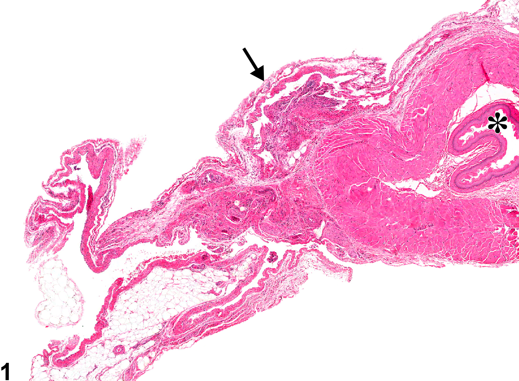
Esophagus - Angiectasis in a female F344/N rat from a chronic study. Dilated vascular spaces (arrow) are visible in the wall of the esophagus (asterisk).
All Images

Esophagus - Angiectasis in a female F344/N rat from a chronic study. Dilated vascular spaces (arrow) are visible in the wall of the esophagus (asterisk).
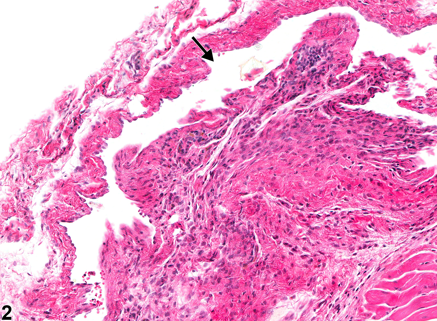
Esophagus - Angiectasis in a female F344/N rat from a chronic study (higher magnification of Figure 1). Dilated vascular spaces (arrow) are visible in the wall of the esophagus.
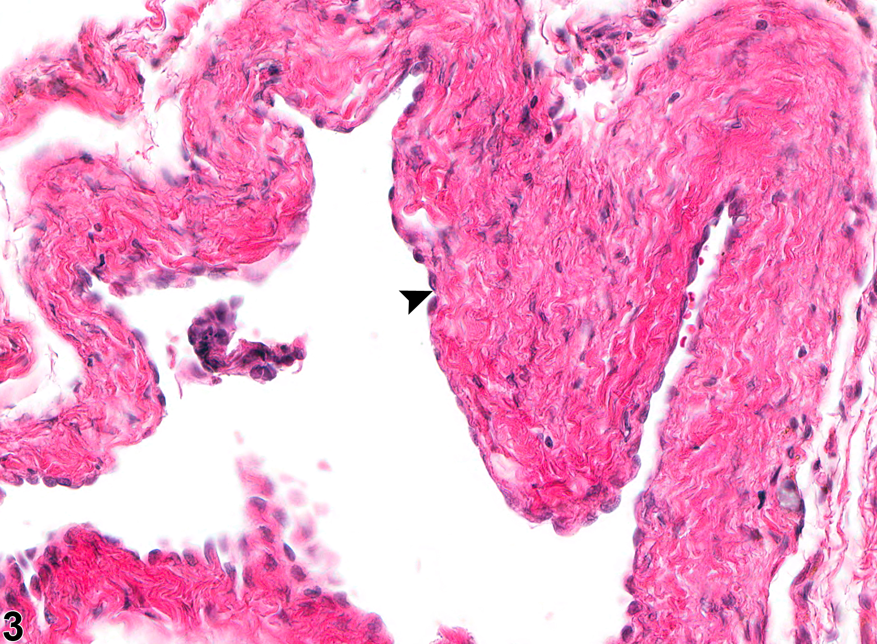
Esophagus - Angiectasis in a female F344/N rat from a chronic study (higher magnification of Figure 1). The dilated vascular spaces are lined by unremarkable endothelial cells (arrowhead).