

Alimentary System
Esophagus - Diverticulum
Narrative
Comment:
Diverticula (Figure 1, arrow) may occur anywhere in the alimentary tract. They are a bulge in a weakened portion of the wall that forms a pocket that is continuous with the lumen. Diverticula are lined by the same type of epithelium as the organ in which they originate, which in the esophagus is squamous epithelium, and the epithelium is continuous with the normal epithelium of the organ. The aperture between the esophagus and the diverticulum is not always present due to the plane of section. Diverticula are occasionally misdiagnosed as adenomas, but the epithelia of diverticula lack the cellular features of neoplasms. Diverticula may become impacted with food, ulcerate, become locally inflamed, and eventually perforate, leading to abscess formation. Diverticula are considered background lesions.
Recommendations:
Whenever present, diverticula should be diagnosed, but it is not necessary to give the lesion a severity grade. Associated lesions, such as inflammation or ulceration, may be diagnosed separately if warranted by severity, but it should be made clear in the pathology narrative that the lesions are associated with the diverticulum.
References:
Bertram TA, Markovits JE, Juliana MM. 1996. Non-proliferative lesions of the alimentary canal in rats GI-1. In Guides for Toxicologic Pathology. STP/ARP/AFIP, Washington, DC, 1-16.
Full Text: https://www.toxpath.org/docs/SSNDC/GINonproliferativeRat.pdfLeininger JR, Jokinen MP, Dangler CA, Whiteley LO. 1999. Oral cavity, esophagus, and stomach. In: Pathology of the Mouse (Maronpot RR, ed). Cache River Press, St Louis, MO, 29-48.
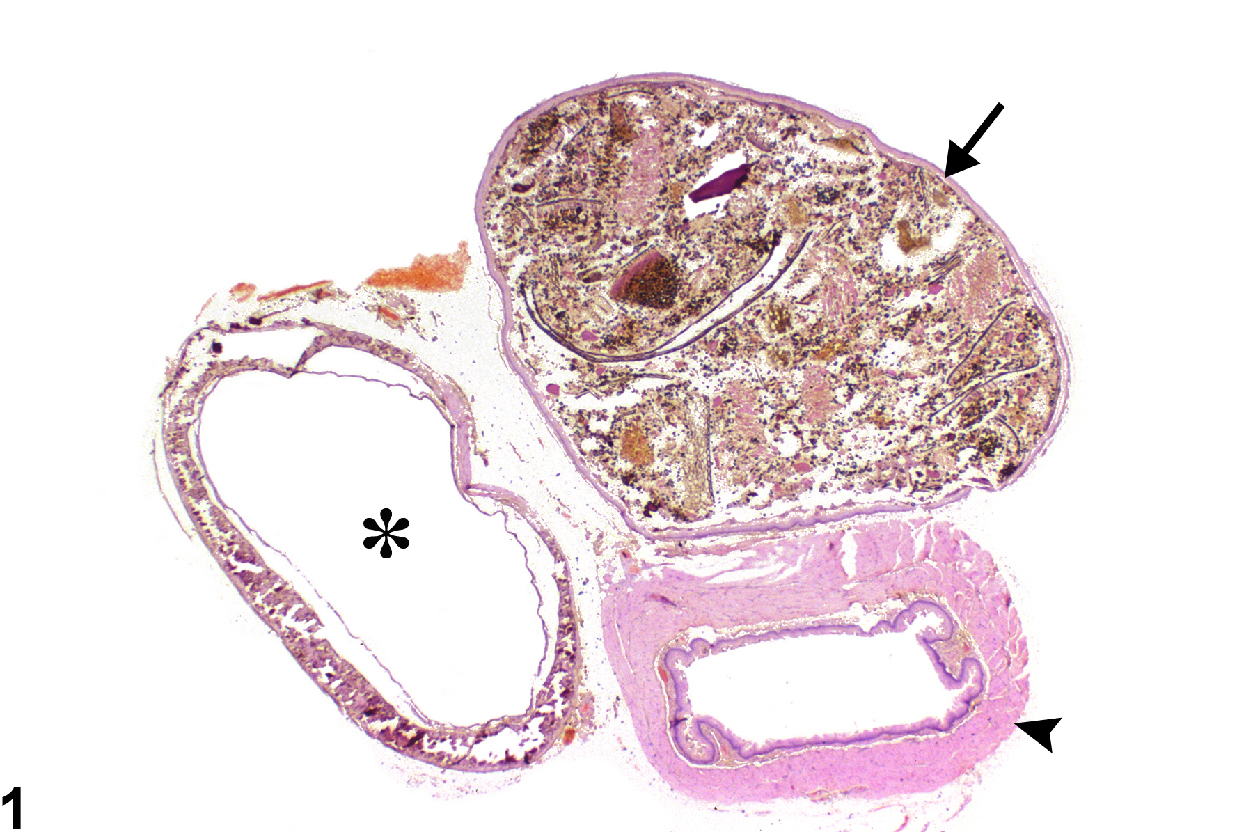
Esophagus - Diverticulum in a female F344/N rat from a chronic study. The diverticulum (arrow) adjacent to the esophagus (arrowhead) is filled with feed material (asterisk = trachea).
All Images

Esophagus - Diverticulum in a female F344/N rat from a chronic study. The diverticulum (arrow) adjacent to the esophagus (arrowhead) is filled with feed material (asterisk = trachea).