

Alimentary System
Esophagus - Edema
Narrative
Comment:
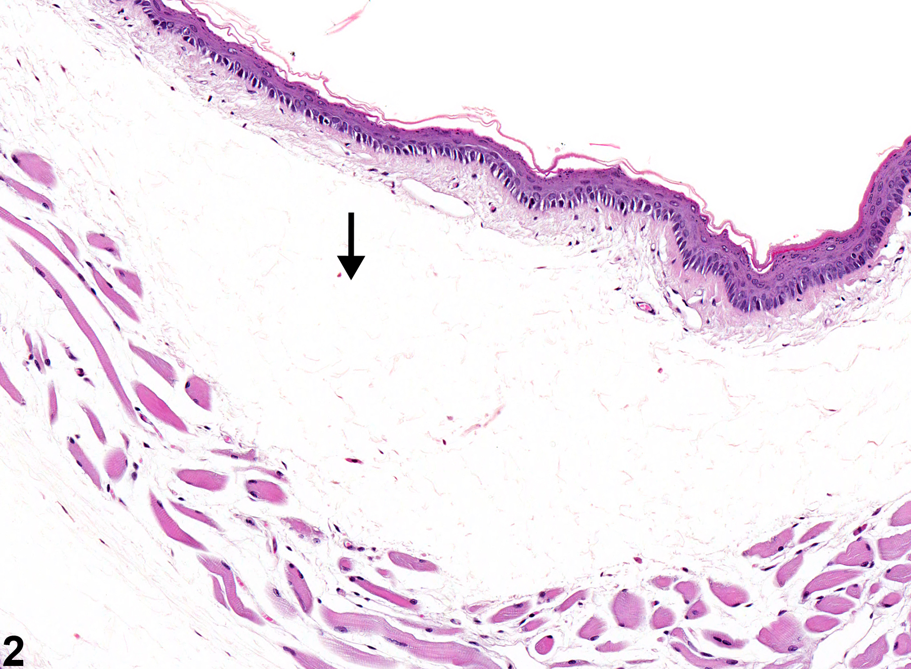
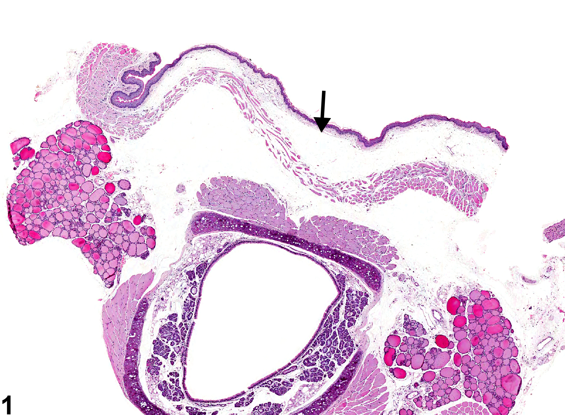
Edema fluid, particularly fluid with a high protein content, appears as homogeneous eosinophilic material in extracellular spaces. It is often lost at processing and may appear as clear spaces in tissues (Figure 1 and Figure 2). Edema is the result of alteration in any of the factors that regulate normal fluid distribution among the plasma, interstitium, and cells, such as increased vascular permeability, increased intravascular hydrostatic pressure, decreased intravascular osmotic pressure, and decreased lymphatic drainage. One common cause of edema in the esophagus is trauma secondary to gavage.
Recommendations:
Edema is diagnosed and graded. Edema as part of an inflammatory process, such as acute inflammation, is ordinarily not recorded separately unless it is a significant component of the lesion.
References:
Mosier DA. 2007. Vascular disorders and thrombosis. In: Pathologic Basis of Veterinary Disease, 4th ed (McGavin MD, Zachary JF, eds). Mosby, St Louis, MO, 63-99.
Esophagus - Edema in a male B6C3F1 mouse from a chronic study. Separation of tissues by clear space is visible (arrow).
All Images

Esophagus - Edema in a male B6C3F1 mouse from a chronic study. Separation of tissues by clear space is visible (arrow).

Esophagus - Edema in a male B6C3F1 mouse from a chronic study (higher magnification of Figure 1). Separation of tissues by clear space is visible (arrow).