

Alimentary System
Esophagus, Muscularis - Degeneration
Narrative
Brown HR, Hardisty JF. 1990. Oral cavity, esophagus and stomach. In: Pathology of the Fischer Rat (Boorman GA, Montgomery CA, MacKenzie WF, eds). Academic Press, San Diego, CA, 9-30.
Abstract: https://www.ncbi.nlm.nih.gov/nlmcatalog/9002563Valentine BA, McGavin MD. 2007. Skeletal muscle. In: Pathologic Basis of Veterinary Disease, 4th ed (McGavin MD, Zachary JF, eds). Mosby, St Louis, MO, 973-1039.
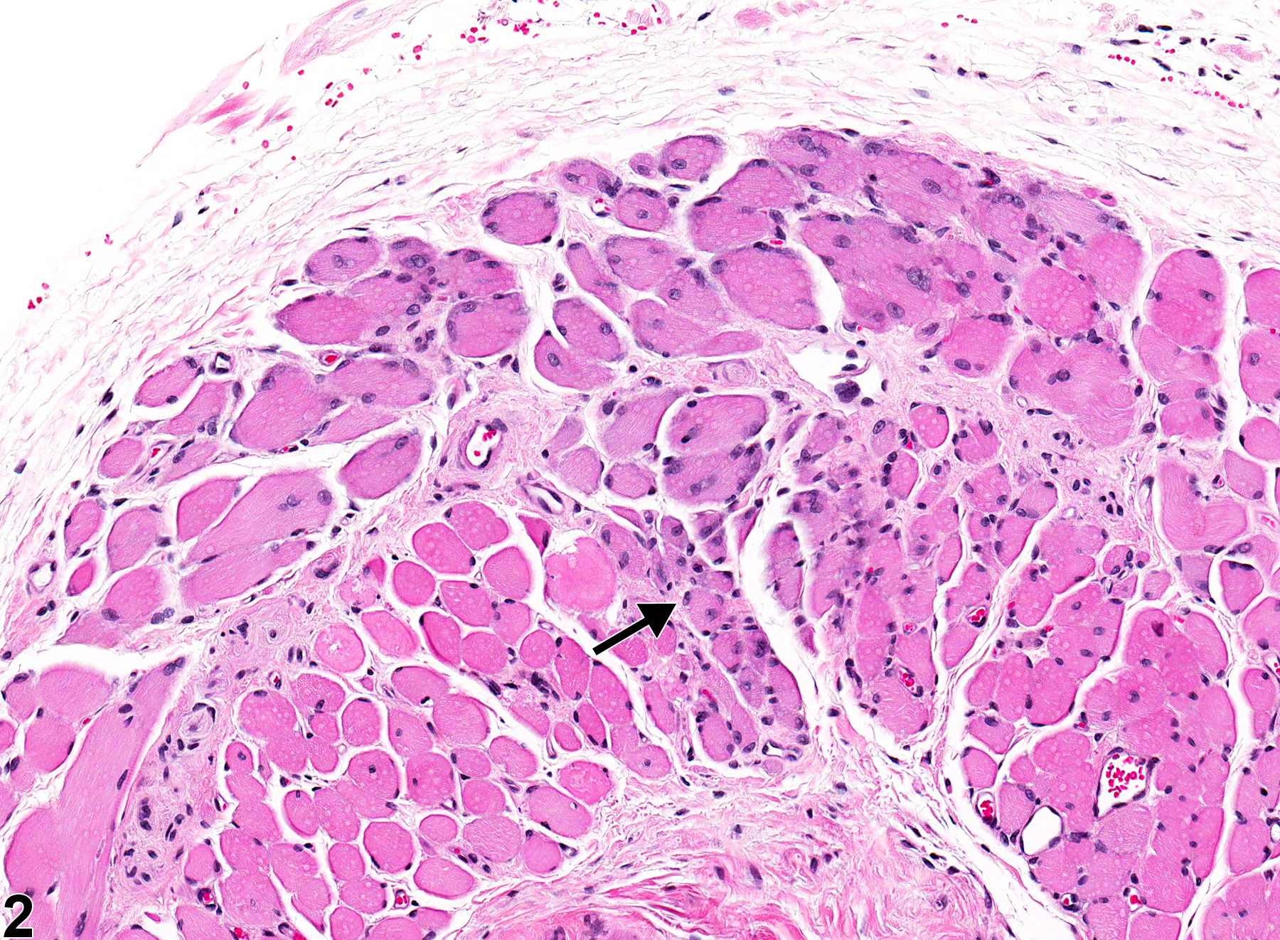
Esophagus, Muscularis - Degeneration in a male F344/N rat from a chronic study. Decreased muscle fiber size (arrow).
All Images

Esophagus, Muscularis - Degeneration in a male F344/N rat from a chronic study. Decreased muscle fiber size (arrow).

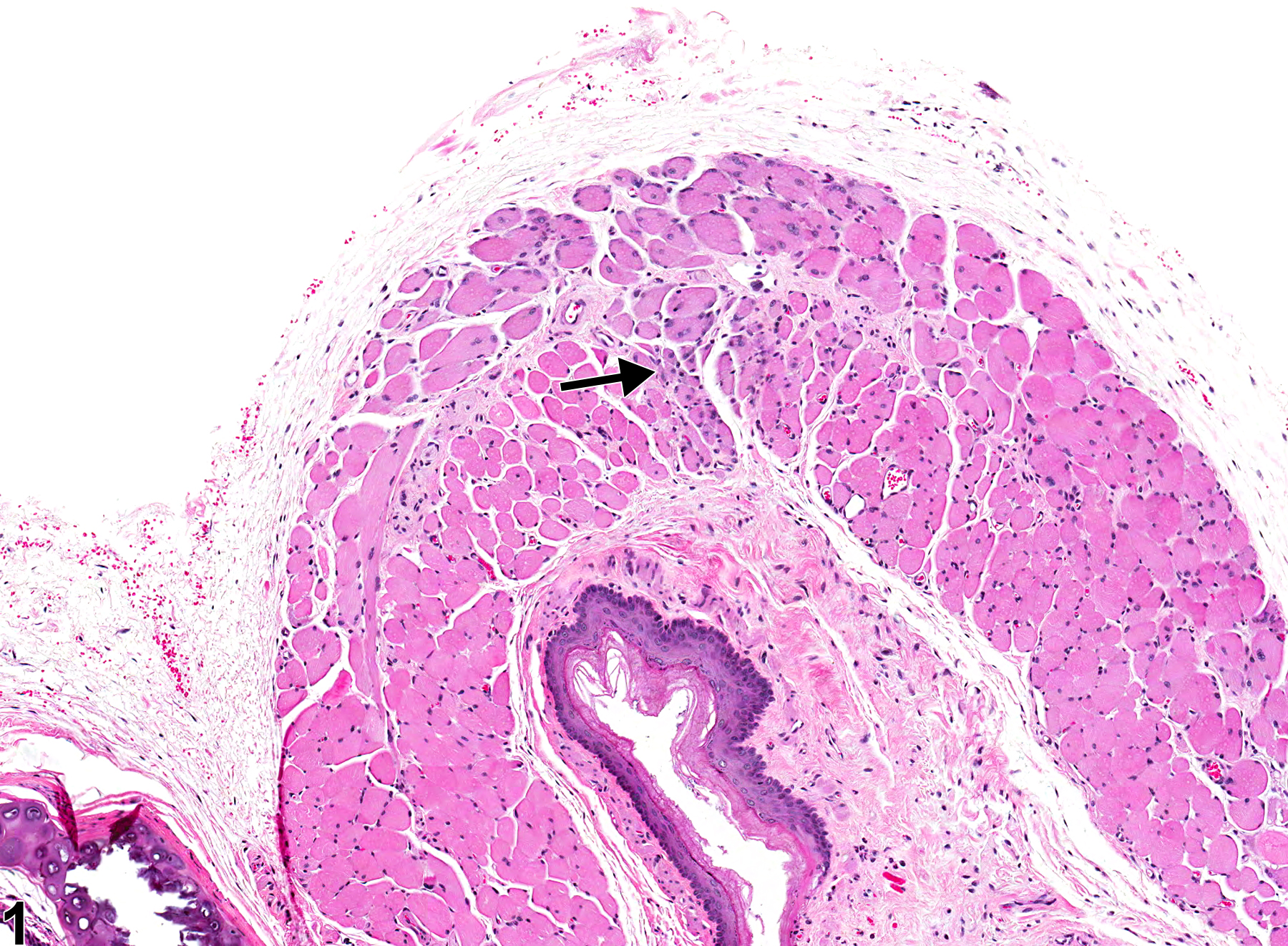
Esophagus, Muscularis - Degeneration in a male F344/N rat from a chronic study (higher magnification of Figure 1). Note the decreased muscle fiber size (arrow).

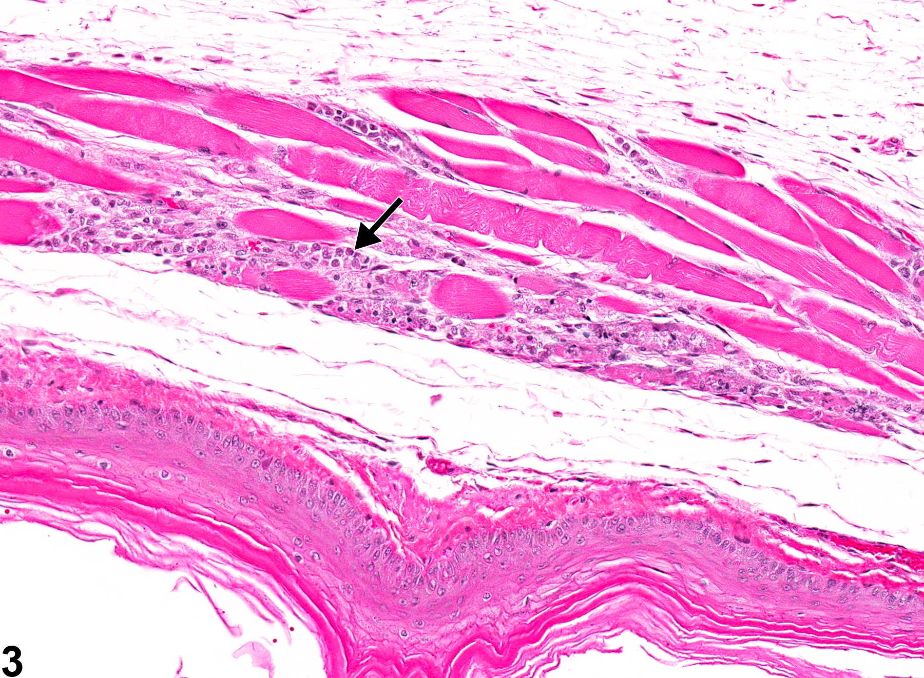
Esophagus, Muscularis - Degeneration in a female Sprague-Dawley rat from a chronic study. There are infiltrates of macrophages amid the muscle fibers (arrow).

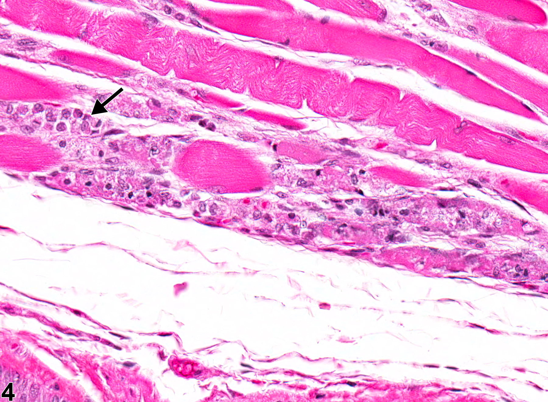
Esophagus, Muscularis - Degeneration in a male F344/N rat from a chronic study (higher magnification of Figure 3). There are infiltrates of macrophages amid the muscle fibers (arrow).