

Alimentary System
Esophagus - Ulcer
Narrative
Bertram TA, Markovits JE, Juliana MM. 1996. Non-proliferative lesions of the alimentary canal in rats GI-1. In Guides for Toxicologic Pathology. STP/ARP/AFIP, Washington, DC, 1-16.
Full Text: https://www.toxpath.org/docs/SSNDC/GINonproliferativeRat.pdf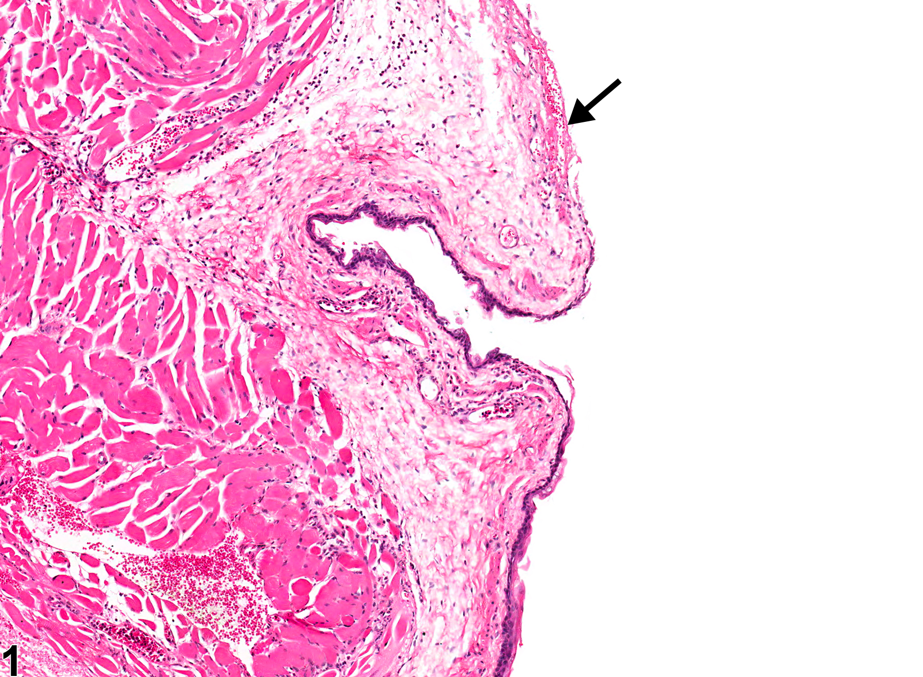
Esophagus - Ulcer in a female F344/N rat from a chronic study. A portion of the epithelium is missing (arrow).
All Images

Esophagus - Ulcer in a female F344/N rat from a chronic study. A portion of the epithelium is missing (arrow).

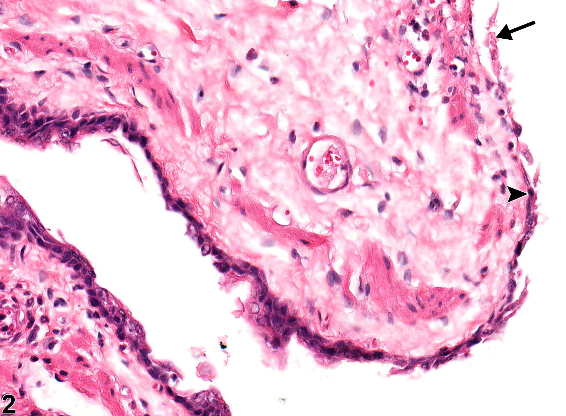
Esophagus - Ulcer in a female F344/N rat from a chronic study (higher magnification of Figure 1). The ulcerated region (arrow) is flanked by a region with regeneration (arrowhead).

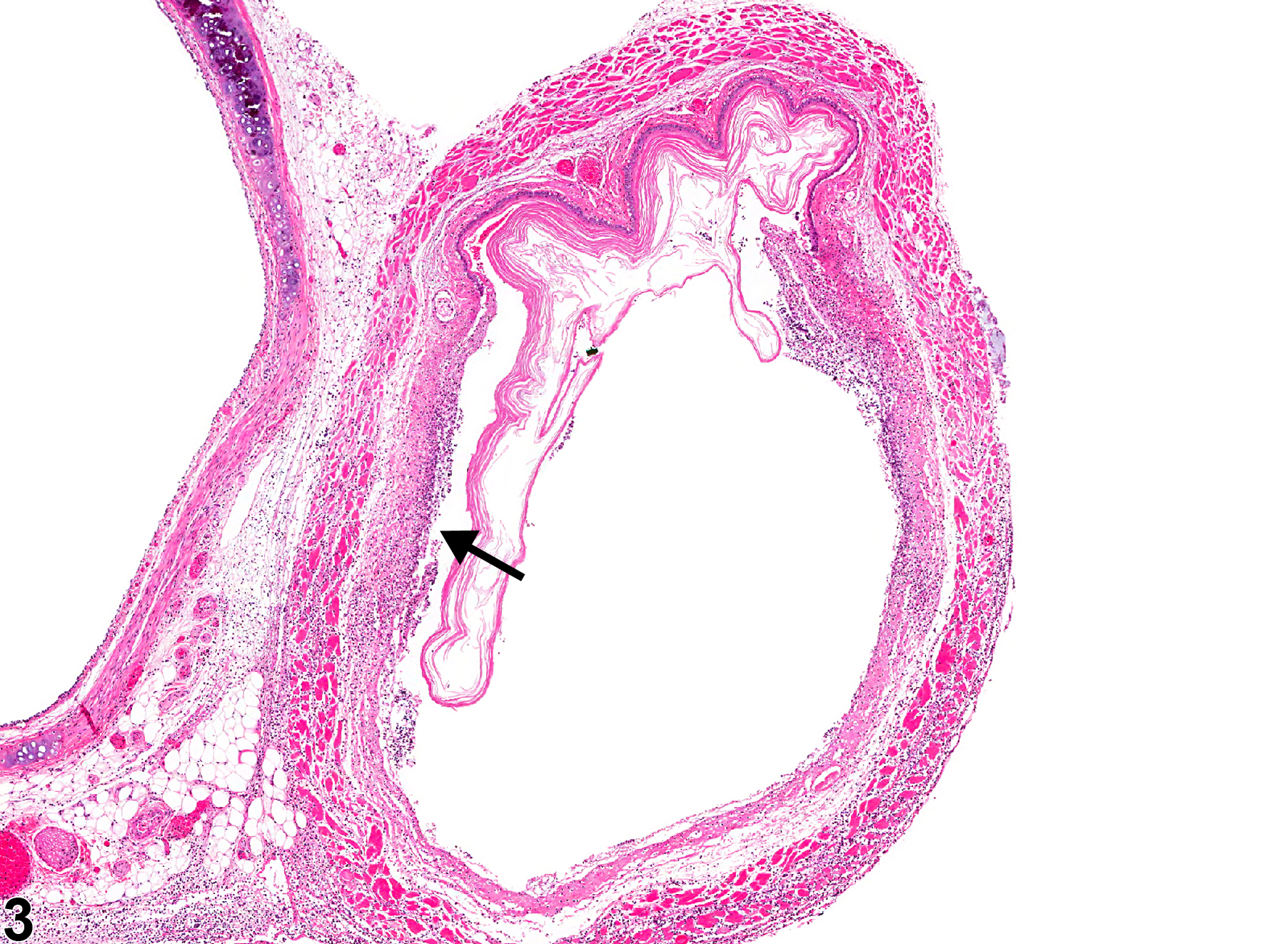
Esophagus - Ulcer in a female F344/N rat from a chronic study. A portion of the epithelium is missing (arrow), and only the overlying keratin remains.

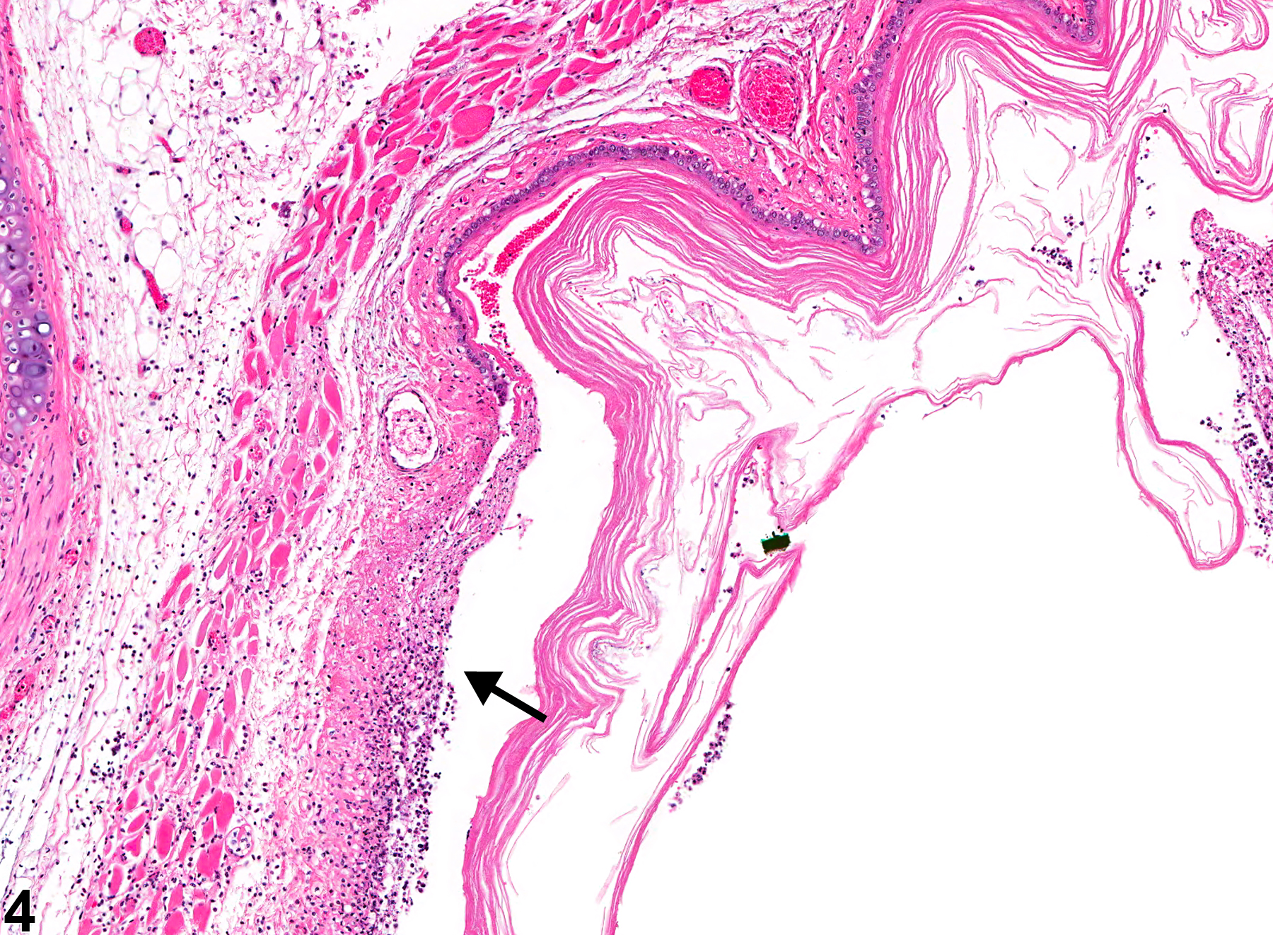
Esophagus - Ulcer in a female F344/N rat from a chronic study (higher magnification of Figure 3). The denuded epithelium is replaced by necrotic debris and inflammatory cells (arrow).

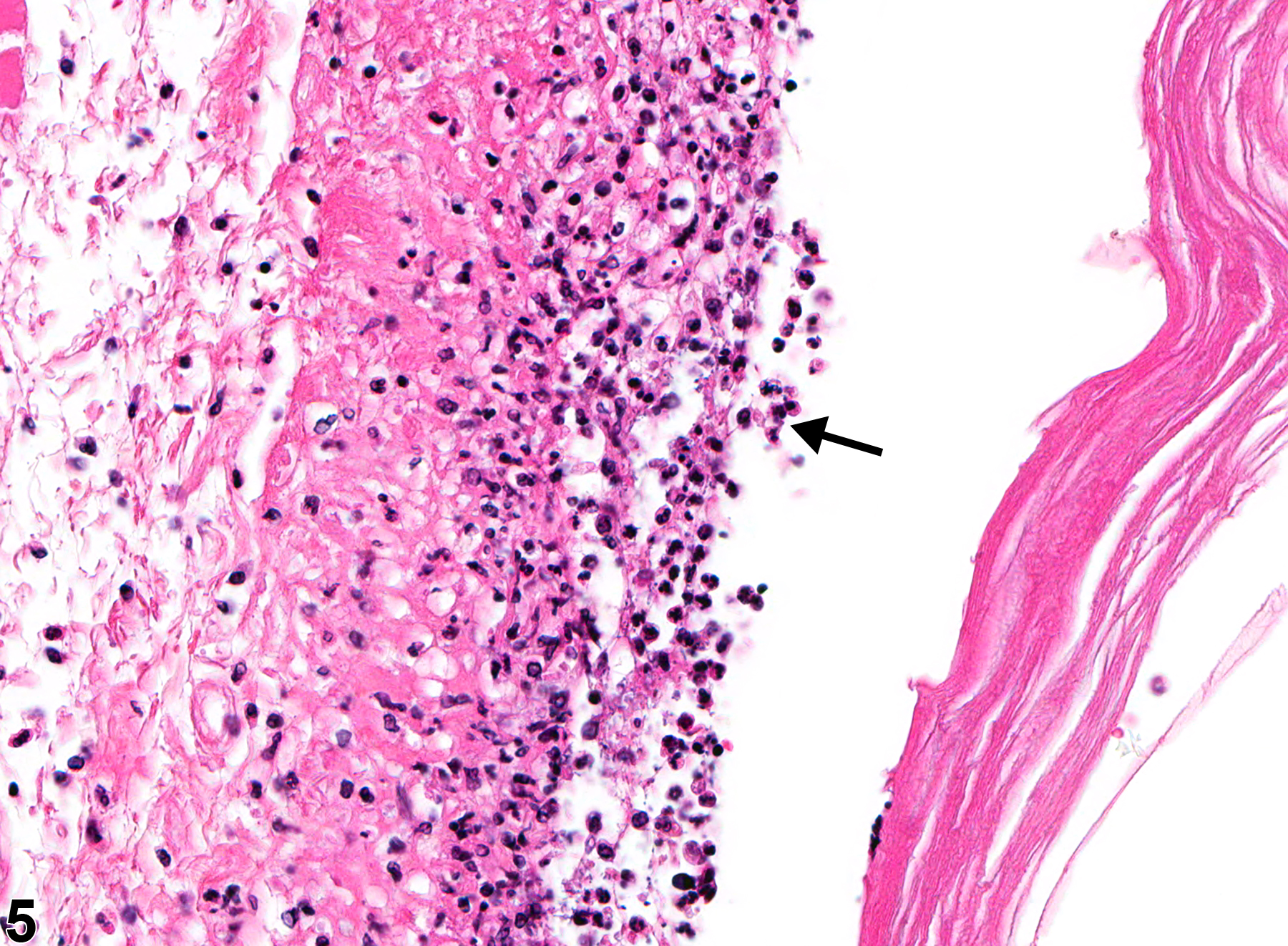
Esophagus - Ulcer in a female F344/N rat from a chronic study (higher magnification of Figure 4). The denuded epithelium is replaced by necrotic debris and inflammatory cells (arrow).