U.S. flag

An official website of the United States government

Dot gov

The .gov means it's official.
Federal government websites often end in .gov or .mil. Before sharing sensitive information, make sure you're on a federal government site.

Https

The site is secure.
The https:// ensures that you are connecting to the official website and that any information you provide is encrypted and transmitted securely.

Skip to Main Navigation
Skip to Page Content
Skip to Atlas Navigation

Stomach, Forestomach - Diverticulum

Image of diverticulum in the forestomach from a female B6C3F1 mouse in a chronic study
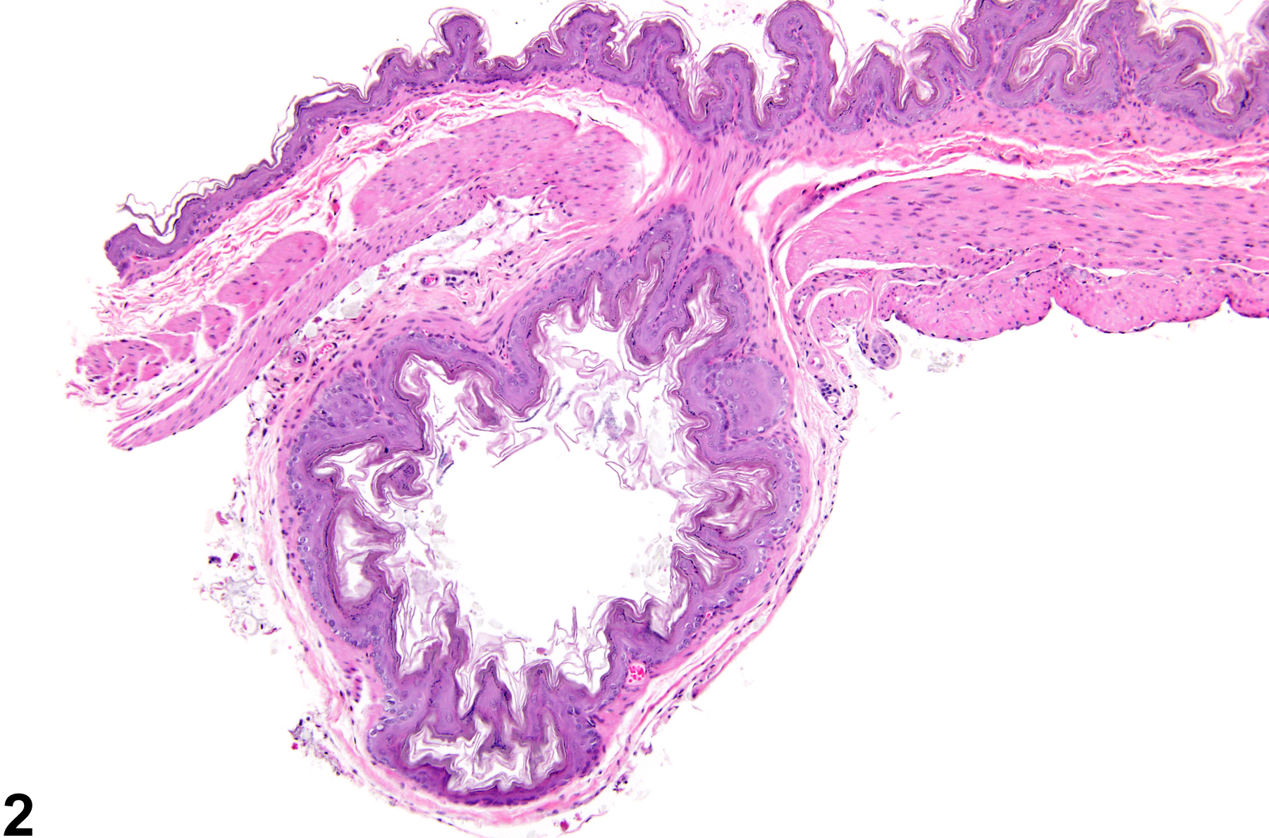
Stomach, Forestomach - Diverticulum in a female B6C3F1 mouse from a chronic study. An out-pouching of the wall of the forestomach filled with keratin, inflammatory cells, and ingesta is present.
Figure 1 of 2
Image of diverticulum in the forestomach from a male B6C3F1 mouse in a chronic study
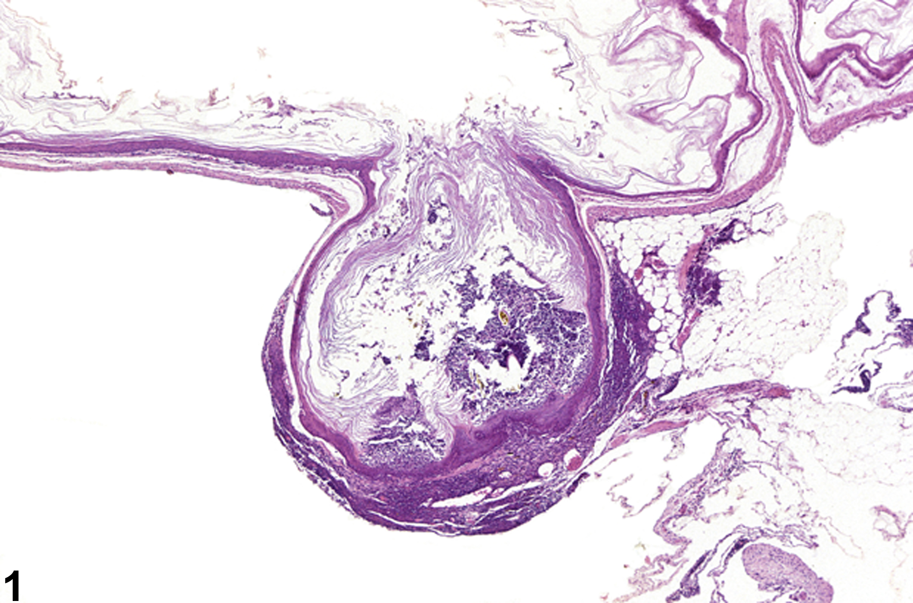
Stomach, Forestomach - Diverticulum in a male B6C3F1 mouse from a chronic study. An out-pouching of the wall of the forestomach is present.
Figure 2 of 2
next arrow

comment:

Diverticula are more common in the forestomach than in the glandular stomach but can occur from the esophagus to the colon. Diverticula are congenital defects characterized by out-pouchings of a portion of the wall in which the normal cell types are present. The wall and lumen of a diverticulum are continuous with the wall and lumen of the forestomach ( Figure 1image opens in a pop-up window and Figure 2image opens in a pop-up window ), although the communication between them is not always visible ( Figure 2image opens in a pop-up window ). Diverticula are occasionally misdiagnosed as a adenomas, but diverticula lack many of the features of neoplasms, such as a mass-like proliferation of cells, cellular atypia, and loss of normal architecture. Diverticula may become impacted with food, ulcerate, become locally inflamed, and eventually perforate, leading to abscess formation. Diverticula are considered incidental lesions.

recommendation:

Diverticulum should be diagnosed but not graded. Associated lesions, such as inflammation or ulcer, may be diagnosed separately if warranted by severity, but the pathologist should make the association with the diverticulum clear in the pathology narrative.

references:

Bertram TA, Markovits JE, Juliana MM. 1996. Non-proliferative lesions of the alimentary canal in rats GI-1. In Guides for Toxicologic Pathology. STP/ARP/AFIP, Washington, DC, 1-16.
Full Text: https://www.toxpath.org/docs/SSNDC/GINonproliferativeRat.pdf

Leininger JR, Jokinen MP, Dangler CA, Whiteley LO. 1999. Oral cavity, esophagus, and stomach. In: Pathology of the Mouse (Maronpot RR, ed). Cache River Press, St Louis, MO, 29-48.
Abstract: http://www.cacheriverpress.com/books/pathmouse.htm