

Alimentary System
Stomach, Forestomach - Ectopic Tissue
Narrative
Bertram TA, Markovits JE, Juliana MM. 1996. Non-proliferative lesions of the alimentary canal in rats GI-1. In Guides for Toxicologic Pathology. STP/ARP/AFIP, Washington, DC, 1-16.
Full Text: https://www.toxpath.org/docs/SSNDC/GINonproliferativeRat.pdfFrantz JD, Betton GR, Cartwright ME, Crissman JW, Macklin AW, Maronpot RR. 1991. Proliferative lesions of the non-glandular and glandular stomach in rats. GI-3. In Guides for Toxicologic Pathology. STP/ARP/AFIP, Washington, DC, 1-20.
Full Text: https://www.toxpath.org/docs/SSNDC/StomachProliferativeRat.pdf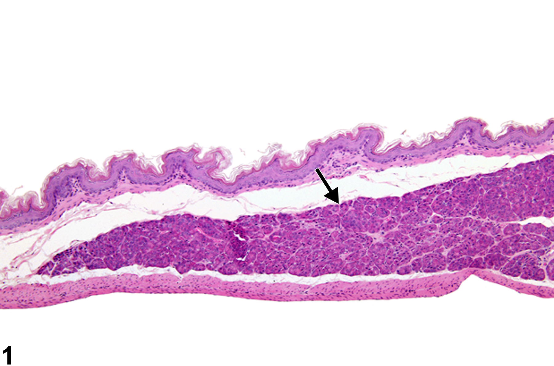
Stomach, Forestomach - Ectopic pancreas in a female B6C3F1 mouse from a chronic study. Ectopic pancreatic acinar cells are present in the submucosa of the forestomach (arrow).
All Images

Stomach, Forestomach - Ectopic pancreas in a female B6C3F1 mouse from a chronic study. Ectopic pancreatic acinar cells are present in the submucosa of the forestomach (arrow).

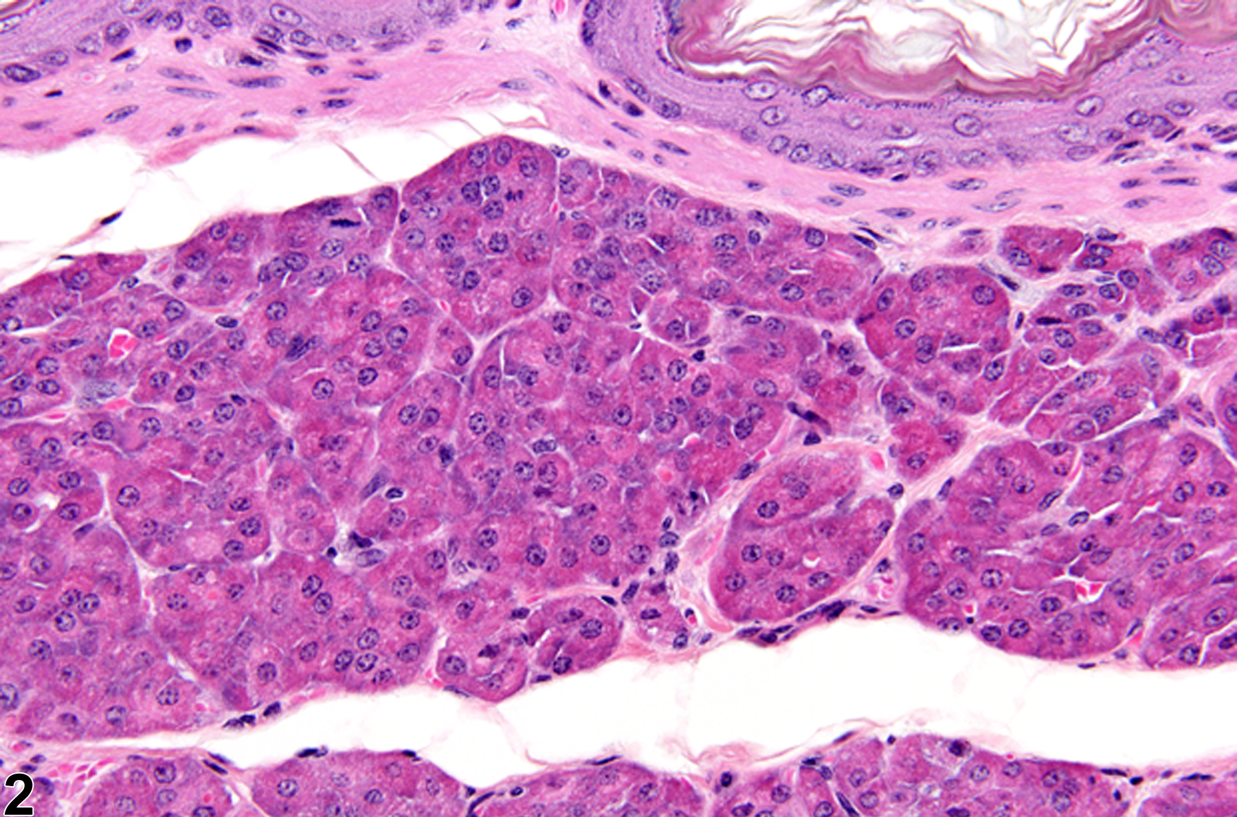
Stomach, Forestomach - Ectopic pancreas in a female B6C3F1 mouse from a chronic study (higher magnification of Figure 1). Ectopic pancreatic acinar cells are present in the submucosa of the forestomach.

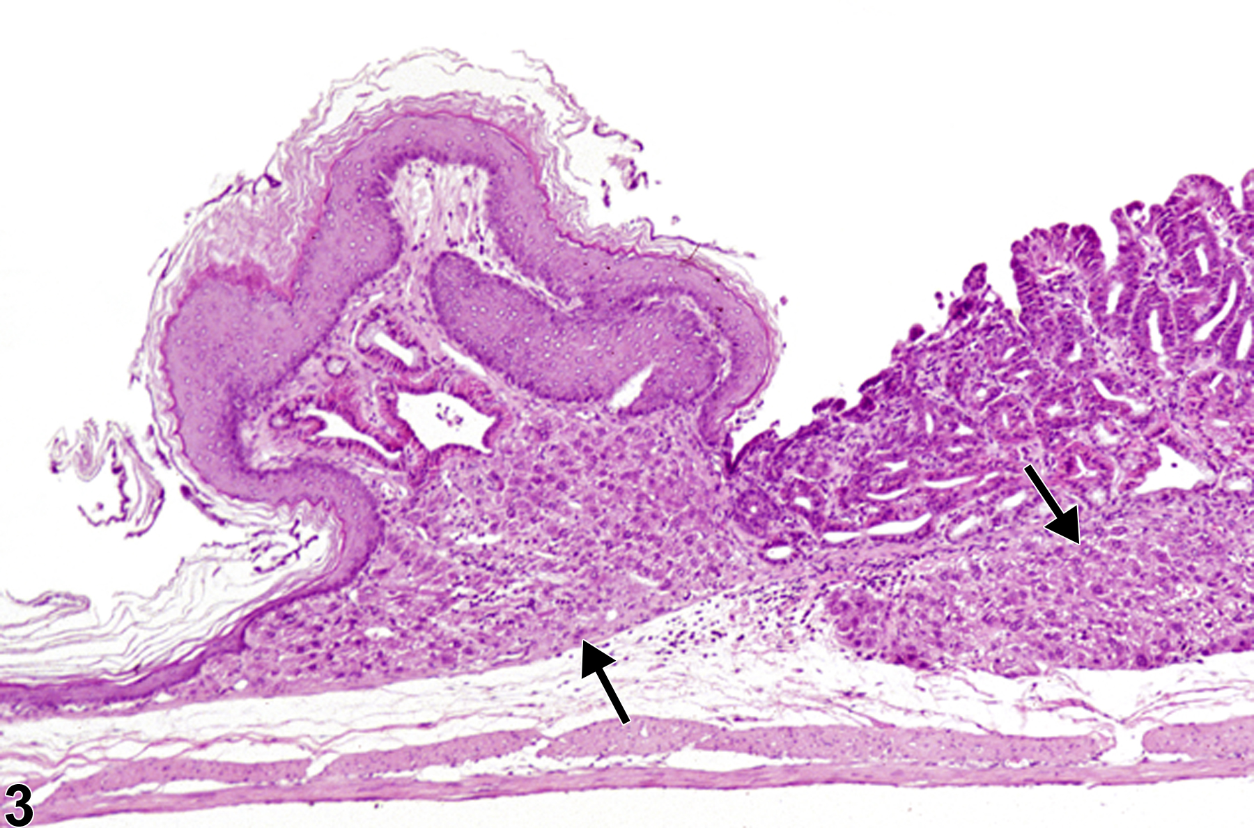
Stomach, Forestomach - Ectopic liver in a female F344/N rat from a chronic study. Ectopic hepatocytes (arrows) are present in the submucosa adjacent to the limiting ridge.

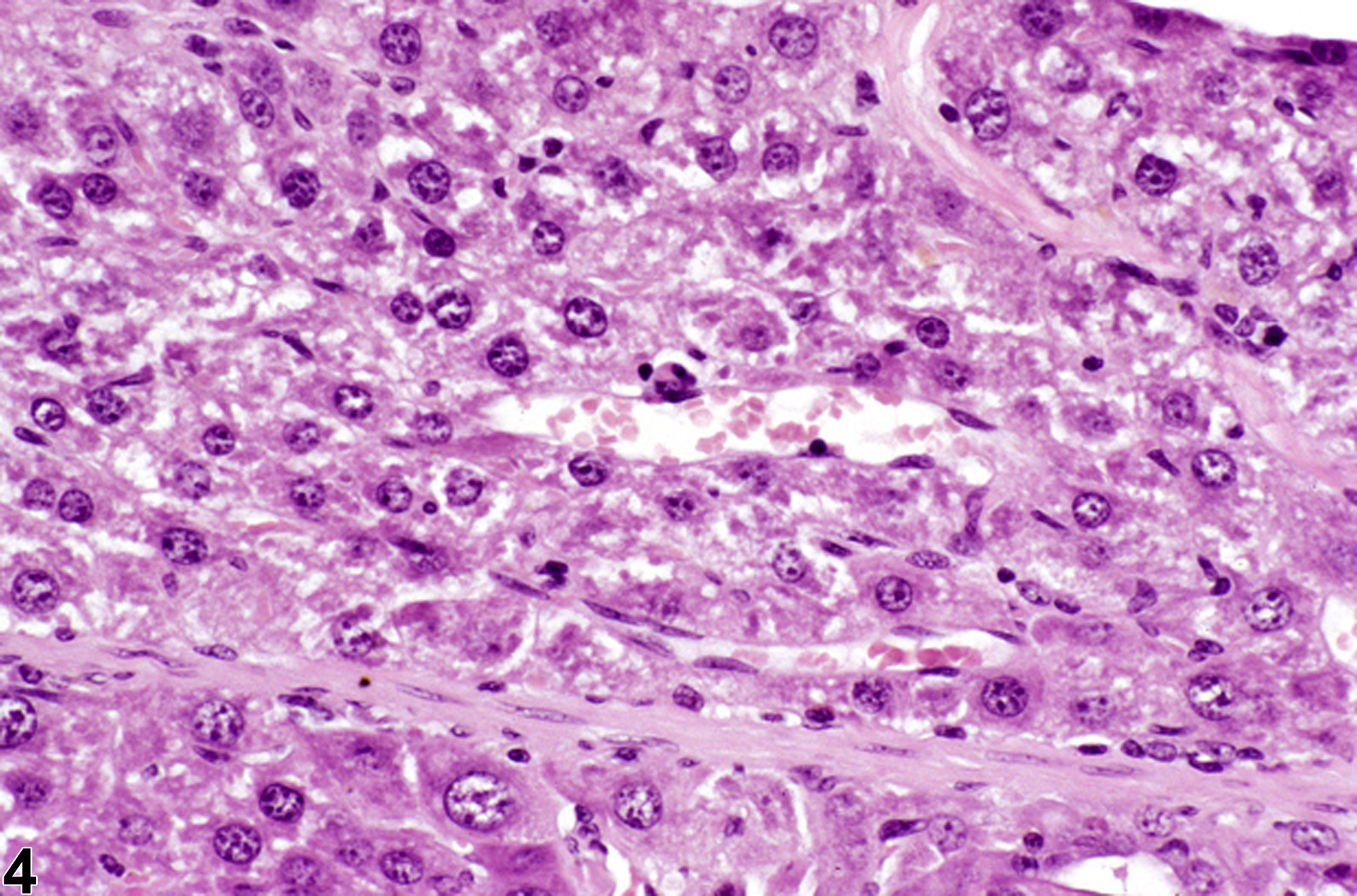
Stomach, Forestomach - Ectopic liver in a female F344/N rat from a chronic study (higher magnification of Figure 3). Ectopic hepatocytes are present in the submucosa adjacent to the limiting ridge.

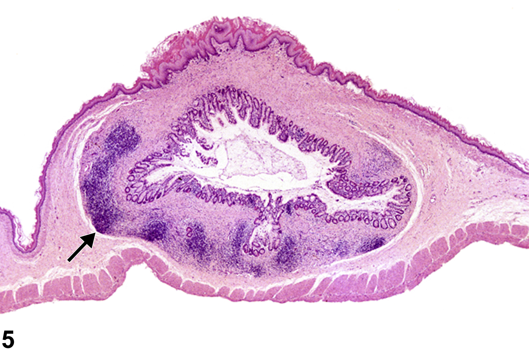
Stomach, Forestomach - Ectopic intestine in a male F344/N rat from a chronic study. The submucosa is expanded by a focus of ectopic intestine with gut associated lymphoid tissue (arrow).