U.S. flag

An official website of the United States government

Dot gov

The .gov means it's official.
Federal government websites often end in .gov or .mil. Before sharing sensitive information, make sure you're on a federal government site.

Https

The site is secure.
The https:// ensures that you are connecting to the official website and that any information you provide is encrypted and transmitted securely.

Skip to Main Navigation
Skip to Page Content
Skip to Atlas Navigation

Stomach, Forestomach - Foreign Body

Image of foreign body in the forestomach from a male F344/N rat in a chronic study
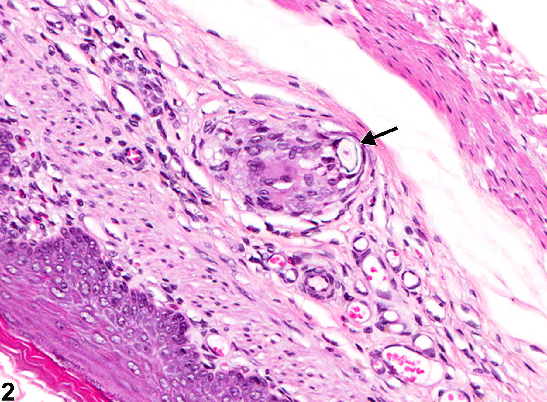
Stomach, Forestomach - Foreign body in a male F344/N rat from a chronic study. A foreign body (hair) (arrow) has penetrated the epithelium and submucosa.
Figure 1 of 2
Image of foreign body in the forestomach from a male F344/N rat in a chronic study
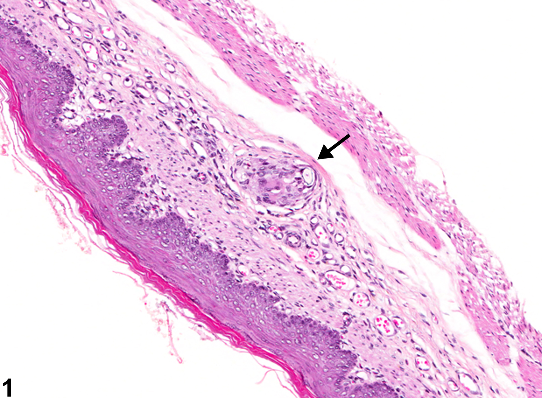
Stomach, Forestomach - Foreign body in a male F344/N rat from a chronic study (higher magnification of Figure 1). A foreign body (hair) (arrow) with associated granulomatous inflammation is present in the submucosa.
Figure 2 of 2
next arrow

comment:

Hair or feed material can become embedded in the wall of the forestomach secondary to injury from a gavage procedure or ulceration from xenobiotics. In Figure 1image opens in a pop-up window and Figure 2image opens in a pop-up window , a small granuloma with a central hair shaft (arrow) is in the submucosa; the overlying mucosa has healed from a previous injury. This is considered a small incidental lesion in a control animal that may have occurred secondary to previous gavage trauma.

recommendation:

A primary foreign body should be diagnosed but not graded. If a foreign body causes a significant inflammatory response, then both the foreign body and inflammation are diagnosed. A foreign body such as hairs or feed material that has been passively pushed into an ulcerated area should not be diagnosed separately but can be described in the narrative.

references:

Brown HR, Hardisty JF. 1990. Oral cavity, esophagus and stomach. In: Pathology of the Fischer Rat (Boorman GA, Montgomery CA, MacKenzie WF, eds). Academic Press, San Diego, CA, 9-30.
Abstract: https://www.ncbi.nlm.nih.gov/nlmcatalog/9002563

Leininger JR, Jokinen MP, Dangler CA, Whiteley LO. 1999. Oral cavity, esophagus, and stomach. In: Pathology of the Mouse (Maronpot RR, ed). Cache River Press, St Louis, MO, 29-48.
Abstract: http://www.cacheriverpress.com/books/pathmouse.htm