

Alimentary System
Stomach, Forestomach, Epithelium - Hyperplasia, [Focal, Diffuse]
Narrative
Betton GR. 1998. The digestive system I: The gastrointestinal tract and exocrine pancreas. In: Target Organ Pathology (Turton J, Hooson J, eds). Taylor and Francis, London, 29-60.
Betton GR, Salmon GK. 1984. Pathology of the forestomach in rats treated for one year with a new H-2 receptor antagonist, SK&F 93479 trihydrochloride. Scand J Gastroenterol Suppl 101:103-108.
Abstract: https://www.ncbi.nlm.nih.gov/pubmed/2888184Boorman GA, Hong HL, Jameson CW, Yoshitomi K, Maronpot RR. 1986. Regression of methyl bromide-induced forestomach lesions in the rat. Toxicol Appl Pharmacol 86:131-139.
Abstract: https://www.ncbi.nlm.nih.gov/pubmed/3764933Chan PC, Mahler J, Peddada S, Lomnitski L, Nyska A. 2003. Forestomach tumor induction by 2,4-hexadienal in F344N rats and B6C3F1 mice. Arch Toxicol 77:511-520.
Abstract: https://www.ncbi.nlm.nih.gov/pubmed/12879212Frantz JD, Betton GR, Cartwright ME, Crissman JW, Macklin AW, Maronpot RR. 1991. Proliferative lesions of the non-glandular and glandular stomach in rats. GI-3. In Guides for Toxicologic Pathology. STP/ARP/AFIP, Washington, DC, 1-20.
Full Text: https://www.toxpath.org/docs/SSNDC/StomachProliferativeRat.pdfFrederick CB, Hazelton GA, Frantz JD. 1990. The histopathological and biochemical response of the stomach of male rats following two weeks of oral dosing with ethyl acrylate. Toxicol Pathol 18:247-256.
Full Text: http://tpx.sagepub.com/content/18/2/247.full.pdfNational Toxicology Program. 2002. NTP TOX-70. Toxicity Studies of p-tert-Butylcatechol (CAS No. 98-29-3) Administered in Feed to F344/N Rats and B6C3F1 Mice. NTP, Research Triangle Park, NC.
Abstract: https://ntp.niehs.nih.gov/go/1385National Toxicology Program. 2007. NTP TR-543. Toxicology and Carcinogenesis Studies of alpha-Methylstyrene (CAS No. 98-83-9) in F344/N rats and B6C3F1 mice (Inhalation Studies). NTP, Research Triangle Park, NC.
Abstract: https://ntp.niehs.nih.gov/go/28010National Toxicology Program. 2010. NTP TR-544. Toxicology and Carcinogenesis Studies of Dibromoacetonitrile (CAS No. 3252-43-5) in F344/N Rats and B6C3F1 Mice (Drinking Water Studies). NTP, Research Triangle Park, NC.
Abstract: https://ntp.niehs.nih.gov/go/32617National Toxicology Program. 2010. NTP TR-558. Toxicology and Carcinogenesis Studies of 3,3’,4,4’-Tetrachloroazobenzene (TCAB) (CAS No. 14047-09-7) in Harlan Sprague Dawley Rats and B6C3F1 Mice (Gavage Studies). NTP, Research Triangle Park, NC.
Abstract: https://ntp.niehs.nih.gov/go/33564Nera EA, Lok E, Iverson F, Ormsby E, Karpinski DF, Clayson DB. 1984. Short term pathological and proliferative effects of butylated hydroxyanisole and other phenolic antioxidants in the forestomach of Fischer 344 rats. Toxicology 32:197-213.
Abstract: https://www.ncbi.nlm.nih.gov/pubmed/6474484Nyska A, Moomaw CR, Lomnitski L, Chan PC. 2001. Glutathione S-transferase pi expression in forestomach carcinogenesis process induced by gavage-administered 2,4-hexadienal in the F344 rat. Arch Toxicol 75:618-624.
Abstract: https://www.ncbi.nlm.nih.gov/pubmed/11808924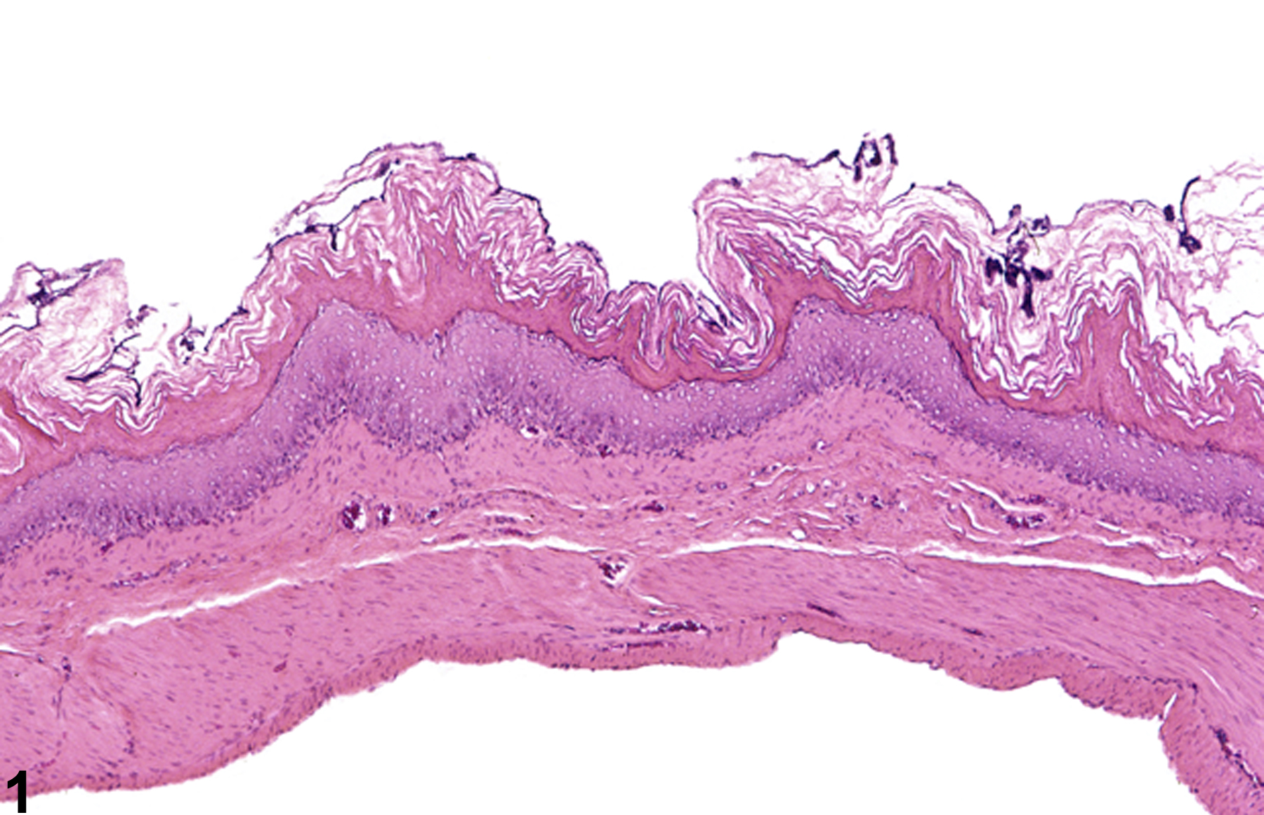
Stomach, Forestomach, Epithelium - Hyperplasia in a male F344/N rat from a subchronic study. The epithelial hyperplasia is diffuse.
All Images

Stomach, Forestomach, Epithelium - Hyperplasia in a male F344/N rat from a subchronic study. The epithelial hyperplasia is diffuse.

Stomach, Forestomach, Epithelium - Hyperplasia in a female F344/N rat from a chronic study. The epithelial hyperplasia is focal and papillary.

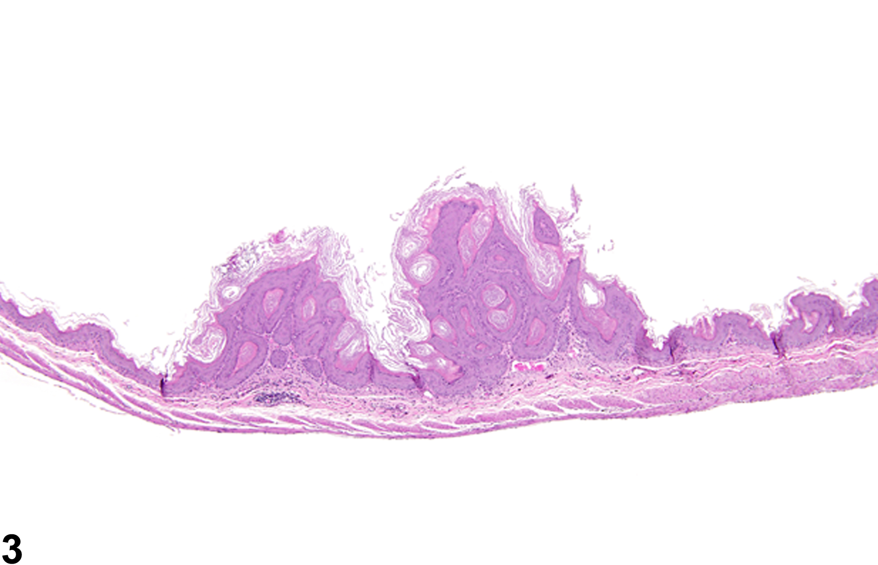
Stomach, Forestomach, Epithelium - Hyperplasia in a male B6C3F1 mouse from a chronic study. Two exophytic, papillary foci of hyperplasia are present.

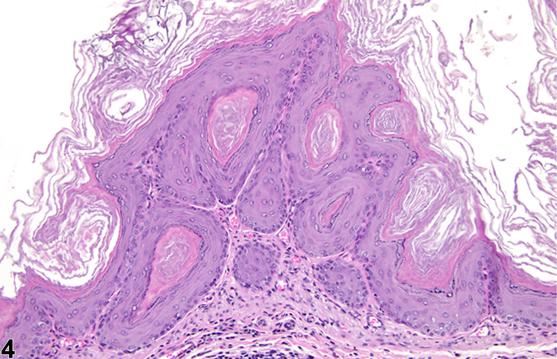
Stomach, Forestomach, Epithelium - Hyperplasia in a male B6C3F1 mouse from a chronic study (higher magnification of Figure 3). A papillary focus of hyperplasia is present.

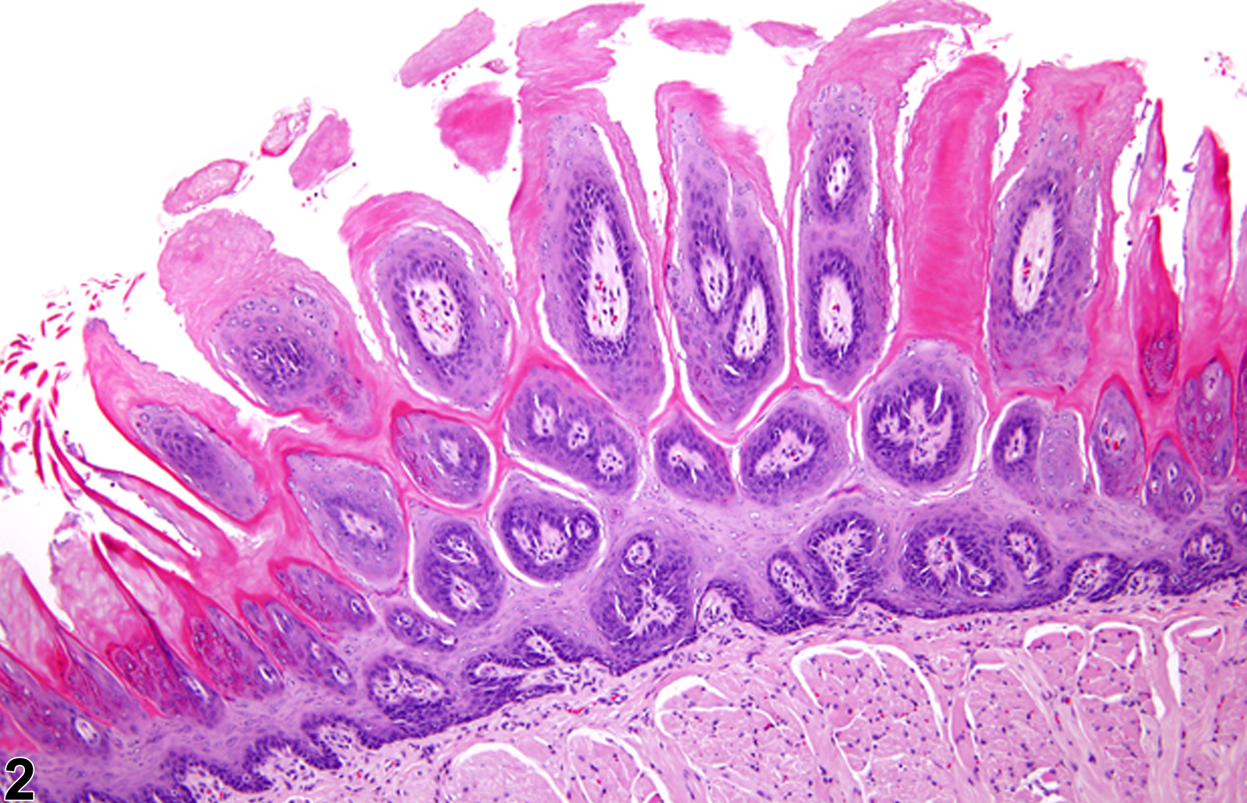
Stomach, Forestomach - Normal limiting ridge in a male F344/N rat from a subchronic study.

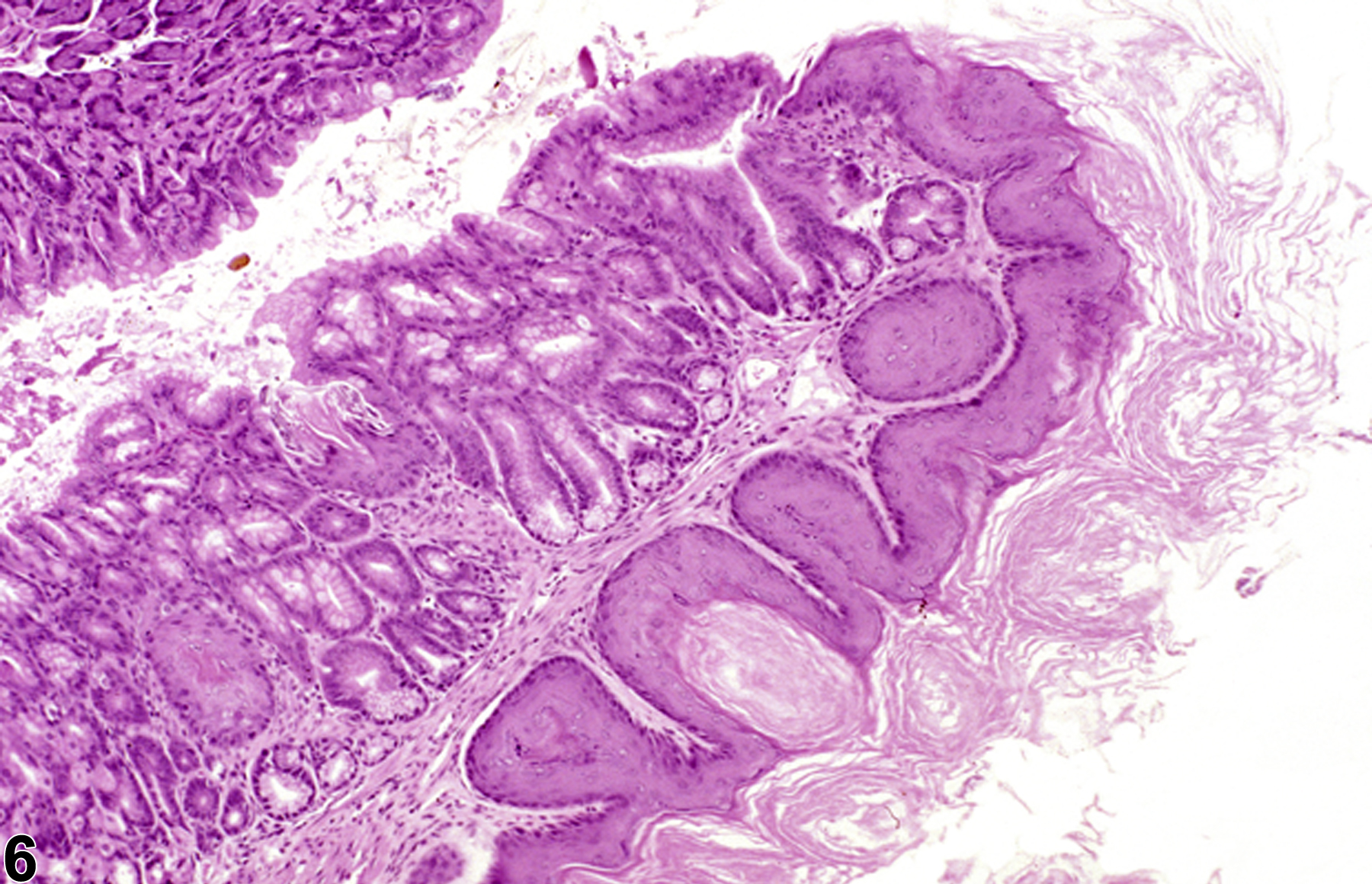
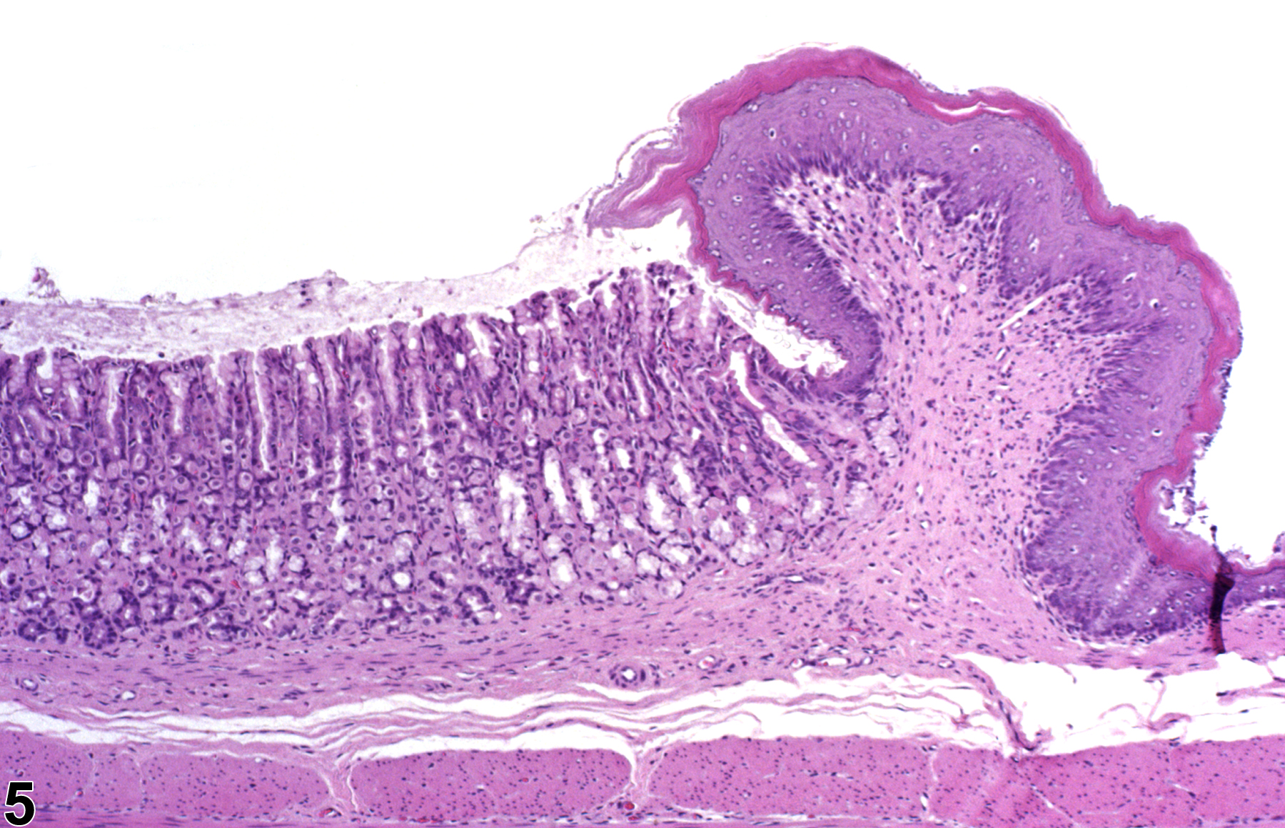
Stomach, Forestomach, Epithelium - Hyperplasia in a male B6C3F1 mouse from a subchronic study. Epithelial hyperplasia of the limiting ridge is present.

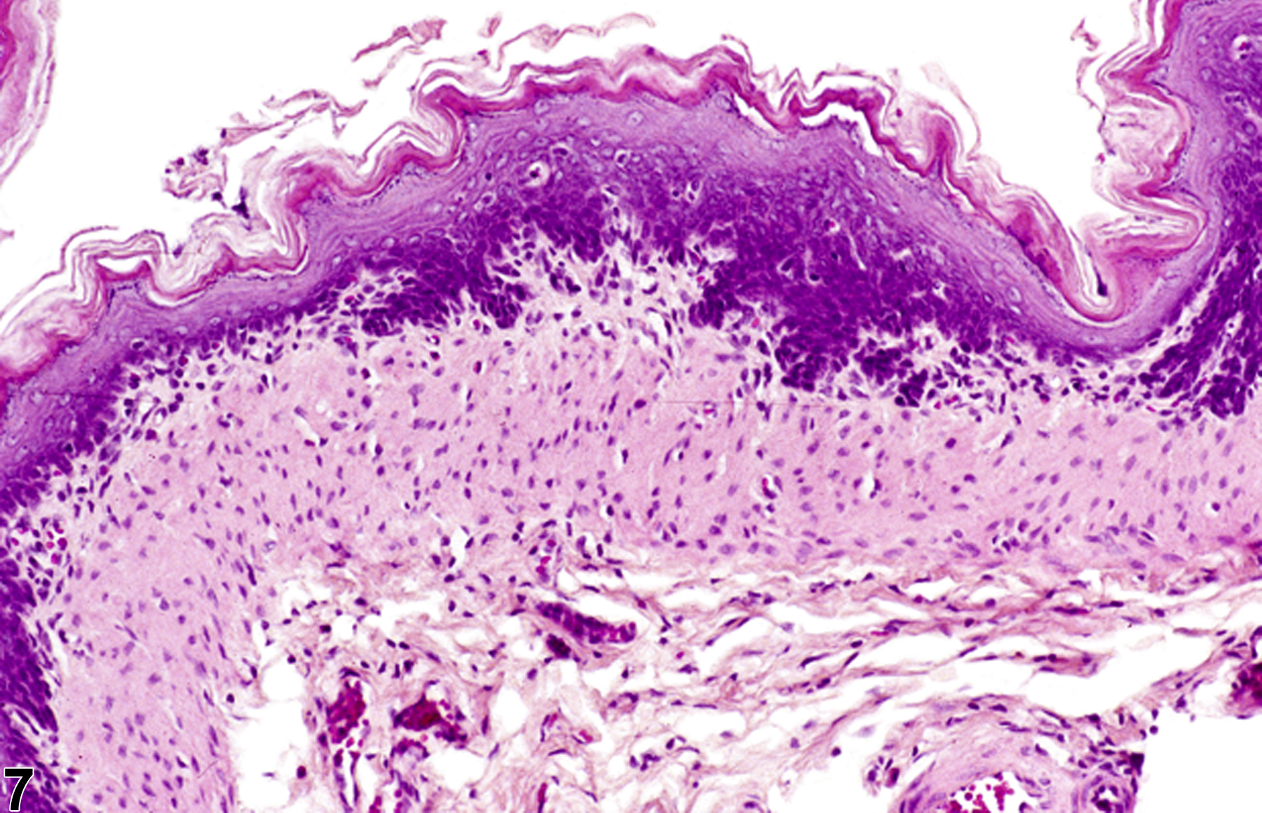
Stomach, Forestomach, Epithelium - Hyperplasia in a male F344/N rat from a chronic study. Hyperplasia of the basal cells is present in the forestomach.

Stomach, Forestomach, Epithelium - Hyperplasia in a male F344/N rat from a chronic study (higher magnification of Figure 7). Hyperplasia of the basal cells is present in the forestomach.