

Alimentary System
Stomach, Forestomach - Inflammation
Narrative
Leininger JR, Jokinen MP, Dangler CA, Whiteley LO. 1999. Oral cavity, esophagus, and stomach. In: Pathology of the Mouse (Maronpot RR, ed). Cache River Press, St Louis, MO, 29-48.
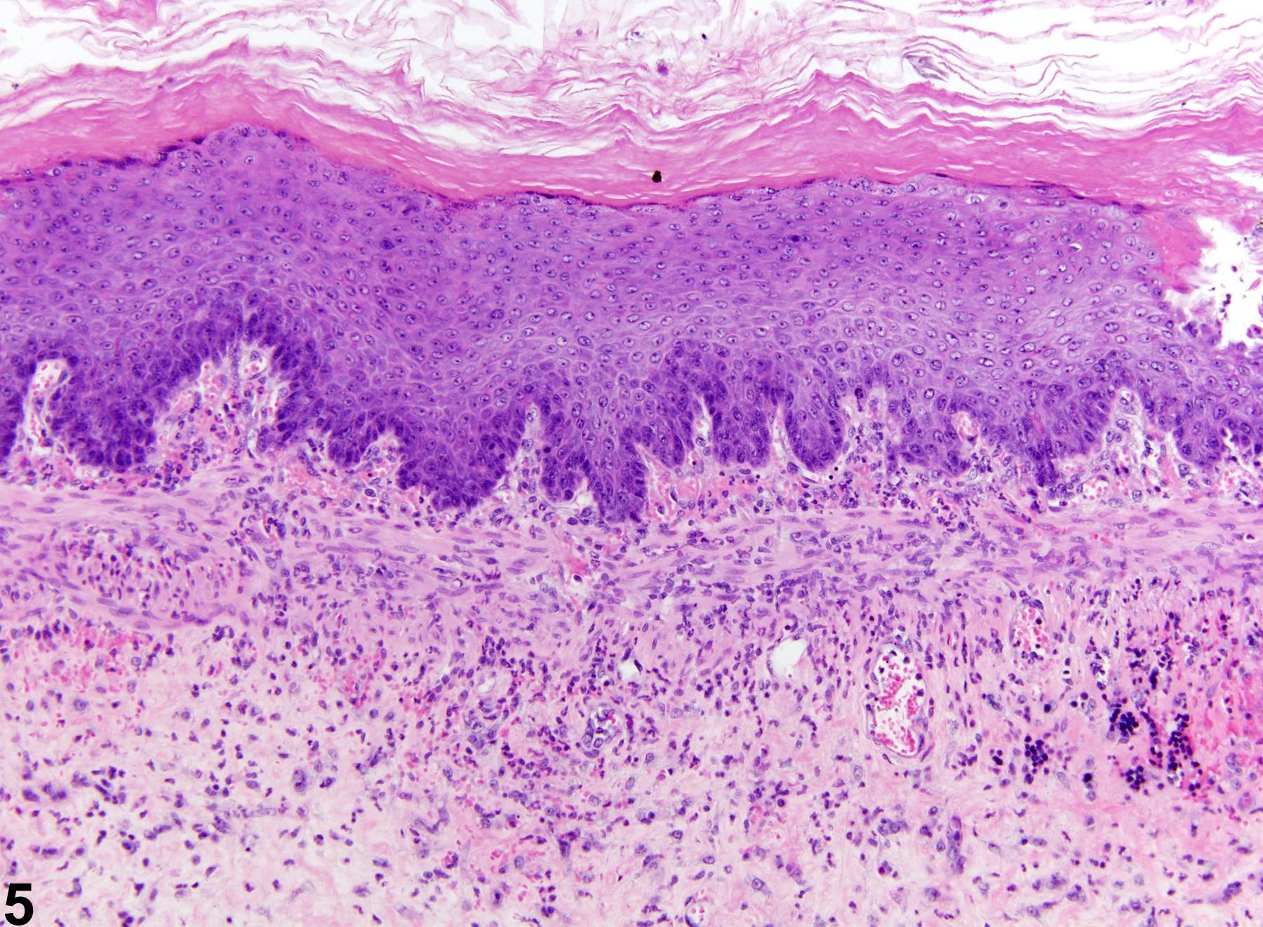
Stomach, Forestomach - Inflammation, Suppurative in a male F344/N rat from a chronic study. This suppurative inflammation is associated with an ulcer.
All Images

Stomach, Forestomach - Inflammation, Suppurative in a male F344/N rat from a chronic study. This suppurative inflammation is associated with an ulcer.

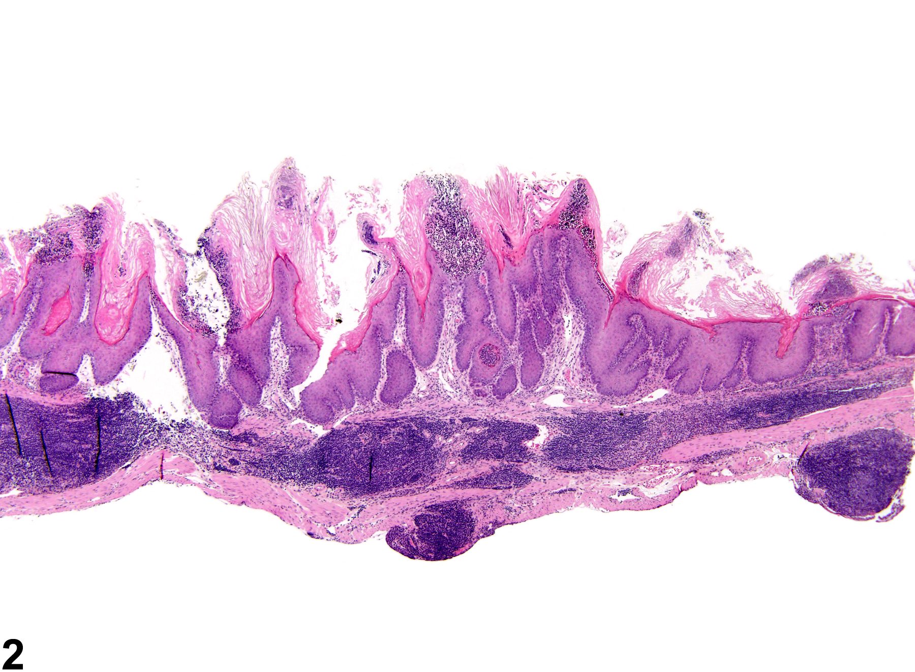
Stomach, Forestomach - Inflammation, Chronic in a female B6C3F1 mouse from a chronic study. Numerous lymphocytes are present in the submucosa and serosa, often forming nodules or follicles similar to gut-associated lymphoid tissue. The overlying epithelium is hyperplastic and contains suppurative inflammation.

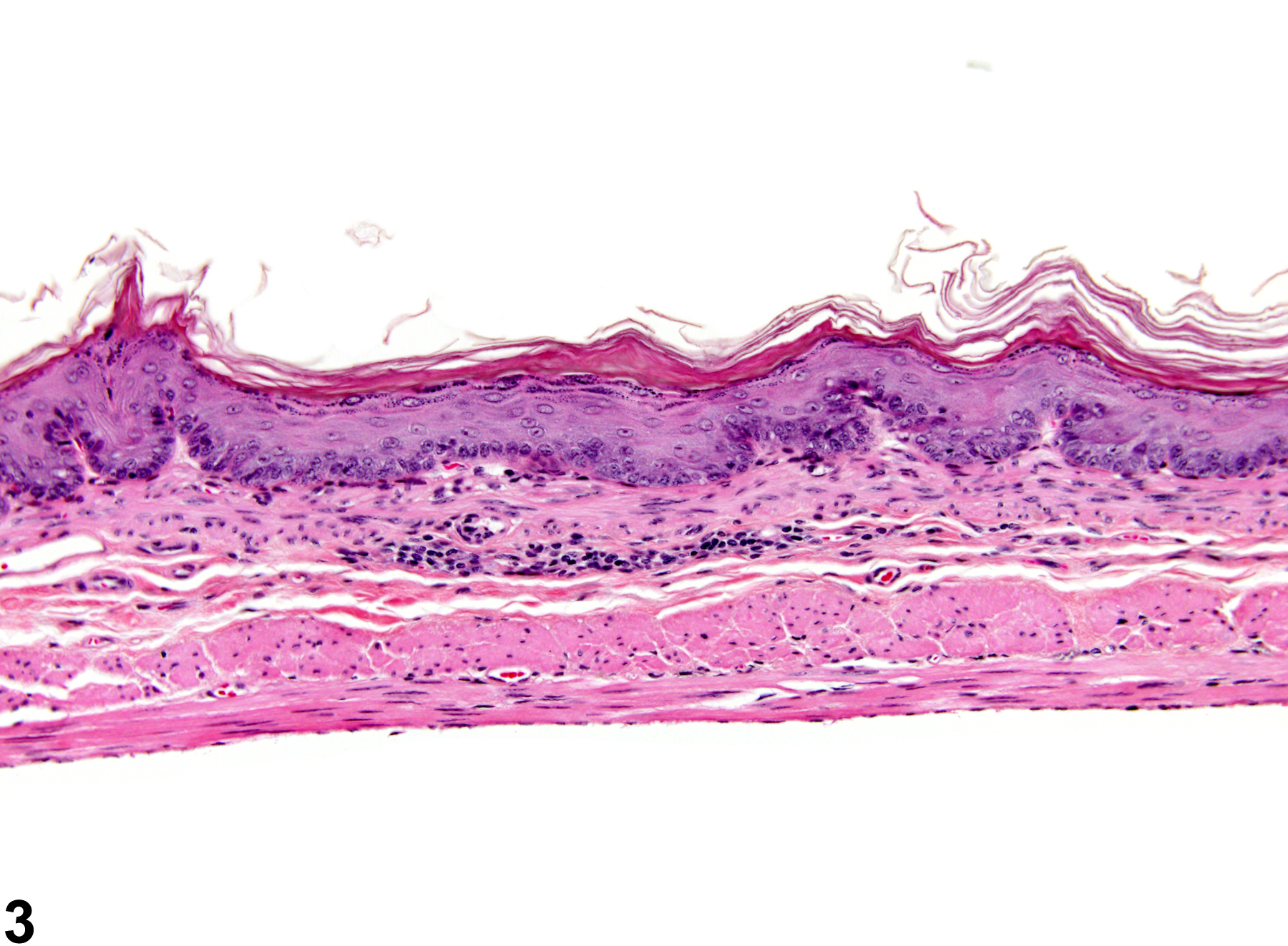
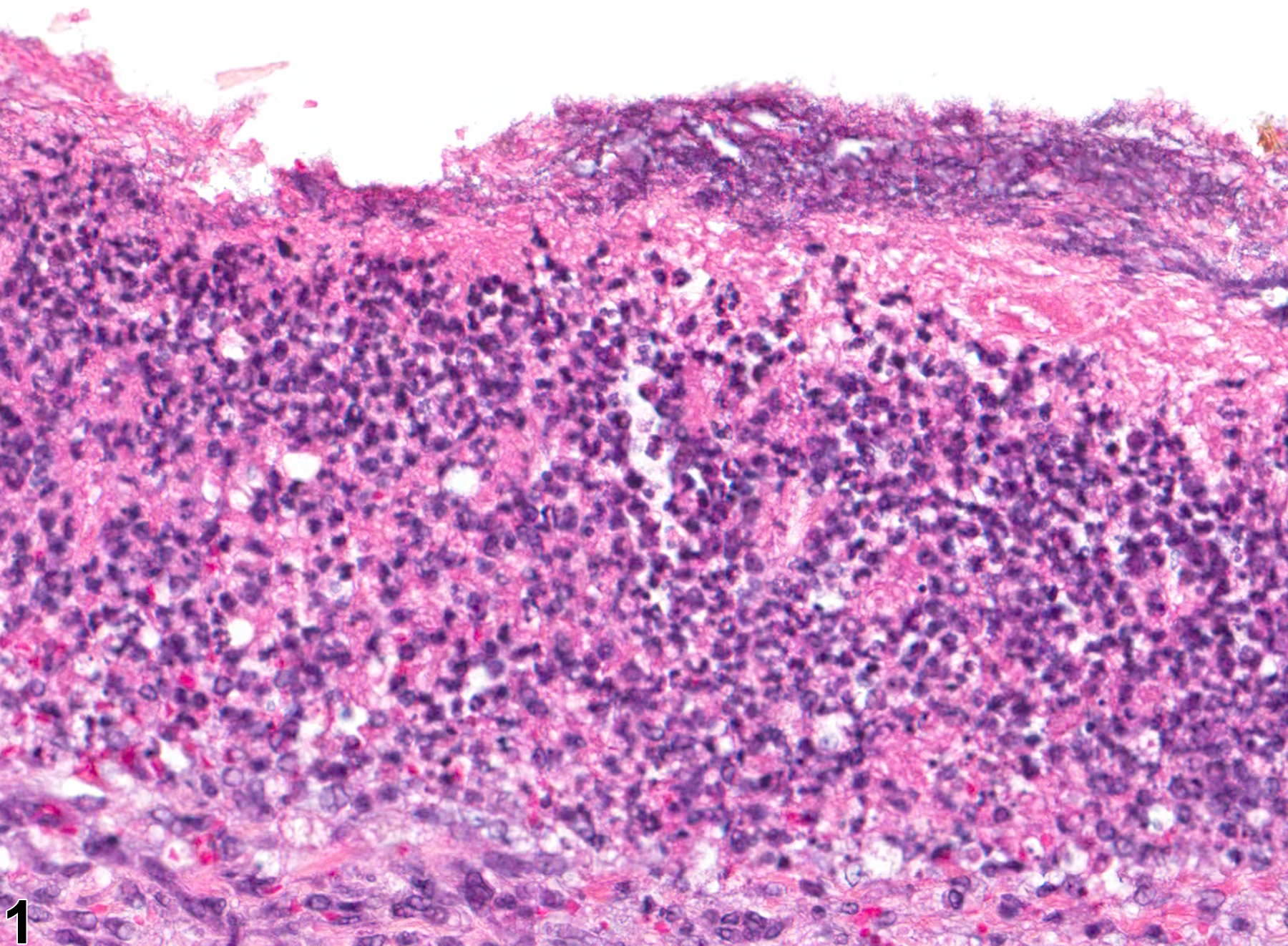
Stomach, Forestomach - Inflammation, Chronic in a female B6C3F1 mouse from a chronic study. Minimal chronic inflammation is present in the submucosa.
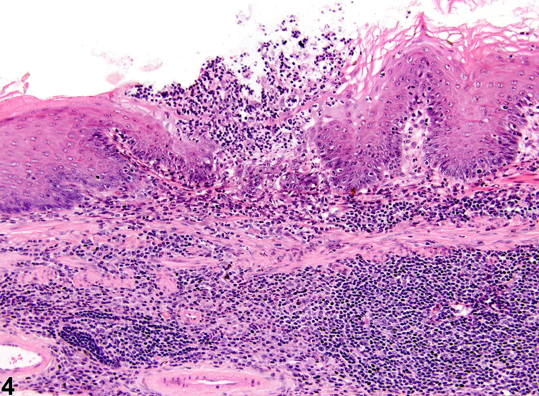
Stomach, Forestomach - Inflammation, Chronic active in a female B6C3F1 mouse from a chronic study. Numerous lymphocytes, macrophages, and neutrophils are present within the submucosa; the overlying squamous epithelium of the stomach is hyperplastic and ulcerated and contains suppurative inflammation.
Stomach, Forestomach - Inflammation, Chronic active in a male F344/N rat from a chronic study. Numerous lymphocytes, plasma cells, and neutrophils are present within the submucosa; the overlying squamous epithelium of the stomach is hyperplastic.